INTRODUCTION
Nasopharyngeal carcinoma (NPC) is the most commonly diag- nosed head and neck malignancy in Southeast Asia, especial- ly in Southern China.1,2 With the application of intensity mod-
ulated radiotherapy (IMRT), the loco-regional control of NPC has been improved dramatically.3-5 Nevertheless, distant me- tastasis (DM) remains as the main failure pattern,6-9 with the DM rate estimated at approximately 20–30%.4,5,10 Bone metas- tasis is one of most common metastatic sites of NPC.8
Several studies have reported on treatment outcomes of NPC with bone metastasis, either for patients with bone me- tastases at primary diagnosis or with recurrent bone disease after definitive chemoradiotherapy (CRT). Therein, the medi- an overall metastasis survival (OMS) time is reported to be 12–23.5 months.11-13 These data indicate that bone metastasis patients may differ greatly in terms of survival; however, meth- ods have not yet to be developed for evaluating prognoses and stratifying these patients into different risk groups before treat- ment, especially those with bone-only metastasis after defini- tive CRT.
Prognostic Evaluation of Nasopharyngeal Carcinoma with Bone-Only Metastasis after Therapy
Tianzhu Lu1,2,3*, Qiaojuan Guo1,2,3*, Xiaofei Cui1,2,3, Zhuhong Chen1,2,3, Shaojun Lin1,2,3, Luying Xu1,2,3, Jin Lin1,2,3, Jingfeng Zong1,2,3, and Jianji Pan1,2,3
1Provincial Clinical College, Fujian Medical University, Fuzhou, Fujian;
2Department of Radiation Oncology, Fujian Provincial Cancer Hospital, Fuzhou, Fujian;
3Fujian Provincial Key Laboratory of Translational Cancer Medicine, Fuzhou, Fujian, China.
Purpose: To evaluate the prognosis of nasopharyngeal carcinoma (NPC) patients who developed bone-only metastasis after pri- mary treatment and the stratification of these patients into different risk groups based on independent prognostic factors.
Materials and Methods: Eighty NPC patients who developed bone-only metastasis after definitive radiotherapy from October 2005 to December 2010 were enrolled. All these patients received palliative treatment for bone metastasis, including chemotherapy and/or radiotherapy. Clinical features, treatment modality, and laboratory parameters were examined with univariate and multi- variate analyses.
Results: The median follow-up time was 15.5 months (range, 2–67 months) for the whole cohort. The median overall metastatic survival (OMS) time and the 2-year estimate OMS rate were 26.5 months and 52%, respectively. Multivariate analysis indicated that patients with short metastases-free interval, multiple bone metastases sites, high serum lactic dehydrogenase levels, and treated with radiotherapy or chemotherapy alone had significantly worse outcomes. Patients were stratified into three different risk groups based on the number of adverse factors present. The OMS curves of the three groups were all significantly different (p<0.001).
Conclusion: Severl prognostic factors were found to be associated with worse outcomes. According to the number of adverse fac- tors present, bone-only metastasis patients can be stratified into three risk groups with significantly different prognoses. Such grouping may help in improving the design of clinical trials and in guiding individualized treatment for NPC patients with bone- only metastasis.
Key Words: Nasopharyngeal neoplasm, neoplasm metastasis, prognosis Yonsei Med J 2016 Jul;57(4):840-845
http://dx.doi.org/10.3349/ymj.2016.57.4.840 pISSN: 0513-5796 · eISSN: 1976-2437
Received: July 20, 2015 Revised: December 4, 2015 Accepted: December 9, 2015
Corresponding author: Dr. Jianji Pan, Provincial Clinical College of Fujian Medical University, Department of Radiation Oncology, Fujian Provincial Cancer Hospital, No.
420 Fuma Road, Fuzhou 350014, Fujian, China.
Tel: 86-591-83638732, Fax: 86-591-83928767, E-mail: [email protected]
*Tianzhu Lu and Qiaojuan Guo contributed equally to this work.
•The authors have no financial conflicts of interest.
© Copyright: Yonsei University College of Medicine 2016
This is an Open Access article distributed under the terms of the Creative Com- mons Attribution Non-Commercial License (http://creativecommons.org/licenses/
by-nc/3.0) which permits unrestricted non-commercial use, distribution, and repro- duction in any medium, provided the original work is properly cited.
In this retrospective study, we analyzed 80 NPC patients with bone-only metastasis after radical CRT in an attempt to investi- gate the prognostic factors affecting survival of this subgroup of patients and to stratify patients into different risk groups based on the presence of risk factors before re-treatment.
MATERIALS AND METHODS
Patients and data collection
Between October 2005 and December 2010, a total of 2139 his- tologically proven NPC patients without DM were treated by definitive radiotherapy with or without chemotherapy at our institution, either two-dimensional radiotherapy (2D-RT) or three-dimensional conformal radiotherapy/IMRT. At time of censorship, a total of 171 patients developed bone metastasis, with or without local-regional recurrence and distant failure at other sites. Among them, 80 patients with bone-only metastasis were retrospectively analyzed in this study, excluding 52 pa- tients with local-regional recurrence and/or co-existed with other types of metastases and another 39 bone metastases pa- tients who had not received any treatment.
Bone-only metastasis was defined as only bone-type metas- tasis without non-skeletal metastasis at the time of their initial diagnosis of metastatic NPC. Bone metastasis was diagnosed based on the presence of symptoms and imaging checks, in- cluding bone scan, computed tomography (CT), magnetic res- onance imaging, or positron emission tomography. All patients were restaged according to the 7th edition of American Joint Committee on Cancer. The median metastases free interval (MFI) was defined as the interval between the date of the first consultation and the date of first diagnosis with DM. Solitary bone metastasis was defined as a single site bone lesion with- in a two month period after diagnosis of bone metastasis. Mul- tiple-site bone metastasis comprised two or more sites of bone metastasis. Serum lactic dehydrogenase (S-LDH) and serum alkaline phosphatase (S-ALP) before treatment were estimat- ed employing the optimized standard method recommended by the German Society of Clinical Chemistry.14 S-LDH >245 IU/L and S-ALP >110 IU/L were considered a sign of high lev- els thereof. Hemoglobin (Hb) levels <11.0 g/dL were consid- ered to indicate anemia according to World Health Organiza- tion standards for cancer patients.15
Treatment of bone metastases
Of the 80 patients, 48 received combined chemotherapy and palliative radiation of the bone, while 10 and 22 patients under- went chemotherapy alone and palliative radiotherapy alone, respectively. Palliative radiation comprised long course radio- therapy (39 patients with 2D-RT and 21 patients with IMRT), with a median dose of radiation of 30 Gy (range 30–66 Gy).
Among patients who underwent palliative radiotherapy, most of them (40 patients) underwent 30 Gy irradiation in 10 frac-
tions; 20 patients received a dose of 36–45 Gy (2–2.5 Gy/frac- tion); and the remaining 10 patients received 46–66 Gy irradia- tion (2 Gy/fraction) for non-spine metastasis sites.
In total, 58 patients underwent platinum-based chemothera- py (range 1–8 cycles; median: 3 cycles). Forty-three patients re-
Table 1. Demographic and Clinical Characteristics of the Cohort (n=80) Characteristic No. of patients (%) Gender
Male 57 (71.2)
Female 23 (28.8)
Age at metastases (yr) Median 50.5 (range 15 to 78)
≤50 40 (50.0)
>50 40 (50.0)
Clinical stage
II 5 (6.3)
III 36 (45.0)
IV 39 (48.7)
No. of metastases sites
Single 27 (33.8)
Multiple 53 (66.2)
Bone metastasis sites
Vertebra 57 (71.3)
Non-vertebra 23 (38.7)
MFI (month)
≤12 30 (37.5)
>12 50 (62.5)
KPS
≤80 39 (48.8)
>80 41 (51.2)
Hb (g/dL)
<11.0 15 (18.8)
≥11.0 65 (81.2)
ALB (g/L)
<40 53 (66.2)
≥40 27 (33.8)
S-LDH (IU/L)
<245 50 (62.5)
≥245 30 (37.5)
S-ALP (IU/L)
<110 65 (81.2)
≥110 15 (18.8)
Treatment
Chemotherapy alone 10 (12.5)
Radiotherapy alone 22 (27.5)
Chemo-radiotherapy 48 (60.0)
Bisphosphonate
<2 cycles 45 (56.2)
≥2 cycles 35 (43.8)
MFI, metastases free interval; KPS, Karnofsky Performance Status; Hb, he- moglobin; ALB, albumin; S-LDH, serum lactic dehydrogenase; S-ALP, serum alkaline phosphatase.
ceived platinum plus gemcitabine, and the other 15 patients used platinum plus paclitaxel. In our series, 64 patients also un- derwent bisphosphonate therapy, ranging from one to six cy- cles (median: 2 cycles).
Follow-up and statistical analyses
Patients were evaluated for response every two cycles during systemic chemotherapy and then every three months until death, based on CT or isotopic bone scan. OMS was measured and calculated from the first day of diagnosis of bone metas- tasis to the date of death or final follow up. Survival data were analyzed with SPSS software, version 18.0 (SPSS Inc., Chicago, IL, USA). Survival curves were created with the Kaplan-Meier method and compared with the log-rank test. Multivariate analyses were performed to test the independent significance of potential prognostic factors by a Cox proportional hazards model. Two-tailed p-values ≤0.05 were considered statistically significant.
RESULTS
Patients characteristics
Sixty-two of the 80 patients developed bone metastasis in the first two years after diagnosis of a primary tumor. The median MFI was 15 months (ranging 3 to 66 months). Twenty-seven and 53 patients presented with solitary and multiple bone me- tastases, respectively. Other clinical characteristics of the pa- tients enrolled are listed in Table 1.
The median follow-up period after the diagnosis of bone metastasis for the entire cohort was 15.5 months (range, 2–63 months).
Survival
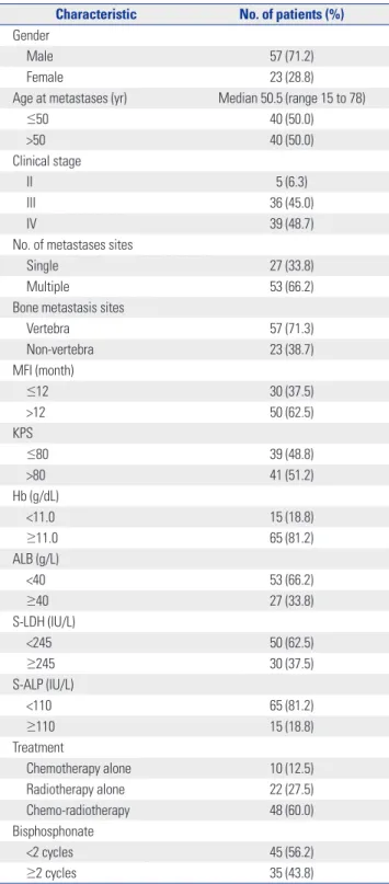
Thirty-nine patients had expired at the time of analysis, with a median OMS time and a 2-year estimate OMS rate to be- 26.5 months and 52%, respectively (Fig. 1A). In particular, patients with CRT had a median overall metastases survival of 40.0 mo- nths, which was significant longer than that of patients treated with radiotherapy alone (17.4 months) (p=0.043) or chemo- therapy alone (12.6 months) (p=0.018). However, the difference in median OMS time between patients in the chemotherapy alone group and the radiotherapy alone group showed no sta- tistical significance (p=0.570) (Fig. 1B).
Univariate and multivariate analysis
Potential prognostic factors, including patient factors [gender, age, Karnofsky Performance Status at diagnosis of bone metas- tasis], disease factors (clinical stage, number of bone metastases sites, bone metastasis sites, and MFI), laboratory factors [Hb lev- el, albumin level (ALB), S-LDH, and S-ALP] and treatment fac- tors (chemotherapy, radiotherapy, bisphosphonates) were ana- lyzed by the log-rank test, as shown in Table 2. In additiona to treatment modality, log-rank test indicated that multiple bone metastases, MFI ≤12 months, Hb <11.0 g/dL, ALB <40 g/L, and S-LDH ≥245 IU/L were associated with poorer OMS.
Multivariable analysis showed that treatment modality, MFI, number of metastases sites, and S-LDH level remained as sig- nificant predictors for OMS. However, Hb and ALB failed to en- ter the final Cox model, as shown in Table 2.
Prognostic evaluation and risk groups
In order to evaluate the prognosis of patients with bone-only metastasis, all included patients were stratified into three dif- ferent risk groups based on the presence of the independently
Fig. 1. (A) The overall metastasis survival of 80 patients who developed bone-only metastasis after definitive radiotherapy. (B) Overall metastasis sur- vival curves according to different treatment arms in patients.
1.0
0.8
0.6
0.4
0.2
0.0
1.0
0.8
0.6
0.4
0.2
0.0
Time (months) Time (months)
Chemo-radiotherapy Radiotherapy alone Chemotherapy alone Chemo-radiotherapy
Radiotherapy alone Chemotherapy alone
0 10 20 30 40 50 60 70 0 10 20 30 40 50 60 70
Overall metastasis survival Overall metastasis survival
A B
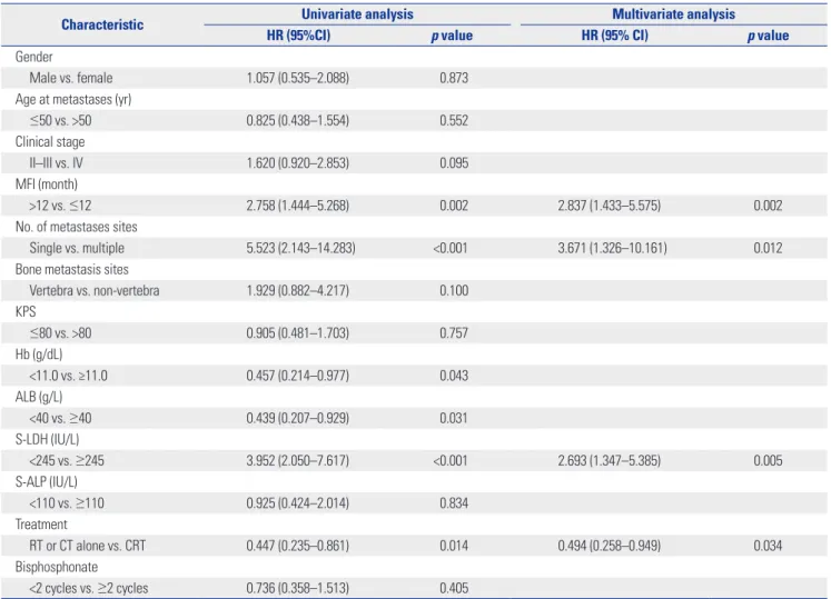
significant prognostic factors (MFI, number of metastases sites, and S-LDH level), except treatment modality, as follows: group A (without any adverse factor or with only one factor), low-risk group; group B (with two adverse prognostic factors), interme- diate-risk group; and group C (with three adverse prognostic fac- tors), high-risk group. There were 42, 26, and 12 patients in these three groups, respectively.
As shown in Fig. 2, the OMS curves for the risk groups were significantly different (p<0.001). All patients in group C had ex- pired within two years. The 2-year estimate OMS rates of group A, B, and C were 75.3%, 39.9%, and 0%, respectively, with the median OMS time of groups B and C to be 21 and 9 months, re- spectively. The median OMS time of group A could not be es- timated, because the follow-up time of this group was not long enough and most patients were still alive.
DISCUSSION
DM has heterogeneity, as different metastatic characteristics Table 2. Univariate and Multivariate Analysis of Variables for Bone-Only Metastasis Patients
Characteristic Univariate analysis Multivariate analysis
HR (95%CI) p value HR (95% CI) p value
Gender
Male vs. female 1.057 (0.535–2.088) 0.873
Age at metastases (yr)
≤50 vs. >50 0.825 (0.438–1.554) 0.552
Clinical stage
II–III vs. IV 1.620 (0.920–2.853) 0.095
MFI (month)
>12 vs. ≤12 2.758 (1.444–5.268) 0.002 2.837 (1.433–5.575) 0.002
No. of metastases sites
Single vs. multiple 5.523 (2.143–14.283) <0.001 3.671 (1.326–10.161) 0.012
Bone metastasis sites
Vertebra vs. non-vertebra 1.929 (0.882–4.217) 0.100
KPS
≤80 vs. >80 0.905 (0.481–1.703) 0.757
Hb (g/dL)
<11.0 vs. ≥11.0 0.457 (0.214–0.977) 0.043
ALB (g/L)
<40 vs. ≥40 0.439 (0.207–0.929) 0.031
S-LDH (IU/L)
<245 vs. ≥245 3.952 (2.050–7.617) <0.001 2.693 (1.347–5.385) 0.005
S-ALP (IU/L)
<110 vs. ≥110 0.925 (0.424–2.014) 0.834
Treatment
RT or CT alone vs. CRT 0.447 (0.235–0.861) 0.014 0.494 (0.258–0.949) 0.034
Bisphosphonate
<2 cycles vs. ≥2 cycles 0.736 (0.358–1.513) 0.405
HR, hazard ratio; CI, confidence interval; MFI, metastases free interval; KPS, Karnofsky Performance Status; Hb, hemoglobin; ALB, albumin; S-LDH, serum lactic dehydrogenase; S-ALP, serum alkaline phosphatase; CT, chemotherapy; RT, radiotherapy; CRT, chemo-radiotherapy.
Fig. 2. Overall metastasis survival curves according to three different risk groups.
1.0
0.8
0.6
0.4
0.2
0.0
Time (months)
Low risk group Intermediate risk group High risk group
0 10 20 30 40 50 60 70
Overall metastasis survival
show different prognoses.8 Bone metastasis is one of the pre- dominant failure sites of NPC, the treatment outcome of which is far from satisfactory.8,11-13 Our series indicated a median OMS time of 26.5 months, with a 2-year OMS rate of 52%. Multivari- ate analysis identified MFI, numbers of bone metastasis, S- LDH level, and treatment modality as significant prognostica- tors. According to the presence of three independent predicting factors, except treatment modality, patients can be well strati- fied into three different risk groups, with OMS curves signifi- cantly different from each other.
Several studies have evaluated the survival and prognostic factors of NPC patients with bone metastasis.11-13,16 A recent report from China indicated a median OMS of 12 months, in which 70 NPC patients with bone metastasis at their first visit were enrolled.12 Among them, 29 co-existed with other meta- static sites, including lung and/or liver, althgouh no further prognostic analysis were performed in this series.12 Another Chinese study by Jin, et al.11 demonstrated that patients who received zoledronic acid (ZA) combined with chemotherapy had significant longer median OMS (23.5 months) than those who received chemotherapy alone (17.5 months). In their study, a total of 307 cases were included, and bone metastasis was diagnosed either at the time of the first visit or during the fol- low-up time after definitive treatment; patients with other types of DM were included as well.11 Besides ZA, chemotherapy cy- cles, vertebral metastases, serum ALP level, and skeletal-relat- ed events were found to be significant prognostic factors for OMS in multivariate analysis.11 Recently, analysis of a relative- ly large sample of 312 NPC patients with an initial diagnosis or developing bone-only metastasis during follow-up time found CRT, number of bone metastasis, and spine metastasis to be prognostic factors. The median OMS thereof was 23.4 months.13 The only other study to evaluate the survival of NPC patients who developed bone-only metastasis after radical treatment was initiated by Cao, et al.16 in 116 cases. They indicated that CRT, age, local recurrence, subsequent metastasis, and dis- ease free interval (DFI) were independent predicting factors for OS; the median OS time was 33.3 months.16 Using a differ- ent and more objective endpoint, the present study estimated a median OMS time of 26.5 months, which was comparable to that reported by Li, et al.13 Also, the patients enrolled in our study were similar to those in Cao, et al.16 Our Cox model indi- cated that MFI (similar to DFI in Cao, et al.’s study), treatment modality, S-LDH, and numbers of metastasis sites were inde- pendent factors that significantly affected treatment outcomes.
Our series indicated that patients could benefit, in terms of survival, from combined treatment, which is consistent with the results reported by Cao, et al.16 and Li, et al.,13 who suggested that chemotherapy or radiotherapy alone is not sufficient to control metastatic tumors. We recommended that long-course radiotherapy could be better, as has been demonstrated by Hartsell, et al.17 and Howell, et al.18 Compared with single frac- tion radiotherapy, long-course radiotherapy could reduce re-
treatment rates. In our cohort, at least one cycle of bisphospho- nate was used in about 80% patients, although no survival benefit was indicated. This result concurred closely with that noted in Li, et al.13 However, Jin, et al.11 found that bisphospho- nate could improve OMS in NPC patients with bone metasta- sis. A possible reason for this inconsistency may be associated with different immune microenvironments.19 Zhang, et al.20 showed that T-cell deficiency reduces the antitumor effects of bisphosphonate, compared with immune-competent mice, in animals with bone metastases.
As have been reported in other studies,13,21,22 we also found that multiple-bone metastases was an adverse prognostic fac- tor of survival. Elevated S-LDH is frequently observed in cancer patients, which could be attributed to the release of enzymes from malignant cells.23 Patients with abnormally elevated S- LDH had significantly worse treatment outcome than those with normal S-LDH level. Other investigators have also identi- fied that elevated S-LDH is associated with poor prognosis in metastatic and loco-regionally advanced NPC.24,25 Another in- dependent predicting factor was MFI. Patients with MFI ≤12 months were associated with poor survival, and similar results have also been presented in other reports.8,16,26 The possible reason could lie in emerging chemotherapy or radiotherapy re- sistant clones within the tumor of those patients who devel- oped DM in a short time after treatment.
In order to evaluate the prognosis of patients with bone-on- ly metastasis, these patients were stratified into three different risk groups, with the 2-year estimate OMS rates for low, inter- mediate, and high risk groupto at 75.3%, 39.9%, and 0% (p<
0.001). All 12 patients in the high risk group were deceased with in the first two years. Among them, seven received CRT; pa- tients who underwent CRT showed relative higher OMS than those who received chemotherapy or radiotherapy alone, al- though the difference showed no statistical significance (data no shown). Considering the unfortunate results of the high-risk group, management should aim to improve end-stage quality of life, and more efficient systematic treatment (i.e., immune treatment) may need to be introduced and evaluated in multi- center studies. For those in the low and intermediate risk groups, more aggressive treatment (i.e., combined CRT) should be con- sidered.
Several limitations should be addressed for our series. First is the retrospective nature of the study. Secondly, our results were concluded from a relatively small sample from a single institu- tion (80 cases); however, they were derived from 2139 patients treated over 2005 and 2010. Accordingly, our results should be validated in a relatively large group of patients collected from multiple centers. Thirdly, the modes of chemotherapy and ra- diotherapy used in our series varied, which might have had a confounding effect. Finally, the present study did not report the incidence of skeletal-related events, as not all the patients had regular follow-up and it was difficult to obtain such information in patients who had died at the time of censorship.
Our results indicated that multiple bone metastases, short MFI, high level S-LDH, and RT or CT alone are associated with short metastasis survival. Different prognostic factors were as- sociated with different outcomes for patients who developed bone-only metastasis after primary treatment. Grouping pa- tients according to the presence of these risk factors could well distinguish patients with different outcomes. Considering it was convenient and efficient, clinicians could use the number of adverse prognostic factors present to evaluate the prognosis of these patients. From both a therapeutic and research point of view, our prognostic grouping may be helpful in improving the design of clinical trials involving NPC patients with bone- only metastasis and in guiding individualized treatment.
ACKNOWLEDGEMENTS
This work was sponsored by National Clinical Key Specialty Construction Program and Key Clinical Specialty Discipline Construction Program of Fujian, People’s Republic of China.
This research was also supported by a grant from the National Natural Science Foundation of China (grant No. 81341108).
REFERENCES
1. Wei WI, Sham JS. Nasopharyngeal carcinoma. Lancet 2005;365:
2041-54.
2. Jemal A, Bray F, Center MM, Ferlay J, Ward E, Forman D. Global cancer statistics. CA Cancer J Clin 2011;61:69-90.
3. Lai SZ, Li WF, Chen L, Luo W, Chen YY, Liu LZ, et al. How does in- tensity-modulated radiotherapy versus conventional two-dimen- sional radiotherapy influence the treatment results in nasopharyn- geal carcinoma patients? Int J Radiat Oncol Biol Phys 2011;80:661-8.
4. Peng G, Wang T, Yang KY, Zhang S, Zhang T, Li Q, et al. A prospec- tive, randomized study comparing outcomes and toxicities of in- tensity-modulated radiotherapy vs. conventional two-dimensional radiotherapy for the treatment of nasopharyngeal carcinoma. Ra- diother Oncol 2012;104:286-93.
5. Lin S, Pan J, Han L, Guo Q, Hu C, Zong J, et al. Update report of na- sopharyngeal carcinoma treated with reduced-volume intensity- modulated radiation therapy and hypothesis of the optimal mar- gin. Radiother Oncol 2014;110:385-9.
6. Wang TJ, Riaz N, Cheng S, Lu JJ, Lee NY. Intensity-modulated radi- ation therapy for nasopharyngeal carcinoma: a review. J Radiat Oncol 2012;1:129-46.
7. Cao CN, Luo JW, Gao L, Yi JL, Huang XD, Wang K, et al. Update re- port of T4 classification nasopharyngeal carcinoma after intensity- modulated radiotherapy: an analysis of survival and treatment tox- icities. Oral Oncol 2015;51:190-4.
8. Teo PM, Kwan WH, Lee WY, Leung SF, Johnson PJ. Prognostica- tors determining survival subsequent to distant metastasis from nasopharyngeal carcinoma. Cancer 1996;77:2423-31.
9. Hui EP, Leung SF, Au JS, Zee B, Tung S, Chua D, et al. Lung metasta- sis alone in nasopharyngeal carcinoma: a relatively favorable prog- nostic group. A study by the Hong Kong Nasopharyngeal Carcino- ma Study Group. Cancer 2004;101:300-6.
10. Lee N, Xia P, Quivey JM, Sultanem K, Poon I, Akazawa C, et al. In- tensity-modulated radiotherapy in the treatment of nasopharyn- geal carcinoma: an update of the UCSF experience. Int J Radiat Oncol Biol Phys 2002;53:12-22.
11. Jin Y, An X, Cai YC, Cao Y, Cai XY, Xia Q, et al. Zoledronic acid combined with chemotherapy bring survival benefits to patients with bone metastases from nasopharyngeal carcinoma. J Cancer Res Clin Oncol 2011;137:1545-51.
12. Zhao CL, Qian GQ, Chen XY, Chen C. Retrograde analysis of clini- cal characteristics of bone metastasis in 1,031 cases of preliminarily diagnosed nasopharyngeal carcinoma. Asian Pac J Cancer Prev 2014;15:3785-8.
13. Li AC, Xiao WW, Wang L, Shen GZ, Xu AA, Cao YQ, et al. Risk fac- tors and prediction-score model for distant metastasis in naso- pharyngeal carcinoma treated with intensity-modulated radio- therapy. Tumour Biol 2015;36:8349-57.
14. Provisional recommendations on IFCC methods for the measure- ment of catalytic concentrations of enzymes. Part 2. IFCC method for aspartate aminotransferase. J Clin Chem Clin Biochem 1977;15:
39-51.
15. Groopman JE, Itri LM. Chemotherapy-induced anemia in adults:
incidence and treatment. J Natl Cancer Inst 1999;91:1616-34.
16. Cao X, Han Y, He L, Xiang J, Wen Z. Risk subset of the survival for nasopharyngeal carcinoma patients with bone metastases: who will benefit from combined treatment? Oral Oncol 2011;47:747-52.
17. Hartsell WF, Scott CB, Bruner DW, Scarantino CW, Ivker RA, Roach M 3rd, et al. Randomized trial of short- versus long-course radio- therapy for palliation of painful bone metastases. J Natl Cancer Inst 2005;97:798-804.
18. Howell DD, James JL, Hartsell WF, Suntharalingam M, Machtay M, Suh JH, et al. Single-fraction radiotherapy versus multifraction ra- diotherapy for palliation of painful vertebral bone metastases- equivalent efficacy, less toxicity, more convenient: a subset analysis of Radiation Therapy Oncology Group trial 97-14. Cancer 2013;119:
888-96.
19. Capietto AH, Faccio R. Immune regulation of bone metastasis.
Bonekey Rep 2014;3:600.
20. Zhang K, Kim S, Cremasco V, Hirbe AC, Collins L, Piwnica-Worms D, et al. CD8+ T cells regulate bone tumor burden independent of osteoclast resorption. Cancer Res 2011;71:4799-808.
21. Koizumi M, Yoshimoto M, Kasumi F, Ogata E. Comparison be- tween solitary and multiple skeletal metastatic lesions of breast cancer patients. Ann Oncol 2003;14:1234-40.
22. Ahn SG, Lee HM, Cho SH, Lee SA, Hwang SH, Jeong J, et al. Prog- nostic factors for patients with bone-only metastasis in breast can- cer. Yonsei Med J 2013;54:1168-77.
23. Schwartz MK. Lactic dehydrogenase. An old enzyme reborn as a cancer marker? Am J Clin Pathol 1991;96:441-3.
24. Turen S, Ozyar E, Altundag K, Gullu I, Atahan IL. Serum lactate de- hydrogenase level is a prognostic factor in patients with locoregion- ally advanced nasopharyngeal carcinoma treated with chemora- diotherapy. Cancer Invest 2007;25:315-21.
25. Jin Y, Ye X, Shao L, Lin BC, He CX, Zhang BB, et al. Serum lactic dehydrogenase strongly predicts survival in metastatic nasopha- ryngeal carcinoma treated with palliative chemotherapy. Eur J Cancer 2013;49:1619-26.
26. Toh CK, Heng D, Ong YK, Leong SS, Wee J, Tan EH. Validation of a new prognostic index score for disseminated nasopharyngeal car- cinoma. Br J Cancer 2005;92:1382-7.