Lab Anim Res 2016: 32(4), 217-223 https://doi.org/10.5625/lar.2016.32.4.217
ISSN 2233-7660 (Online)
Antioxidant and hepatoprotective effects of fermented red ginseng against high fat diet-induced hyperlipidemia in rats
Myeong-Hwan Kim
#, Eun-Jin Lee
#, Jeong-Mu Cheon, Ki-Jun Nam, Tae-Ho Oh, Kil-Soo Kim*
College of Veterinary Medicine, Kyungpook National University, Daegu, 41566, Korea
This study was performed to investigate the antioxidant and hepatoprotective effects of fermented red ginseng (Panax ginseng C.A. Meyer; FRG) on high-fat diet-induced hyperlipidemia in rats. Sprague-Dawley rats were divided into four groups of seven: normal control, NC; high-fat diet control, HFC; high-fat diet–
0.5% FRG, HF-FRGL; and high-fat diet–1% FRG, HF-FRGH. All rats were fed a high-fat diet for eight weeks, except those in the NC group, while rats in the FRG treatment groups received drinking water containing 0.5% or 1% FRG. After eight weeks of treatment, levels of alanine aminotransferase (ALT), aspartate aminotransferase (AST), total cholesterol (TC), triglycerides (TG), low-density lipoprotein-cholesterol (LDL-C), and high-density lipoprotein-cholesterol (HDL-C) in the serum were measured. The concentration of the oxidative stress marker malondialdehyde (MDA), and activity of antioxidant enzymes including superoxide dismutase (SOD), catalase (CAT), and glutathione peroxidase (GSH-Px) in rat liver were evaluated. Histological analysis of the liver was performed using hematoxylin and eosin. The high-fat diet markedly increased serum levels of ALT, AST, TC, TG, and LDL-C and hepatic MDA levels, while administration of FRG to the hyperlipidemic rats resulted in a significant decline in the levels of these parameters. Furthermore, the decline in the levels of serum HDL-C and hepatic SOD, CAT, and GSH-Px induced by the high-fat diet was attenuated by FRG treatment. In addition, histopathological analysis of liver sections suggested that FRG treatment also provided protection against liver damage. These results suggested that FRG improved lipid profiles, inhibited lipid peroxidation, and played a protective role against liver injury in hyperlipidemic rats.
Keywords: Fermented red ginseng, antioxidants, hepatoprotective effects, high fat diet, hyperlipidemia Received 5 September 2016; Revised version received 28 November 2016; Accepted 30 November 2016
Hyperlipidemia and oxidative stress are known risk factors for the development of various diseases such as cancer, heart disease, atherosclerosis, digestive disease, autoimmune disease, and aging [1]. Reactive oxygen species (ROS), which are oxygen-containing compounds produced by general metabolic pathways, are known to induce oxidative damage to proteins, lipids, and DNA [2,3]. Yang et al. [4] suggested that appropriate therapy of increasing antioxidant intake in subjects with abnormally elevated lipid levels can attenuate the course of the disease. However, currently available pharmacological
agents for metabolic disorders have a number of limitations, such as various adverse side effects and high rates of secondary failure [5]. Hence, biomedical researchers have been interested in discovering new agents including medicinal herbs that are capable of reducing serum lipid levels.
Plant products are generally considered to be less toxic and thus, cause fewer side effects than drugs manufactured by chemical synthesis. The potential therapeutic and preventive benefits of plant-based medications have been the subject of extensive studies, and many natural
#
These authors contributed equally to this work.
*Corresponding author: Kil-Soo Kim, College of Veterinary Medicine, Kyungpook National University, 80 Daehakro, Buk-gu, Daegu 41566, Korea
Tel: +82-53-950-7792; Fax: +82-53-950-5955; E-mail: [email protected]
This is an Open Access article distributed under the terms of the Creative Commons Attribution Non-Commercial License (http://creativecommons.org/licenses/
by-nc/3.0) which permits unrestricted non-commercial use, distribution, and reproduction in any medium, provided the original work is properly cited.
constituents have been discovered possessing significant antiglycemic, hypolipidemic, and antihypertensive properties [6,7].
Ginseng (root of Panax ginseng C.A. Meyer, family Araliaceae) is one of the most commonly used health products or natural remedy medicinal herbs in Asian countries [8]. Red ginseng (RG) is produced by steam processing P. ginseng to enhance its biological activities [9]. It has been reported that wild ginseng alleviates metabolic disorders [10]. Moreover, it can be further processed into fermented red ginseng (FRG) by treating with edible microorganisms and enzymes that increase the saponin content to maximize its efficacy [11].
Previous reports have shown that FRG reduced blood glucose levels in streptozotocin-induced diabetic rats [12,13]. Furthermore, Korean red ginseng fermented using Monascus purpureus (FRG) exhibited hepato- protective, hypolipidemic, and antioxidative activities in rats [14]. Although many studies have examined the properties of ginseng, no research has been conducted to evaluate the protective effects of FRG against liver injury in hyperlipidemic rats. Therefore, this study is performed to investigate the effects and possible action mechanisms of aqueous extract of FRG in the liver of rats with hyperlipidemia.
Materials and Methods
Preparation of fermented Korean red ginseng
Fermented Korean red ginseng was prepared using the following procedure: 300 g of six-year-old white dried Korean ginseng (P. ginseng Meyer) provided by Samkwang Company (Daejeon, Korea) was immersed in 2 L of distilled water, and steamed for 24 h. A further 4 L of distilled water was added, and the ginseng solution was steamed at 90
oC for 48 h to produce red ginseng (RG) extracts. The RG extracts were used to make FRG extracts by the addition of red yeast rice (M. purpureus, KCCM12002) and fermentation for 12 h at 40
oC. The FRG was stored 20
oC until further use. The voucher specimen was deposited at the veterinary toxicology laboratory of Kyungpook National University.
Animals
Twenty-eight adult male Sprague-Dawley rats (eight weeks of age) were obtained from Koatech Co. (Osan, Korea). All rats were provided free access to standard rodent chow (AIN-76A diet, # 100000; Dyets Inc.,
Bethlehem, PA, USA) and filtered tap water, and were housed in an air-controlled room under standard conditions (temperature, 22±2
oC; relative humidity, 50±
10%; 12 h light/dark cycle) throughout the study. All animal experiments were performed in accordance with the Guidelines for Animal Care and Use of Kyungpook National University.
Experimental design
After one week of acclimatization, the rats were randomly divided into four dietary groups (n=7 in each group). Rats in the normal control (NC) group received regular rodent chow, while those in the other three groups were fed with a high-fat diet (45% kcal fat, Research Diets, New Brunswick, NJ) for eight weeks.
Group I (NC) and Group II (high-fat diet control; HFC) animals received filtered tap water and served as the normal and hyperlipidemic control group, respectively, whereas Group III (HF-FRGL) and Group IV (HF- FRGH) animals received drinking water containing 0.5 and 1% FRG for eight weeks, respectively. The rats were allowed free access to food and water during the experimental period. Their weight was measured once a week and diet intakes were calculated by subtracting the remainder from the supply. The gained weight during the experimental period was divided by cumulative dietary intake to obtain the food efficiency ratio (FER); the formula for FER is as follows: FER=weight gained (g)/
diet intake (g) ×100. At the end of the experiments, all rats were anesthetized by ethyl ether after fasting for 12 h. Blood samples were collected via the posterior vena cava and centrifuged at 1,000 ×g for 15 min at 4
oC.
After blood sampling, the liver was dissected and rinsed in physiological saline, blotted on filter paper, and weighed. Serum and liver samples were frozen at −80
oC, and were used to measure the biochemical parameters.
Measurement of blood biochemical parameters and lipid profiles
Serum levels of alanine aminotransferase (ALT),
aspartate aminotransferase (AST), total cholesterol (TC),
triglyceride (TG), low-density lipoprotein-cholesterol
(LDL-C), and high-density lipoprotein-cholesterol
(HDL-C) were measured using commercial kits (Asan
Pharmaceutical Co., Seoul, Korea) with an automatic
chemistry analyzer (Hitachi 7180; Hitachi, Tokyo,
Japan).
Measurement of hepatic malondialdehyde and antioxidant enzymes
Liver samples were perfused with cold normal saline to completely remove all blood cells, cut into small pieces, placed in 0.2 M phosphate buffer (pH 7.4), and homogenized to obtain 20% homogenate. The homogenate was centrifuged at 3,000 rpm for 15 min. Malondi- aldehyde (MDA) levels, an index of lipid peroxidation, were determined by measuring thiobarbituric acid reactive substances (TBARS) according to the modified method of Draper and Hadley [15]. In addition, superoxide dismutase (SOD), catalase (CAT), and glutathione peroxidase (GSH-Px) activities were determined using the OxiSelect
TMkit from Cell Biolabs, Inc. (San Diego, CA, USA).
Histological analysis
Liver tissues were removed, fixed with 10% neutral formalin duffer, and embedded in paraffin. The paraffin block samples were cut into 5 µm sections and stained with hematoxylin and eosin (H&E) for histological observation using a light microscope (Olympus BX50;
Olympus, Tokyo, Japan).
Statistical analysis
The results are presented as the mean±standard deviation (SD). Data were analyzed by SAS software (version 9.3; SAS Institute Inc., Cary, NC, USA).
Statistically significance differences between the control and FRG-treated groups were analyzed by Student’s t- test and analysis of variance (ANOVA). P values <0.05 were considered statistically significant.
Results
Effects of FRG on body weight gain and FER
The body weight gain and FER of rats after eight weeks are presented in Table 1. To determine the effect of FRG on diet efficiency, weight gains in animals were monitored weekly. The body weight gain of rats in the HFC group was higher than that in the NC group (P<0.05). However, FRG-treated animals (0.5 and 1.0%) exhibited lower body weight gain compared to the animals in the HFC group. In addition, the observed change in body weight coincided with a decrease in FER.
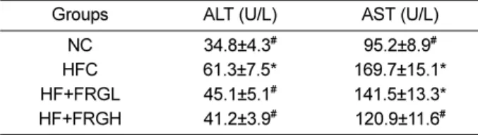
Effects of FRG on serum ALT and AST levels
Changes in serum levels of ALT and AST of each group are presented in Table 2. The serum levels of ALT and AST in the HFC group increased significantly compared to those in the NC group. On the other hand, serum ALT and AST levels were significantly lower in the FRG-treated groups than the HFC group. Although serum AST level in the FRGL group tended to be lower than in the HFC group, the differences were not significant.
Effects of FRG on serum lipid profiles
Figure 1 shows the effects of FRG on the levels of serum TC, TG, LDL-C, and HDL-C in all experimental groups. Serum TC, LDL-C, and TG levels in the HFC group were significantly (P<0.05) higher than the NC group, while the opposite was observed for serum HDL- C level. Treatment of hyperlipidemic rats with FRG resulted in a significant decline in serum levels of TC, LDL-C, and TG, with an increase in serum HDL-C level.
Serum TC levels were 57.3 and 54.7% lower in the FRGL and FRGH groups, respectively, compared to
Table 1. The body weight gain and food efficiency ratio in rats treated with FRG
Group Body weight gain (g/8 weeks)
Food efficiency ratio (%)
NC 190.5±17.3
#0,21±0.02
#HFC 231.6±16.7* 0.28±0.02*
HF+FRGL 201.2±17.1
#0.24±0.02
#HF+FRGH 194.1±16.2
#0.23±0.02
#All values are mean SD of 7 rats for each group.
NC: normal control. HFC: high fat diet control, HF+FRGL: high fat diet+FRG 0.5%, HF+FRGH: high fat diet+FRG 1.0%
Food efficiency ratio; body weight gain/food intake.
*Mean values were significantly different from NC group: P<0.05.
#
Mean values were significantly different from HFC group: P<0.05.
Table 2. The levels of serum ALT and AST in rats treated with FRG
Groups ALT (U/L) AST (U/L)
NC 34.8±4.3
#95.2±8.9
#HFC 61.3±7.5* 169.7±15.1*
HF+FRGL 45.1±5.1
#141.5±13.3*
HF+FRGH 41.2±3.9
#120.9±11.6
#All values are mean SD of 7 rats for each group.
NC: normal control. HFC: high fat diet control, HF+FRGL: high fat diet+FRG 0.5%, HF+FRGH: high fat diet+FRG 1.0%
ALT: alanine aminotransferase, AST: aspartate aminotransferase
*Mean values were significantly different from NC group: P<0.05.
#
Mean values were significantly different from HFC group: P<0.05.
those in the HFC group. Furthermore, serum TG level of the FRGH group was 82.4% lower (P<0.05) than that of the HFC group. The TG levels in the FRGL group was also shown to be reduced by 89.4%, but was not statistically significant. Additionally, the serum LDL-C level was 76.6 and 65.0% lower (P<0.05) in the FRGL and FRGH groups, respectively, compared to the HFC group. In contrast, serum HDL-C levels increased significantly by 133.9 and 178.2% (P<0.05) in the FRGL and FRGH groups, respectively, compared to those in the HFC group.
Effects of FRG on hepatic lipid peroxidation profile Figure 2 shows the effects of FRG on lipid peroxide levels in hepatic tissue. The amount of MDA in the HFC group was 0.98±0.09 nmole/mg protein, which was 169.0% higher than that in the NC group. Treatment with FRGL and FRGH significantly reduced MDA levels compared to the HFC group by 14.3 and 27.6%, respectively.
Effects of FRG on hepatic antioxidant enzyme activities The antioxidant activities of hepatic SOD, CAT, and GSH-Px are shown in Table 3. The activities of SOD, CAT, and GSH-Px in the HFC group decreased significantly (P<0.05) compared to the NC group by 57.1, 59.5, and 67.6%, respectively. However, FRGL treatment led to a significant increase (P<0.05) in the
activities of SOD (64.3%) and CAT (86.3%) compared to the HFC group. The FRGL treatment also increased the activity of GSH-Px relative to that of the HFC group, but not significantly. On the other hand, activities of hepatic SOD, CAT, and GSH-Px increased significantly (P<0.05) upon FRGH treatment compared to those of the HFC group.
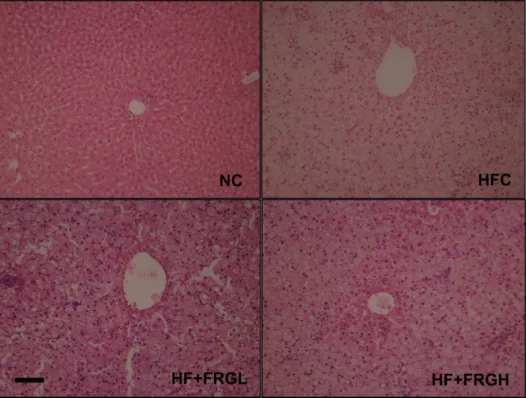
Histopathologic analysis of liver sections
Liver sections of rats in the NC group showed normal distinct hepatic cells, sinusoidal spaces, and a central vein. However, liver sections of the HFC group showed presence of extensive lipid-droplet vacuoles in hepatocytes, disarrangement of hepatic cells, necrotic changes of central lobes, and hepatic lobule damage. In the FRG-
Figure 2. Alterations in hepatic malondialdehyde (MDA) level in rats treated with FRG. Data shown are mean±S.E.M. (n=5).
*Mean values were significantly different from NC group:
P<0.05.
#Mean values were significantly different from HFC group: P<0.05.
Table 3. The hepatic antioxidant enzyme activities in hyperlipidemic rats treated with FRG
Groups SOD
(U/mg protein)
CAT (U/mg protein)
GSH-Px (U) NC 124.7±14.3
#39.5±4.2
#118.5±10.2
#HFC 71.2±7.0* 23.5±2.4* 80.1±8.5*
HF+FRGL 80.2±7.8
#34.1±3.5
#89.6±9.1*
HF+FRGH 89.4±8.2
#36.2±3.8
#103.4±9.8
#All values are mean SD of 7 rats for each group.
NC: normal control. HFC: high fat diet control, HF+FRGL: high fat diet+FRG 0.5%, HF+FRGH: high fat diet+FRG 1.0%
ALT: alanine aminotransferase, AST: aspartate aminotransferase, GLU: glucose
*Mean values were significantly different from NC group: P<0.05.
#