Yonsei Med J http://www.eymj.org Volume 54 Number 6 November 2013 1538
A Case of Cerebellar Infarction Caused by Acute Subclavian Thrombus Following Minor Trauma
Hyeyoung Park,
1,2Hee-Jin Kim,
3Myoung-Jin Cha,
4Jong Yun Lee,
1Im-Seok Koh,
1and Hyo Suk Nam
51Department of Neurology, National Medical Center, Seoul;
2Department of Medicine, The Graduate School, Yonsei University, Seoul;
3Department of Neurology, Samsung Medical Center, Sungkyunkwan University, Seoul;
4Department of Neurology, Samsung Changwon Hospital, Sungkyunkwan University School of Medicine, Changwon;
5Department of Neurology, Yonsei University College of Medicine, Seoul, Korea.
Received: October 30, 2012 Revised: November 28, 2012 Accepted: November 29, 2012
Corresponding author: Dr. Hyo Suk Nam, Department of Neurology,
Yonsei University College of Medicine, 50 Yonsei-ro, Seodaemun-gu, Seoul 120-752, Korea.
Tel: 82-2-2228-1617, Fax: 82-2-393-0705 E-mail: [email protected]
∙ The authors have no financial conflicts of interest.
© Copyright:
Yonsei University College of Medicine 2013 This is an Open Access article distributed under the terms of the Creative Commons Attribution Non- Commercial License (http://creativecommons.org/
licenses/by-nc/3.0) which permits unrestricted non- commercial use, distribution, and reproduction in any medium, provided the original work is properly cited.
Subclavian steal syndrome caused by an acute thrombus is very rare. We present a case of cerebellar infarction with proximal subclavian artery thrombosis. A 56-year-old woman was admitted for sudden vertigo. One day prior to admission, she received a shoulder massage comprised of chiropractic manipulation. On ex- amination, her left hand was pale and radial pulses were absent. Blood pressure was weak in the left arm. Downbeat nystagmus and a right falling tendency were observed. Brain MRI showed multiple acute infarctions in the left cerebellum. The findings of Doppler ultrasonography in the left vertebral artery were compatible with a partial subclavian artery steal phenomenon. Digital subtraction angiography demonstrated a large thrombus in the left subclavian artery. After heparin infusion, thrombus size markedly decreased. Cerebellar infarction caused by acute subclavi- an thrombosis following minor trauma is rare, but the thrombus can be successful- ly resolved with anticoagulation.
Key Words: Subclavian steal syndrome, brain infarction, thrombosis
INTRODUCTION
Subclavian steal syndrome is most commonly caused by atherosclerosis. Howev- er, an acute thrombus and trauma can cause this syndrome in very rare cases.1,2 Here, we report a case of acute cerebellar infarction presented with subclavian steal, developed by the thrombus in the subclavian artery.
CASE REPORT
A 56-year-old woman was admitted to the emergency room for sudden onset verti- go and right sway on awakening. She had received an intense chiropractic manip- ulation of her shoulders the day before admission. Her medical history included
Case Report
http://dx.doi.org/10.3349/ymj.2013.54.6.1538pISSN: 0513-5796, eISSN: 1976-2437 Yonsei Med J 54(6):1538-1541, 2013
Cerebellar Infarction by Subclavian Thrombus
Yonsei Med J http://www.eymj.org Volume 54 Number 6 November 2013 1539
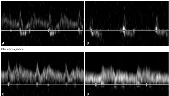
the left posterior inferior cerebellar artery (Fig. 1B). Dop- pler ultrasonography of the left vertebral artery showed bidi- rectional wave. Marked mid-systolic deceleration was seen with a sharp peak immediately before deceleration and re- turn of forward direction during diastole. A provocative hy- peremic cuff test (occlusive inflation of a blood pressure cuff for three minutes with subsequent release) of the left arm aggravated reverse flow. Therefore, these findings were com- patible with a partial subclavian artery steal phenomenon (Fig. 2A and B). Digital subtraction angiography (DSA) showed a large thrombus in the proximal left subclavian ar- tery as well as retrograde flow through the left vertebral ar- diabetes mellitus, which had been managed for six years.
On examination, blood pressure was 137/71 mmHg and 113/56 mmHg in the right and left arms, respectively. Her left hand was pale and cold with an absent radial pulse on the left (Fig. 1A). An audible subclavian bruit was present on the left side. Downbeat nystagmus and a right falling tendency during tandem gait were observed. No motor and sensory deficits were shown. No musculoskeletal abnor- malities that could have been implicated in compression of the subclavian artery were evident. Diffusion-weighted MRI revealed multiple acute infarctions in the left cerebellar hemi- sphere and vermis, compatible with the medial territory of
Fig. 1. (A) The left hand of the patient was pale and cold. (B) Acute infarctions in the left cerebellar hemisphere and vermis are shown in diffusion-weighted MRI.
Fig. 2. (A) Initial Doppler ultrasonography of the left vertebral artery shows bidirectional wave. (B) A provocative hyperemic cuff test on the left arm aggra- vates flow reversal, which is compatible with a partial subclavian steal syndrome. (C) After anticoagulation, follow-up Doppler ultrasonography shows that a systolic deceleration is still seen, but there is no retrograde flow. (D) No augmented flow reversal is noted in the hyperemic cuff test, which is compatible with a pre-steal phenomenon.
A
C A
B
D B
Before anticoagulation
After anticoagulation
Hyeyoung Park, et al.
Yonsei Med J http://www.eymj.org Volume 54 Number 6 November 2013 1540
ymptomatic. Reported prevalence of subclavian steal syn- drome is between 0.6% and 6.4%.4,5 Because of the abun- dant collateral blood supply to the head, neck and shoulder, neurological symptoms are infrequent. Among the patients with subclavian artery steal syndrome, only 5.3% suffer neurological symptoms.6
In this patient, the mechanism of stroke might be an em- bolic occlusion of the medial territory of the posterior inferior cerebellar artery because these lesions do not correspond to border zone cerebellar infarction.7 Moreover, initial Doppler ultrasonography showed only a partial steal, which would be consistent with an embolism from a thrombus in the left sub- clavian artery that caused posterior circulation infarction.
Nevertheless, a hemodynamic mechanism due to the sub- clavian steal could not completely be ruled out.
The question of why there is thrombus in the subclavian artery is uncertain. Our patient did not have a history of co- agulopathy and there was no laboratory evidence of a coag- ulation disorder either. One day prior to the stroke event, the patient had chiropractic manipulation involving pulling her shoulders back forcefully. According to the Virchow triad, formation of a thrombus depends on constituents of the blood, vessel, and hemorrheology. The finding of a narrower inter-scalene triangle in the left on neck CT suggests an un- favorable anatomical structure for which repetitive strong posturing may induce thrombus formation. Our patient did not have a vertebral artery dissection, this can occurr after chiropractic maneuvering, which can also induce posterior circulation ischemia.8
It is noteworthy that serial Doppler ultrasonographies were useful for monitoring thrombus resolution in the sub- tery. No vertebral artery dissection was observed (Fig. 3A).
Neck CT showed a narrowed space between the anterior and lateral scalene muscles in the left side, more so than on the right. Blood tests for coagulation abnormalities, including protein C, S, lupus anticoagulant, and anti-cardiolipin anti- body, were normal. After two days of treatment with heparin, warmth returned to her left arm. Follow-up Doppler ultraso- nography showed improvement of the steal phenomenon. A systolic deceleration was still seen, but there was no retro- grade flow and no augmented reversal flow was noted in the hyperemic cuff test, which was consistent with pre-steal phenomenon (Fig. 2C and D). Follow-up DSA at 10 days after admission revealed markedly decreased thrombus size (Fig. 3B).
DISCUSSION
In this case, a cerebellar infarction was caused by acute sub- clavian thrombosis following minor trauma. After anticoag- ulation, the thrombus successfully resolved without compli- cation.
The subclavian steal syndrome is a well known phenom- enon that is comprised of a stenosis or occlusion of the sub- clavian artery.3 Because the subclavian artery is close to the origin of the vertebral artery, subclavian artery stenosis can cause retrograde blood flow through the ipsilateral vertebral artery. Along with ischemic symptoms of the ipsilateral up- per arm, neurological symptoms, including dizziness and blurred vision, could occur due to impaired circulation to the posterior brain.2 Subclavian artery stenosis is usually as-
Fig. 3. (A) Digital subtraction angiography shows a large thrombus in the proximal left subclavian artery. (B) Ten days after admission, a follow-up study shows markedly decreased thrombus size.
A B
Cerebellar Infarction by Subclavian Thrombus
Yonsei Med J http://www.eymj.org Volume 54 Number 6 November 2013 1541
REFERENCES
1. Bornstein NM, Norris JW. Subclavian steal: a harmless haemody- namic phenomenon? Lancet 1986;2:303-5.
2. Ochoa VM, Yeghiazarians Y. Subclavian artery stenosis: a review for the vascular medicine practitioner. Vasc Med 2011;16:29-34.
3. Cho HJ, Song SK, Lee DW, Choi HY, Heo JH. Carotid-subclavian steal phenomenon. Neurology 2007;68:702.
4. Hennerici M, Klemm C, Rautenberg W. The subclavian steal phe- nomenon: a common vascular disorder with rare neurologic defi- cits. Neurology 1988;38:669-73.
5. Osiro S, Zurada A, Gielecki J, Shoja MM, Tubbs RS, Loukas M.
A review of subclavian steal syndrome with clinical correlation.
Med Sci Monit 2012;18:RA57-63.
6. Fields WS, Lemak NA. Joint Study of extracranial arterial occlu- sion. VII. Subclavian steal--a review of 168 cases. JAMA 1972;
222:1139-43.
7. Kumral E, Kisabay A, Ataç C. Lesion patterns and etiology of ischemia in superior cerebellar artery territory infarcts. Cerebro- vasc Dis 2005;19:283-90.
8. Chen WL, Chern CH, Wu YL, Lee CH. Vertebral artery dissection and cerebellar infarction following chiropractic manipulation.
Emerg Med J 2006;23:e1.
9. Kliewer MA, Hertzberg BS, Kim DH, Bowie JD, Courneya DL, Carroll BA. Vertebral artery Doppler waveform changes indicat- ing subclavian steal physiology. AJR Am J Roentgenol 2000;174:
815-9.
10. Babic S, Sagic D, Radak D, Antonic Z, Otasevic P, Kovacevic V, et al. Initial and long-term results of endovascular therapy for chronic total occlusion of the subclavian artery. Cardiovasc Inter- vent Radiol 2012;35:255-62.
clavian artery. Before anticoagulation, a partial subclavian steal waveform was seen on Doppler ultrasonography. The systolic flow reversal in the partial subclavian steal is related to the pressure gradient between the vertebral artery and ipsi- lateral arm distal to the stenosis. A partial steal can be con- verted to a near or complete steal through a provocative hy- peremic cuff test. After anticoagulation, the Doppler changed from a partial steal to a pre-steal wave pattern. In the pre- steal, the vertebral artery flow is always antegrade, but with a transient sharp deceleration of blood flow after the first systolic peak.9
Patients with subclavian steal syndrome caused by ath- erosclerotic stenosis have been successfully treated with sur- gery.5 Recent treatment with percutaneous transluminal an- gioplasty is also promising.10 However, when the cause of the subclavian steal is an acute thrombus, as in this case, sur- gical or interventional treatment poses a high risk for recur- rent embolism. Anticoagulation seems to be the most rea- sonable therapy to prevent recurrent embolization from a subclavian thrombus, and it may be helpful in thrombus resolution.
In summary, we report a patient with cerebellar infarc- tions caused by acute subclavian thrombus following minor trauma. In a patient with cerebellar infarction and discordant blood pressure readings between arms, subclavian thrombus should be suspected.
ACKNOWLEDGEMENTS
This work was supported by a Korea Science and Engi- neering Foundation grant funded by the Korea government (2009-0069165) and a faculty research grant from Yonsei University College of Medicine for 2011 (6-2011-0095).