Ⅰ. Introduction
Tissue engineered bone (TEB) is a new breakthrough against the disadvantage of autogenous bone graft requiring secondary operation site for harvesting enough amount of bone into defect after bony lesion removal as well as avoid the possibili- ty of complications like inflammation or infection from xenogenic or synthetic bone graft in the department of oral and maxillofacial surgery1. Due to the low frequency of adherent bone marrow stromal cells (BMSC) and mesenchymal stem cells (MSC), usually 4-6 weeks in vitro culture time is neces- sary for expansion after cell harvesting. MSC have a multipo- tency to differentiate into osteoblast, adipocyte, chondrocyte, myoblast etc and the capacity of differentiation is considered to be various according to the tissues obtained or donor age2,3. It is hard to define the relation between age and osteoblast dif-
ferentiation due to the different experimental conditions to har- vest MSC and the large variations in samples from human donors. However, in clinics, there are patients with broad ranges of age, so the prediction of MSC frequency and differ- entiation capacity related with the age of the patient should be established.
Chromatin remodeling is related with telomere length regula- tion and epigenetic status of mammalian cells4,5. Highly methy- lated chromatin areas tend to be transcriptionally less active although the mechanism is not fully understood. DNA methyl- transferases transferring methyl groups to the DNA, mostly at CpG sites, to convert cytosine to 5-methylcytosine suppresses the gene expression similar to the histone deacetylation so reg- ulates the epigenetic status in mammalian cells6-8. Until now, histone acetylation and DNA methylation is the best under- stood epigenetic modifications of chromatin, with an important roles in gene expression and cancer9. Trichostatin A (TSA) is a potent, reversible inhibitor of histone deacetylase (HDAC) class I and II families and is known to induce osteoblast differ- entiation from human bone marrow stromal cells (hBMSC). 5- Azacitidine, a DNA methyltransferase inhibitor, has been used as an adjuvant for adipocyte or myoblast transdifferentia- tions10,11.
최 진 영
110-749 서울시 종로구 연건동28
서울대학교 치과대학 구강악안면외과학교실 Jin-Young Choi
Department of Oral and Maxillofacial Surgery, School of Dentistry, Seoul National University
28, Yeongeon-dong, Jongno-gu, Seoul, 110-749, Korea Tel: +82-2-2072-3992 Fax: +82-2-766-4948 E-mail: [email protected]
Osteoblast differentiation of human bone marrow stromal cells (hBMSC) according to age for bone tissue engineering
Gin-Ah Song1,2, Hyun-Mo Ryoo1, Jin-Young Choi2
1Department of Molecular Genetics, School of Dentistry and Dental Research Institute, BK21 Program,
2Department of Oral and Maxillofacial Surgery, School of Dentistry, Seoul National University, Seoul, Korea
Tissue engineered bone (TEB) can replace an autogenous bone graft requiring an secondary operation site as well as avoid complications like inflam- mation or infection from xenogenic or synthetic bone graft. Adult mesenchymal stem cells (MSC) for TEB are considered to have various ranges of differentiation capacity or multipotency by the donor site and age. This study examined the effect of age on proliferation capacity, differentiation capacity and bone morphogenetic protein-2 (BMP-2) responsiveness of human bone marrow stromal cells (hBMSC) according to the age. In addition, to evaluate the effect on enhancement for osteoblast differentiation, the hBMSC were treated with Trichostatin A (TSA) and 5-Azacitidine (5-AZC) which was HDAC inhibitors and methyltransferase inhibitors respectively affecting chromatin remodeling temporarily and reversibly. The young and old group of hBMSC obtained from the iliac crest from total 9 healthy patients, showed similar proliferation capacity. Cell surface markers such as CD34, CD45, CD90 and CD105 showed uniform expression regardless of age. However, the young group showed more prominent transdifferentia- tion capacity with adipogenic differentiation. The osteoblast differentiation capacity or BMP responsiveness was low and similar between young and old group. TSA and 5-AZC showed potential for enhancing the BMP effect on osteoblast differentiation by increasing the expression level of osteogenic master gene, such as DLX5, ALP. More study will be needed to determine the positive effect of the reversible function of HDAC inhibitors or methyltransferase inhibitors on enhancing the low osteoblast differentiation capacity of hBMSC.
Key words:Mesenchymal stem cells, Osteoblast differentiation, Histone deacetylase inhibitors, DNA methyltransferase inhibitors, Aging
[paper submitted 2010. 4. 12 / revised 2010. 6. 10 / accepted 2010. 6. 23]
Abstract (J Korean Assoc Oral Maxillofac Surg 2010;36:243-9)
In this study, we studied the effect of age on proliferation capacity, osteoblast differentiation capacity and bone morpho- genetic protein (BMP)-2 responsiveness of hBMSC according to the age. Further, we examined the effect of TSA and 5-azaci- tidine on osteoblast differentiation of hBMSC according to age.
Ⅱ. Materials and Methods 1. Isolation and cultivation of hBMSC
About 10 mL volume of bone marrow from 9 healthy adult was aspirated from iliac crest during oral and maxillofacial surgery following informed consent, according to institutional review board approval of the Seoul National University Dental Hospital. The characteristics of BMSC donors are listed in Table 1. After bone marrow aspiration, centrifugation with Ficoll-Paque PLUS (GE Healthcare, Uppsala, Sweden) was performed and separated mononuclear cells with stem cell population were seeded into polystyrene plastic 175 cm2tissue culture flasks and cultured in mesenchymal stem cell growth medium (MSCGM) (Lonza, Vervieres, Belgium) at 37℃ in 5% humidified CO2. After two days, non-adherent cells were removed by media replacement and adherent cells were expanded in MSCGM. Media exchange was performed every 3-4 days during about 2 weeks. For passaging, the cells were detached with 0.05% trypsin/EDTA solution (Lonza, Vervieres, Belgium) and total cell number was counted manu- ally by hemocytomer and re-seeded with a density of 1,000 cells/cm2.
2. Cell proliferation assay
hBMSC, with serial dilution started from 1×10³cells, were seeded into 96-well culture plates and incubated for 12 hours for standard curve. And respective hBMSC, 1×10³cells, were
seeded into 96-well plates and cultured in MSCGM. Cell counting was performed enzymatically using Cell Counting kit (CCK) 8 assay (Dojindo, Tokyo, Japan) after incubation time as indicated.
3. Fluorescence-activated cell sorting (FACS) analysis of isolated hBMSC
For cell surface marker characterization of hBMSC, cells were detached and washed with phosphate-buffered saline (PBS), then cells were incubated in blocking solution (5% fetal bovine serum in PBS) for 20 min. After washing with PBS, the cell pellets (1×10⁵cells per antibody) were resuspended in 100 μL PBS, 1 μL primary antibody was added and incubated for 30 min. Monoclonal primary antibodies (fluorescein isoth- iocyanate-conjugated or Alexa Fluor 488-conjugated mouse anti-human) recognizing surface markers CD34, CD45, CD90 (Becton Dickinson, San Jose, USA) and CD105 (Invitrogen, Carlsbad, CA, USA) were used. After three washing steps with PBS, the cells were re-suspended in 400 μL PBS and analyzed using BD FACSCalibur and BD FACSCalibur software (Becton Dickinson, San Jose, USA).
4. Evaluation of Multipotency
Multipotency was monitored by in vitro differentiation of MSC to adipogenic, osteogenic lineages according to Pittenger et al.12. Adipogenic differentiation was induced by adipogenic induction medium, MSCGM with 5 μM Indome-thacin, 0.5 mM isobutyl-methyl-xanthine (IBMX), 1 μM dexamethasone and 10 μg/mL insulin purchased from Sigma chemical (St.
Louis, MO, USA). Medium was changed every 2-3 days for 3 weeks. Oil-red O staining (Sigma Chemical, St. Louis, MO, USA) was performed to visualize lipid droplets. Osteogenic differentiation was induced by cultivation of MSC in osteogenic induction medium, MSCGM with 10 mM sodium β-glycerophosphate, 1 nM dexamethasone, and 50 μg/mL 1- ascorbic acid 2-phosphate purchased form Sigma Chemical (St. Louis, MO, USA). Medium was changed every 2-3 days for 3 weeks. Osteogenic differentiation was visualised by Alizarin red S staining (Sigma chemical, St. Louis, MO, USA) of matrix mineralization associated with osteoblasts.
5. Alkaline phosphatase (ALP) staining
Two days after hBMSC seeding, cells were treated with a recombinant human bone morphogenetic protein (rhBMP)-2 (30, 60 ng/mL) for a further 48 hours and the growth medium Table 1.Characteristics of hBMSC donors from iliac crest
Subject Age/Sex
Y121/M
Y2 25/F
Y3 26/F
M139/F
M2 39/F
M3 33/F
E154/F
E2 61/F
E3 70/M
(hBMSC: human bone marrow stromal cell, M: male, F: female)
was changed after one or two days. After a further 4 to 5 days of culture following rhBMP-2 treatment, cells were washed twice with PBS, fixed with 2% paraformaldehyde, and stained for alkaline phosphatase according to the manufacturer’s instructions (Sigma Chemical, St. Louis, MO, USA).
6. Gene expression analysis with a semiquantitive reverse transcription polymerase chain reaction (RT-PCR)
RNA was isolated using TRIzol (Invitrogen, Carlsbad, CA, USA). The concentration and purity of the RNA preparations were determined by measuring the absorbance of RNA at 230, 260 and 280 nm. The RNA integrity was analyzed on a 1%
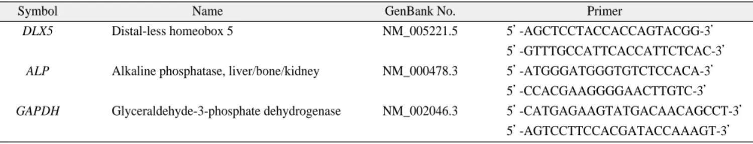
native agarose gel for checking 18S and 28S band, and cDNAs were synthesized with Superscript II Reverse Transcriptase (Invitrogen, Carlsbad, CA, USA). About 100 to 150 ng of total RNA was used for analysis. We performed a polymerase chain reaction (PCR) using HiPi PCR 5x Master Mix kit from ELPIS bio (Daejeon, Korea) according to the manufacturer’s instruc- tions. All primers were synthesized by Integrated DNA Technologies, Inc. (IDT) (Coralville, IA, USA). The primer sets for the analysis are listed in Table 2. The expression level of glyceraldehyde-3-phosphate dehydrogenase (GAPDH) of samples was used for control.
Ⅲ. Results
1. Proliferation and cell surface marker characterization of hBMSC
The numbers of total harvested BMSC and cell growth rate according to the age was important for providing the pre- dictable standard and generally acceptable information. We used the hBMSC from healthy donor’s iliac crest between young and old age patients. A total of 9 patients with two males and seven females provided their bone marrow 10 mL
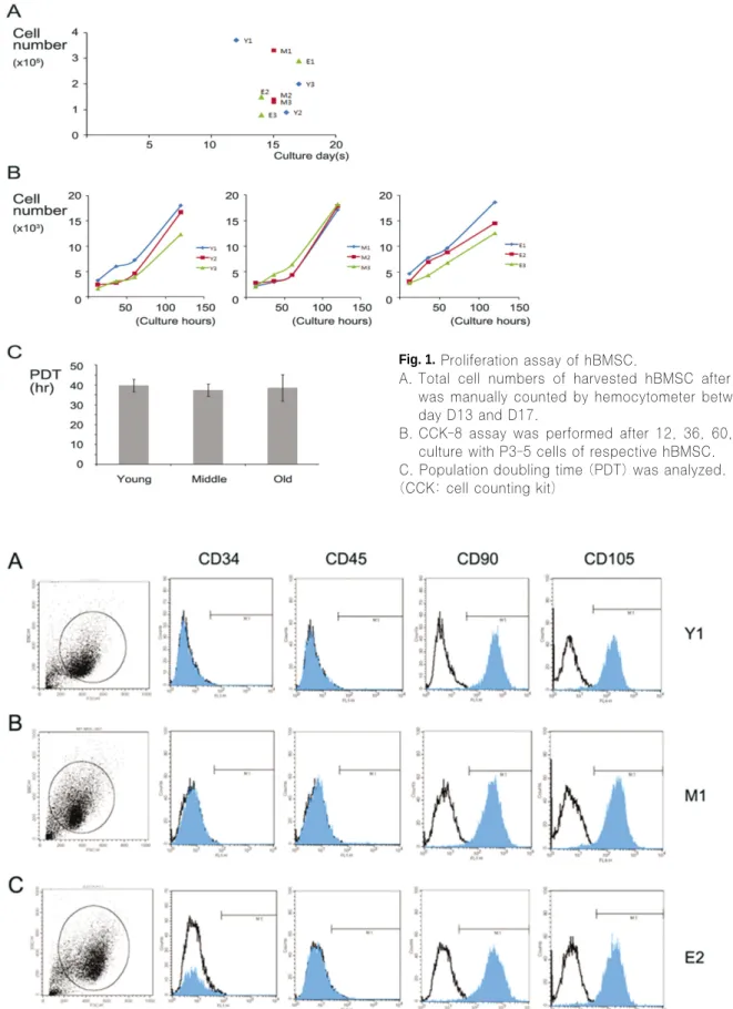
with aspiration from iliac crest under general anesthesia. First, we counted the expanded cells after P0 culture using hemocy- tometers before reseeding cells to P1 culture.(Fig. 1.A) The time of passage from P0 to P1 was determined by colony for- mation and cell density. Average first passage time was taken at 13 days to 17 days from the day of initial seeding of bone marrow cells immediately after Ficoll centrifugation. We har- vested the amount of cells from 1×10⁵to 4×10⁵broadly at this time. Human samples showed the large scale of variations in total harvested BMSC and no consistent character between young and old group was found. Then, we seeded the cells at the stage of P3-5 for cell proliferation study using CCK-8 study.(Fig. 1.B) There was no prominent difference in cell growth rate between young and old BMSC. BMSC from old donors over age of sixty showed the similar cell population doubling time (PDT) with young donor groups.(Fig. 1.C)
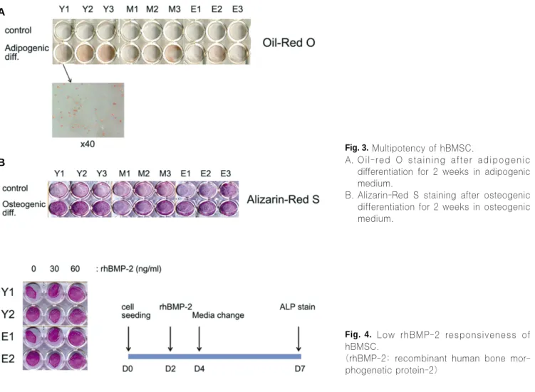
Next, FACS analysis of the immunophenotypic surface pro- file for CD34, CD45, CD90 and CD105 of isolated hBMSC was performed to find the discrepancy of the cell surface marker expression between young and old donors.(Fig. 2) FACS analysis provides the most direct approach for testing for cell surface marker differences. hBMSC isolated in this study were positive for the markers CD90, CD105, but nega- tive for CD34, CD45 according to the criteria for MSC. Most of the cells formed one major group having equal size (x-axis of the each most left panel of the Fig. 2) and equal signal (y- axis) independently to age groups which could be seen homo- geneous. Interestingly, BMSC from old donors showed the similar pattern of cell surface marker expression with young donor cells in all cell surface markers studied. Further, the BMSC from early passage such as P2 showed the similar homogeneous group with cells from late passage such as P5 or P6 (data not shown). Thus, age did not affect on obtainable total cells of BMSC, cell proliferation and cell surface marker characters of those cells.
Table 2.Primer sets for RT-PCR
Symbol Name GenBank No. Primer
DLX5 Distal-less homeobox 5 NM_005221.5 5’-AGCTCCTACCACCAGTACGG-3’
5’-GTTTGCCATTCACCATTCTCAC-3’
ALP Alkaline phosphatase, liver/bone/kidney NM_000478.3 5’-ATGGGATGGGTGTCTCCACA-3’
5’-CCACGAAGGGGAACTTGTC-3’
GAPDH Glyceraldehyde-3-phosphate dehydrogenase NM_002046.3 5’-CATGAGAAGTATGACAACAGCCT-3’
5’-AGTCCTTCCACGATACCAAAGT-3’
(RT-PCR: reverse transcription polymerase chain reaction)
Fig. 2.Surface marker expression of hBMSC between young and old aged group.
FACS of the immunophenotypic surface profile for CD34, CD45, CD90 and CD105 from isolated hBMSC was analyzed.
Black line empty histograms represent the fluorescence from negative-control cells incubated without antibody: blue colored histograms represent the counts of singals incubated with the relevant cell surface antibody.
(FACS: fluorescence-activated cell sorting)
Fig. 1.Proliferation assay of hBMSC.
A. Total cell numbers of harvested hBMSC after P0 culture was manually counted by hemocytometer between culture day D13 and D17.
B. CCK-8 assay was performed after 12, 36, 60, 120 hours culture with P3-5 cells of respective hBMSC.
C. Population doubling time (PDT) was analyzed.
(CCK: cell counting kit)
2. Multipotency of hBMSC
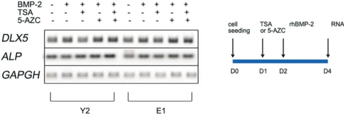
BMSC have a character to differentiate into osteoblast but also into chondroblast, adipocyte, myoblast etc by in vitro and in vivo studies. In contrast to pluripotent embryonic stem cells, adult stem cells have been considered to be multipotent, being more restricted in their differentiation capacity and only giving rise to cell types related to their tissues of origin. For example, bone marrow origin cells showed readily differentiation to osteoblast whereas fat tissue origin cells showed easily differ- entiation to adipocyte. In the aspect of age, there are conflict- ing reports on the effects of age on osteogenic capacity of BMSC. Thus, we evaluated the capacity of the multipotency with adipogenesis using adipogenic medium.(Fig. 3.A) We treated the BMSC with generally accepted adipogenic formula for 2 weeks and Oil-red O staining was performed for compar- ison. Staining method such as Oli-red O, Alizarin red S and ALP is useful for evaluating the differentiation in the case of lots of samples. Despite the sample variations, we found that hBMSC from young donors showed the higher adipogenic capacity than cells from old donors. Like adipogenesis,
Alizarin-Red S staining showed the higher mineralization ten- dency of young donor’s BMSC compared to old donor’s cells, but osteogenic differentiation was not prominent compared to adipogenesis.(Fig. 3.B)
3. Low BMP-2 responsiveness of hBMSC
BMP-2 promotes the differentiation of osteoprogenitor cells, and also induces an osteogenesis in MSC from rats and mice.
But, compared to the results from animal models, rhBMP-2 are relatively inefficient in inducing human MSC to undergo the osteogenesis, and are much less effective in promoting bone formation in human clinical trials13,14. Here, we checked the BMP responsiveness according to age by the ALP activity.
(Fig. 4) We performed the ALP staining after four to five days from the rhBMP-2 treatment to catch the relatively early response. Old donor BMSC showed similar ALP staining level with young donor cells. Also all hBMSC showed the low response to rhBMP-2 uniformly since the dose increase of rhBMP-2 could not make the staining level stronger.
Fig. 3.Multipotency of hBMSC.
A. Oil-red O staining after adipogenic differentiation for 2 weeks in adipogenic medium.
B. Alizarin-Red S staining after osteogenic differentiation for 2 weeks in osteogenic medium.
A
B
Fig. 4. Low rhBMP-2 responsiveness of hBMSC.
(rhBMP-2: recombinant human bone mor- phogenetic protein-2)
4. Osteogenic master gene expressions after TSA, 5- azacytidine treatments on hBMSC
HDAC inhibitors and methyltransferase inhibitors can affect reversibly on chromatin remodeling which is changed by growth or aging in natural human life. Chromatin state of his- tone acetylation and demethylation of highly methylated chro- matin part such as CpG islands can cause easily gene expres- sion to permit an osteogenesis. To test this possibility of HDAC inhibitor and methyltransferase inhibitor, we used the pretreatment with representative drug, TSA and 5-azacytidine for 24 hours before BMP-2 treatment.(Fig. 5) TSA alone or 5- azacitidine alone plus BMP-2 slightly stimulated the distal-less homeobox 5 (DLX5) or ALP level in all samples compared to BMP-2 only group. The use of TSA and 5-azacytidine together showed slightly increased expression level of osteogenic mas- ter gene, DLX5 and ALP mRNA level synergistically. The osteogenesis showed hardly prominent differences between young and old groups, but the drug effect was slightly positive in all groups, did not show significant difference between young and old groups.
Ⅳ. Discussion
In this study, we investigated the effects of donor age on hBMSC proliferation and differentiation potentials towards adipogenic and osteogenic lineages. Although there were many reports about murine BMSC data, the controversy on several points was still remained in hBMSC works. Most of all, no standardized practice exists for the tissue collection method, in vitro cell expansion and grouping of donor age of BMSC. As generally accepted, BMSC in this study and many others have
used the iliac bone marrow aspiration method, Ficoll separa- tion and adherent marrow stromal cells characters. We selected healthy adult of twenties, thirties and over sixties for evalua- tion of donor age effects.
Within the BMSC containing partial MSC, we found that old donor’s BMSC showed no more declines in proliferation capacity compared to the young group across all the three test- ed donor age groups. Due to similar population doubling time (PDT) from all tested donor age groups, we could harvest simi- lar cell amount from old donor and young donor during same cell expansion period. In addition, cell surface characters such as positive marker CD90, CD105 and negative marker CD34, CD45 showed no difference between young and old adult.
Especially CD105, known surrogate stem cell marker, prolifer- ation-associated and hypoxia-inducible protein, abundantly expressed in angiogenic endothelial cells showed marked vari- ations between samples but we could find no significant differ- ence from all hBMSC related with age. The effect of age on the proliferation and cell surface marker expression of hBMSC was not critical between young and old groups.
Also our data showed old donor’s BMSC showed more declines in differentiation capacity by donor age’s increasing despite of the maintained multipotency. Old BMSC differenti- ated to adipocyte successfully and more prominently compared to osteogenic differentiations. But this result was not in agree- ment with previous work which found that donor age affected less on adipogenic differentiation than osteogenic differentia-
tion15,16. No increase in adipogenic potential was observed with
increasing age, but the relative differences between differentia- tion potentials would favor adipogenesis over osteogenesis.
Some studies addressed the hypothesis that age related decreases in bone regeneration were due to BMSC aging,
Fig. 5.Osteogenic marker gene expression of hBMSC after TSA, 5-AZC pretreatment by semiquantitive RT-PCR.
(TSA: trichostatin A, 5-AZC: 5-azacytidine, DLX5: distal-less homeobox 5, ALP: Alkaline phosphatase, GAPDH:
glyceraldehyde-3-phosphate dehydrogenase)
resulting in a decreased osteogenic potential with a concurrent increase in adipogenic potential. In our study BMSC main- tained their multipotency even in old group but exhibited decreased potential with aging for both osteogenic and adi- pogenic differentiation.
For osteogenic differentiation of BMSC, ostegenic media and rhBMP-2 treatment have been used but there were some limitation due to the low responses of the differentiation or mineralization of BMSC17-19. BMP treatment provided marked- ly successful result of ectopic bone formation, bone healing after fracture or bone formation of critical sized defects in ani- mal experiments. However, many clinical trials have been reported low or unsuccessful responses of BMP treatment in human. Our data showed no increase by rhBMP-2 treatment in vitro in human samples by ALP activity although small increases were observed by mRNA level. More helpful method for BMP treatment on BMSC was necessary.
The concern about epigenetic condition on gene expression level may lead to this solution because chromatin remodeling factor such as HDAC inhibitor and methyltransferase inhibitor have attracted attention due to its temporary, reversible effects on chromatin loosening. Many researchers have provided the evidence supporting that HDAC inhibitors such as TSA stimu- late osteogenic differentiation of hBMSC. And 5-azacitidine, methyltransferase inhibitor, has been used optionally to differ- entiate BMSC to adipocyte or myoblast up to now. Our results showed more highly expressed osteogenic marker gene such as DLX5, ALP after TSA, 5-azacitine pretreatment.
Ⅴ. Conclusions
This study has showed that hBMSC from old donors have identical proliferation and cell surface marker character with young donors BMSC. And TSA, 5-azacitidine showed the pos- sibility of increase of the BMP effect on osteoblast differentia- tions. Based on the results of this study and many other previ- ous studies, it appears that the osteoblast differentiation of hBMSC is considered to be less successful than animal cells.
Future studies should be necessary for positive effect of the reversible inhibitory function of the HDAC inhibitors or methyltransferase inhibitors on enhancing the low capacity of osteoblast differentiation of hBMSC.
References
1. Conget PA, Minguell JJ. Phenotypical and functional properties of human bone marrow mesenchymal progenitor cells. J Cell Physiol 1999;181:67-73.
2. Zhou S, Greenberger JS, Epperly MW, Goff JP, Adler C, Leboff MS, et al. Age-related intrinsic changes in human bone-marrow- derived mesenchymal stem cells and their differentiation to os- teoblasts. Aging Cell 2008;7:335-43.
3. Ma D, Ma Z, Zhang X, Wang W, Yang Z, Zhang M, et al. Effect of age and extrinsic microenvironment on the proliferation and osteogenic differentiation of rat dental pulp stem cells in vitro. J Endod 2009;35:1546-53.
4. Benetti R, Garcia-Cao M, Blasco MA. Telomere length regulates the epigenetic status of mammalian telomeres and subtelomeres.
Nat Genet 2007;39:243-50.
5. Gonzalo S, Jaco I, Fraga MF, Chen T, Li E, Esteller M, et al.
DNA methyltransferases control telomere length and telomere re- combination in mammalian cells. Nat Cell Biol 2006;8:416-24.
6. Schroeder TM, Westendorf JJ. Histone deacetylase inhibitors promote osteoblast maturation. J Bone Miner Res 2005;20:2254- 63.
7. de Boer J, Licht R, Bongers M, van der Klundert T, Arends R, van Blitterswijk C. Inhibition of histone acetylation as a tool in bone tissue engineering. Tissue Eng 2006;12:2927-37.
8. Jeon EJ, Lee KY, Choi NS, Lee MH, Kim HN, Jin YH, et al.
Bone morphogenetic protein-2 stimulates Runx2 acetylation. J Biol Chem 2006;281:16502-11.
9. Atkinson SP, Keith WN. Epigenetic control of cellular senes- cence in disease: opportunities for therapeutic intervention.
Expert Rev Mol Med 2007;9:1-26.
10. Hu E, Tontonoz P, Spiegelman BM. Transdifferentiation of my- oblasts by the adipogenic transcription factors PPAR gamma and C/EBP alpha. Proc Natl Acad Sci U S A 1995;92:9856-60.
11. Ogawa A, Ohba K, Uchida Y, Wada K, Yoshioka T, Muraki T.
New adipogenic cell lines derived from C3H10T1/2. In Vitro Cell Dev Biol Anim 1999;35:307-10.
12. Pittenger MF, Mackay AM, Beck SC, Jaiswal RK, Douglas R, Mosca JD, et al. Multilineage potential of adult human mes- enchymal stem cells. Science 1999;284:143-7.
13. Fang H, Yang X, Chen A, Luo Y. Effect of rhBMP-2 and os- teogenic revulsants on proliferation and differentiation of bone marrow stromal cells in rats. J Huazhong Univ Sci Technolog Med Sci 2007;27:561-3.
14. Osyczka AM, Diefenderfer DL, Bhargave G, Leboy PS.
Different effects of BMP-2 on marrow stromal cells from human and rat bone. Cells Tissues Organs 2004;176:109-19.
15. Justesen J, Stenderup K, Eriksen EF, Kassem M. Maintenance of osteoblastic and adipocytic differentiation potential with age and osteoporosis in human marrow stromal cell cultures. Calcif Tissue Int 2002;71:36-44.
16. Kretlow JD, Jin YQ, Liu W, Zhang WJ, Hong TH, Zhou G, et al.
Donor age and cell passage affects differentiation potential of murine bone marrow-derived stem cells. BMC Cell Biol 2008;
9:60.
17. Mizuno D, Agata H, Furue H, Kimura A, Narita Y, Watanabe N, et al. Limited but heterogeneous osteogenic response of human bone marrow mesenchymal stem cells to bone morphogenetic protein-2 and serum. Growth Factors 2010;28:34-43.
18. Diefenderfer DL, Osyczka AM, Reilly GC, Leboy PS. BMP re- sponsiveness in human mesenchymal stem cells. Connect Tissue Res 2003;44 Suppl 1:305-11.
19. Fleet JC, Cashman K, Cox K, Rosen V. The effects of aging on the bone inductive activity of recombinant human bone morpho- genetic protein-2. Endocrinology 1996;137:4605-10.