© 2011 The Korean Nutrition Society
서 론
현대 사회의 산업화 및 기타 생활환경의 변화는 평균수명 증가와 함께 암의 발생률도 높였다. 2007년 통계청 자료에 따 르면 우리나라의 경우, 암 발생률은 10만 명당 137.5명으로 남녀 모두에서 1위를 기록하고 있고 그 중 간암으로 인한 사 망률은 성인 남자에게서 1위를 차지하고 있다.1) 그에 따른 눈 에 보이지 않는 간접적인 경제적인 손실 또한 막대하기 때문
에 암 등의 질병을 예방할 수 있는 물질에 대한 연구가 많이 발표되고 있다. 그 중 식품의 항산화 효과에 대해서는 널리 알려져 있으나 식품을 통한 항산화 기전 연구는 다양하게 진 행되고 있지 못한 실정이다.
생체 내에서 생성되는 활성산소는 인체의 노화와 질병을 유발하는 주요 원인으로서 세포와 조직에 해로운 독성을 일 으켜 질병을 유발하는 것으로 알려져 있다. 특히 지질과산화 물 (lipid peroxides, LPO)은 활성산소 매개체가 세포자체의 국소적인 방어 기전을 초과함으로써 발생하는 세포선상의 주된 형태로 OH-, O2, H2O2 등이 지질과산화를 유발하는 것 으로 보고되었다.2,3) 초기의 free radical이 생체막에 있는 불포 화 지방산의 α-methylene기를 공격하면 lipid radical (R・)이 형성되고, lipid radical은 산소와 결합하여 peroxiradical (ROO・)이 형성되며, 이것은 다른 α-methylene기를 공격하 DOI 10.4163/kjn.2011.44.6.488
산화스트레스가 유도된 인체 간암세포 (HepG2)에서 Sulforaphane과 Diallyl Sulfide, Capsaicin, Gingerol의 항산화효과 비교연구*
이소연 · 위해리 · 이명숙
§성신여자대학교 식품영양학과, 비만과학연구소
Comparison of the Antioxidant Effects of Diallyl Sulfide, Capsaicin, Gingerol and Sulforaphane in H
2O
2-Stressed HepG2 Cells
*Lee, SoyounㆍWi, HaeriㆍLee, Myoungsook
§Department of Food and Nutrition and Research Institute of Obesity Sciences, Sungshin Women’s University, Seoul 136-742, Korea
ABSTRACT
Oxygen is necessary to sustain life, yet cellular oxygen metabolism creates destructive elements called free radicals.
Free radicals are chemically unbalanced and carrying free electrons that can damage molecules, potentially damaging the cell itself. For this reason, many antioxidant products, including supplements and functional foods, are being devel- oped. In particular, natural products are rich sources of pharmacologically active compounds. The purpose of this study was to investigate the antioxidant effects of target biomaterials in Korean traditional spices such as diallyl sulfide (DAS), capsaicin (CAP), and gingerol (GGR), and to investigate the response of the antioxidant defense system to oxi- dative stress by hydrogen peroxide (H2O2) compared to sulforaphane (SFN) in HepG2 cells. After the analysis of the cell viability using Cell Counting kit-8 (CCK-8) assay, we determined that the optimum levels were 200 μM DAS, 25 μM CAP, 50 μM GGR, and 12.5 μM SFN. Antioxidant enzymes were measured and protein expression was detected by Western blotting. All treatments showed a significant decrease in antioxidant enzyme activity such as superoxide dis- mutase, catalse, and glutathione peroxidase in HepG2 cells. Additionally, DAS, CAP, GGR and SFN increased the anti- oxidant system-related transcription factor Nrf2 which was found to be regulated by the activation of MAPK-JNK in this study. In conclusion, these results indicate the protective effects of DAS CAP, GGR, and SFN against H2O2-in- duced oxidative stress. (Korean J Nutr 2011; 44(6): 488 ~ 497)
KEY WORDS: antioxidant, HepG2 cells, diallyl sulfide, capsaicin, gingerol, sulforaphane.
접수일:2011년 11월 14일 / 수정일:2011년 11월 29일 채택일:2011년 12월 13일
*This work was supported by Sungshin Women’ University Re- search grant of 2009.
§To whom correspondence should be addressed.
E-mail: [email protected]
여 lipid hydroperoxide (ROOH)와 새로운 lipid radical이 된다. 생체지질은 일단 연쇄개시반응으로 지질 라디칼이 생 성되면 연쇄전파반응이 연속적으로 일어나고 지질의 변화가 가속적으로 진행되는 것이 특징이다.4) 특히 호기성 생물, 건강 한 세포는 계속적으로 산화과정을 통하여 에너지를 만들어 야 하므로 ROS (reactive oxygen species)의 생성이 불가피 하며 세포막의 지질과 단백질, 핵산의 형태를 변화시키게 되 는 것이다. 이러한 생체분자의 산화적 손상은 노화와 함께 동 맥경화, 허혈성심질환, 신경성 질환, 암 등을 포함하여 여러 가 지 질병도 초래하게 되는 것이다.5-8)
간세포를 비롯하여 생체 내 세포는 지질과산화물의 체내 축적을 억제시키기 위해 1차 방어기전으로 효소계 산화방어 기전을 보유하고 있는데 superoxide dismutase (SOD), H2O2
를 제거하는 catalase 및 glutathione peroxidase (GPx) 등 을 말하며 세포막과 세포내 여러 부분에서 특이적으로 작용 하여 조직의 과산화를 억제한다.9-11) 그러나 어떠한 환경에서 이들 방어기작의 효용성이 감소하게 되면 세포가 손상되고, 암화과정도 촉진될 수 있다.12) 생체내에서는 산화스트레스에 의하여 민감하게 발현하는 생체지표로 p38, ERK, JNK등의 Mitogen-activated protein kinase (MAPK)를 제안할 수 있
다.13,14) 특히 간세포에서의 산화스트레스 신호에 반응하는 상
위단계 표적단백질이 MAPK라면 하위단계 표적단백질들로 는 NF-E2- related factor 2 (Nrf2)와 같은 전사인자를 그 예 로 들 수 있다.15) 최근 브로컬리의 표지물질인 sulforaphane 이 산화스트레스로부터 세포를 보호하는 phaseII 무독화 효 소인 glutathione S-transferases (GSTs)의 주요조절인자인 Nrf2를 활성화한다고 보고되었다.16) GSTs는 reduced gluta- thione (GSH)의 family효소로 diphenols, quinones, isothio- cyanates, peroxides, vicinal dimercaptans 등의 발암물질 로부터 간, 표피 등의 조직이 암화되는 과정에서의 민감성을 조절한다.17) 과일이나 채소의 β-carotene, 식물의 D-limonene 및 D-carvone, sulforaphane (4-methylsulfinyl butyl iso- thiocyanate) 등이 이와 관련되어 있다고 한다.
그러므로 본 연구에서는 human hepatic carcinoma인 HepG2 세포에 H2O2산화처리를 한 후 한식의 주요양념인 마늘, 고추, 생강의 항산화 지표물질인 diallyl sulfide, gingerol, capsaicin 를 처치하여 sulforaphane의 항산화효소활성 및 항산화기전 을 비교하여 간 손상의 보호효과를 확인하고자 한다.
연 구 방 법
세포배양 및 처치
한국세포주은행 (KCLB; Korean Cell Bank, 서울대학교 의
과대학 암연구소)에서 인체 간암세포 (Human hepatocellular liver carcinoma cell line)를 구입하여 10% FBS (fetal bovine serum)과 항생제 (penicillin-streptomycin)가 첨가된 RPMI- 1640 media로 배양한다. 세포는 일정한 습도를 유지하는 37℃의 incubator에서 공기 (95%)와 CO2 (5%)의 혼합기체를 계속적으로 공급하면서 배양시키고 2일 마다 배지를 교환해 주고 3~4일 마다 계대 배양하며 trypan-blue를 이용한 생존 률측정도 실시한다. Pilot study에서는 일정수준의 confluency 에 도달한 상태에서 독성이 강한 산화스트레스인자로 H2O2를 처치하고 마늘, 고추, 생강의 항산화 지표물질인 diallyl sulfide (DAS), capsaicin (CAP), gingerol (GGR), sulforaphane (SFN) 을 농도별로 각각 함께 처치하여 생존율 (CCK-8)을 측정한 다음 기작규명을 위한 실험적인 농도를 결정하였다. 간암세포 내에서 항산화효소활성 증가, 지질과산화 생성억제 효과 및 기전적규명을 위한 처치농도는 DAS는 200 μmol/L, CAP는 25 μmol/L, GGR은 50 μmol/L, SFN은 12.5 μmol/L으로 결 정되었다.
Cell proliferation
TBHP와 diallyl sulfide, capsaicin, gingerol, sulforaphane 을 동시 혹은 각각 처치한 후 세포의 성장률은 CCK-8로 측정 하며 시약은 살아있는 세포에서 NAD가 NADH로 생성되는 원리를 이용하여 WST-8 시약이 이를 재산화 시키는 과정에 생기는 색의 변화로 450 nm에서 흡광도를 측정함으로서 세 포증식률을 알아보았다.
Superoxide dismutase (SOD) 효소활성
Superoxide dismutase (SOD) 활성도 측정은 Cayman SOD assay kit으로 흡광도 450 nm에서 측정하여, Superoxide radical을 50% dismutation 하는데 필요한 SOD량을 1 unit 으로 하여 분당 활성정도를 단백질 1 mg 단위로 나타내었다.18)
Catalase (CAT) 효소활성
Catalase activity 활성도 측정은 Cayman Catalase assay kit를 이용하여 흡광도 540 nm에서 측정하였으며 결과값은 1 nmol의 Formaldehyde를 생성하는데 필요한 Catalase의 양 을 1 nmol로 하여 분당 활성정도를 단백질 1 mg 단위로 나타 내었다.19)
CAT activity (nmol/min/mL) = Fomaldehyde μM
× dilution factor 20 min
Glutathione (GSH) 효소 level
GSH (glutathione; L-γ.-glutamyl-L-cysteinylglycine)은
단백질, DNA의 합성과 세포막의 이중층에 걸쳐있는 수송단 백질을 산화로부터 보호하는데 중요한 역할을 하는 효소이 다. Glutathione peroxidase는 GSH를 환원제로 사용하여 세포의 과산화지질을 낮추는 역할을 한다. 이때 산화된 GSH 는 GSSH라고 하며 total GSH과 산화된 GSH의 차이로, 환 원된 GSH의 양을 GSH kit (Arborassay, MI, USA)로 측정 하였다.20) 항산화물질이 처치된 cell lysate (4×10⁶)에서의 세 포질에서 2-vinylpyridine를 처리하여 free GSH 반응을 정지 시킨 다음 산화된 GSH를 측정하여 total GSH 수준과의 차 이로 free GSH를 흡광도 405 nm에서 측정하며 이를 단백질 량 1 mg 단위로 보정한다.
GSH = Total GSH levels - Oxidized GSH levels
Glutathione Peroxidase (GPx) activity
GPx 효소 활성은 GPx assay kit (Cayman, MI, USA)에 의 해 측정되었다. 이 분석법은 GPx에 의해 GSH가 산화된 형 태인 GSSG가 환원될 때 소비되는 NADPH의 증감도를 흡 광도를 이용하여 측정하는 원리이다. 실험에 사용되는 시약은 NADPH, glutathione, glutathione peroxidase 및 assay buffer (5 mM EDTA, 50 mM Tris-HCL, pH 7.6)의 혼합물로 구성 되었다. 반응의 개시는 GPx cumen hydroperoxide의 첨가에 의해 이뤄졌으며 GPx 활성은 NADPH 산화 비율로서 340 nm 에서 흡광도를 1분마다 적어도 5회씩 측정하였다. 측정된 결 과는 한 unit의 GPx가 1분 동안 NADPH를 NADP + 로 산 화시키는 양을 1 nm을 기준으로 하여 계산되었다.21)
GPx activity (nmol/min/mL) = △A340/min 0.19 mL
× × dilution factor 0.00373 μM-1 0.02 mL
LPO product
총 지질과산화물 생성정도는 생성시 불안정한 LPO가 fer- rous ions과 쉽게 반응하여 ferric ion을 생성하고 이를 thio- cyanate ion과 반응하는 정도를 detect하는 산화환원반응의 원리를 이용한 LPO kit (Calbiochem, california, USA)로 측정하였다. 항산화물질이 처치된 cell lysate (5×10⁶)에서 클 로로포름으로 추출된 LPO를 500 nm 흡광도계를 이용하여 측정하였고 결과는 standard curve에 의하여 계산된 수치를 단백질 1 mg 당 nmol/L/mg로 나타내었다.22)
Western blotting
DAS, CAP, GGR, SFN에 의한 항산화 관련 단백질 발현을 관찰하기 위하여 western blotting 실험을 시행하였다. DAS, CAP, GGR, SFN을 처치하거나 혹은 처치하지 않은 HepG2
세포를 배양한 후 lysis buffer (50 mM Tris, pH 8.0, 1% Triton x-100, 150 mM NaCl, 1 mM EDTA, 0.1% SDS, 0.5% Sodium deoxycholate)를 처리하여 수거하였다. 수거한 세포액을 14,000 g에서 20분간 원심분리하여 상층액을 실험에 사용하였다.
분리된 상층액 (총 단백질량, 20 μg)을 8%겔 SDS-PAGE를 이용하여 전기영동으로 분리된 단백질은 nitrocellulose mem- brane에 transfer하였다. Transfer된 nitrocellulose mem- brane을 5% 탈지분유에 2시간 동안 blocking한 다음, 1차 항 체 (caspase-3, cytochrome c, bcl-2, NFkB, p-ikB, MAPK (JNK, ERK, p35), NrF2; Santa Cruze Biotechnology)를 membrane과 반응시키고 TBS (Tris-buffered saline/Tween 20, TBS)로 washing하고 1차 항체에 대한 특이적 2차 항체 를 실온에서 1 시간 동안 반응시켰다. Membrane washing 후 ECL용액 (Biorad)으로 발색 후 X-선 필름 (Kodak XAR)에 노 출시켜 현상하였으며, Bio Image J Analyzer (NIH, Bethesda, USA)로 결과를 분석하였다.
통계처리
본 실험에서 얻은 모든 결과는 SPSS SW을 이용하여 분석 하였다. 평균 ± 표준편차 (mean ± SD)로 구하였고, ANOVA test 후 p < 0.05 수준에서 각 처치군 간 혹은 배양시간 별 유 의차를 나타내었다.
연 구 결 과
SFN과 DAS, CAP, GGR 처치에 따른 HepG2 세포증식률 비교
HepG2 세포에 H2O2 산화스트레스를 유도한 후 각각의 chemical을 처리 한 다음 생존률을 측정하였고 결과는 Fig.
1에 나타내었다. 그 결과 H2O2만을 처리한 모든 군의 생존률 이 감소하였으며 DAS의 세포회복효과는 12시간에서 100, 200 μM의 효과가 높았으며, CAP의 세포회복효과는 12시간 에서 50 μM, 24시간에서 25 μM의 효과가 높았다. GGR의 세포회복효과는 12시간 및 24시간 모두에서 50 μM의 효과 가 가장 좋았다. SFN의 경우 모든 시간에서 12.5 μM의 효과 가 높았기 때문에 이와 유사한 범위의 생존율을 보인 농도 (DAS 200 μM, CAP 25 μM, GGR 50 μM, SFN 12.5 μM) 를 선정하여 각 chemical의 항산화 효소 활성도 및 기전규 명을 하고자하였다.
SFN과 DAS, CAP, GGR의 항산화효소활성 비교
HepG2 세포에 DAS 200 μM, CAP 25 μM, GGR 50 μM, SFN 12.5 μM을 처리하여 12시간 및 24시간 후에 항산화 기 능을 가지는 SOD, CAT, GSH, GPx 효소 활성을 측정하여 그
영향을 알아보았다. 그 결과 H2O2만을 처리한 세포에서 SOD 효소활성이 대조군에 비해 75.2%로 감소한 반면에 H2O2와 함께 DAS, CAP, GGR, SFN를 처리한지 12시간배양 후 세포 에서의 SOD 효소 활성은 각각 SFN (129.9%) > GGR (120.5%)
> DAS (107.8%) > CAP (105.9%) 순으로 증가되었다 (Fig. 2A).
그러나 24시간배양 후에는 H2O2처리 군보다 CAP, GGR, SFN 만 108.2%, 114.5%, 104.7%로 SOD효소활성이 증가되었고 DAS는 H2O2처리군 수준으로 감소하였다. CAT 효소의 경우
Fig. 1. Cell viability of DAS, CAP, GGR and SFN in H2O2 induced Human Hepatoma HepG2 Cells. DAS: diallyl sulfide, CAP: capsaicin, GGR: gingerol, SFN: sulforaphane.
1.5
1.0
0.5
0.0
1.5
1.0
0.5
0.0 H2O2 (200 uM)
DAS (um) H2O2 (200 uM)
CAP (um) - + + +
- - 100 200 0 hr
- + + + - - 25 50 0 hr - + + +
- - 100 200 12 hr
- + + + - - 25 50 12 hr - + + +
- - 100 200 24 hr
- + + + - - 25 50 24 hr
*** ***
*** ***
NS NS
DAS CAP
c d
b b b
c
b d
a a a
c a
d
a Control a
N.control 100 uM 200 uM
Control N.control 25 uM 50 uM
Cell viability-OD Cell viability-OD
1.5
1.0
0.5
0.0
1.5
1.0
0.5
0.0 H2O2 (200 uM)
GGR (um) H2O2 (200 uM)
SFN (um) - + + +
- - 100 200 0 hr
- + + + - - 125 25 0 hr - + + +
- - 100 200 12 hr
- + + + - - 125 25 12 hr - + + +
- - 100 200 24 hr
- + + + - - 125 25 24 hr
***
***
***
***
NS NS
GGR SFN
c c
b b
b b
d c
a c a
b
b a
a Control a
N.control 50 uM 100 uM
Control N.control SFN 12.5 uM SFN 25 uM
Cell viability-OD Cell viability-OD
70 60 50 40 30 20 H2O2
Treat - + + + + + - - DAS CAP GGR SFN 12 hr
- + + + + + - - DAS CAP GGR SFN 24 hr
****
*
NSNS NS
NSNS c
b a a a c
c a bc a ab d
Control N.control DAS CAP GGR CAT activity nmol/min/mg protein SFN
B
Fig. 2. Antioxidant effects of DAS, CAP, GGR and SFN on the activities of SOD (A) and CAT (B) in H2O2 induced Human Hepatoma HepG2 Cells. DAS: diallyl sulfide, CAP: capsaicin, GGR: gingerol, SFN:sulforaphane. Data are presented as mean ± SD. a, b, c: Signifi- cant difference between the groups were tested by ANOVA. *: p < 0.05, **: p < 0.01, ***: p < 0.001: Significantly different each groups compared to negative control (only H2O2).
1.5
1.0
0.5 H2O2
Treat - + + + + + - - DAS CAP GGR SFN 12 hr
- + + + + + - - DAS CAP GGR SFN 24 hr
*** *
*** **
**NS *
NS e
d c b a f
c b ab a a
d Control
N.control DAS 200 CAP 25 GGR 50 SFN 12.5
SOOD activity U/mg protein
A
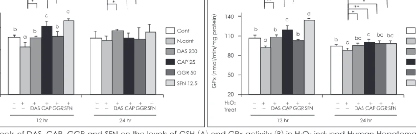
H2O2을 처리시 대조군의 76.8%로 활성이 감소하였으나 GGR 과 SFN을 12시간 배양후 CAT활성이 111.6%, 123.3%로 증가 하였고 24시간 배양후에는 GGR만 CAT 활성이 118.6% 증가 하였다 (Fig. 2B). GSH 수준을 측정한 결과는 Fig. 3A에 나타 내었으며 H2O2만을 처리한 세포에서는 대조군의 90.4% 수준 으로 측정되었다. 그러나 DAS, CAP, GGR, SFN를 12시간배 양 후 각각 110.4%, 124.0%, 112.4%, 130.9%로 GSH 수준이 증가하였다. 24시간배양 시 DAS만 112.4%로 GSH가 증가되 었다. GPx 활성을 측정한 결과 H2O2처치가 대조군의 86.3%
로 감소하였으나 DAS, CAP, GGR, SFN를 함께 처리시 12시 간 배양 후 각각 118.2%, 130%, 111.9%, 146%로 GPx 활성이 증가되었다 (Fig. 3B). 24시간 배양 후에는 12시간배양보다 약 하지만 모든 군에서 H2O2처치군보다 전반적인 증가를 보였다.
본연구의 결과로는 GPx의 활성은 GSH의 농도에 매우 의존적 이었고 전반적으로 SFN의 항산화효과가 가장 뛰어났다.
SFN과 DAS, CAP, GGR의 LPO생성 비교
HepG2 세포에 H2O2만을 10분간 처리한 세포에서 DAS 200 μM, CAP 25 μM, GGR 50 μM, SFN 12.5 μM을 함께 12시 간 및 24시간배양 후에 LPO생성을 알아보았다 (Fig. 4).
H2O2 10분 처치후 12시간, 24시간배양 시 모두 대조군에 비하 여 약 125% 정도 LPO가 생성되었다. 그러나 DAS, CAP, GGR, SFN를 12시간 배양 후에는 SFN과 DAS가, 24시간 배양 후 GGR만 대조군 수준으로 LPO가 저하되었다.
SFN과 DAS, CAP, GGR의 항산화관련 단백질발현 비교
H2O2로 산화스트레스를 유발한 HepG2 세포에서 DAS, CAP, GGR, SFN를 각각 200 μM, 25 μM, 50 μM, 12.5 μM 30분간 처리하여 western blotting analysis로 분석하였다 (Fig. 5 and 6). 우선 산화 스트레스로 인한 전사 인자의 발현 정도를 알아보기 위해 nuclear factor kB (NF-kB)발현을 측정하였다. H2O2만을 처리한 세포에서는 NF-kB 발현이 유 의적으로 증가하였으나 SFN > DAS = CAP = GGR 순으로 발현량이 감소되었다 (Fig. 5B). 이 결과를 정확하게 확인하기 위하여 인산화된 IkBα (p-IkBα)와 IkBα를 측정하여 p-IkB α/IkBα의 비율을 분석하였는데 H2O2 처리 세포에서 대조군 에 비해 발현이 증가하는 반면에 SFN > GGR > CAP > DAS 순으로 유의적인 발현량 감소가 관찰되었다 (Fig. 5A). 이같 은 산화스트레스 시그널의 up-stream 유도인자를 확인하기 위하여 MAPK 발현을 확인한 결과 JNK의 인산화형태인 pJNK가 GGR을 제외한 SFN > DAS = CAP 순으로 증가하 였고 ERK와 p38은 차이가 없었다 (Fig. 6A). 이같은 JNK의 활성화는 Nrf2를 발현시키므로 이를 확인한 결과 DAS, CAP,
14.0 11.0 8.0
5.0 H2O2
Treat - + + + + + - - DAS CAP GGR SFN 12 hr
- + + + + + - - DAS CAP GGR SFN 24 hr
*
***
**
NSNS NS c
b c a b
b Cont
N.cont DAS 200 CAP 25 GGR 50 SFN 12.5
GSH (uM/mg protein)
A
140 110 80 50
20 H2O2
Treat - + + + + + - - DAS CAP GGR SFN 12 hr
- + + + + + - - DAS CAP GGR SFN 24 hr
**
****
** *
*
* b
c d
b c
b bc bc bc
a a
b Cont
Ncont DAS CAP GGR (nmol/min/mg protein)GPx SFN
B
Fig. 3. Effects of DAS, CAP, GGR and SFN on the levels of GSH (A) and GPx activity (B) in H2O2 induced Human Hepatoma HepG2 Cells. DAS: diallyl sulfide, CAP: capsaicin, GGR: gingerol, SFN: sulforaphan. Data are presented as mean ± SD. a, b, c: Significant difference between the groups were tested by ANOVA. *: p < 0.05, **: p < 0.01, ***: p < 0.001: Significantly different each groups compared to negative control (only H2O2).
3.0 2.5 2.0 1.5
1.0 H2O2
Treat
- + + + + + - - DAS CAP GGR SFN 12 hr
- + + + + + - - DAS CAP GGR SFN 24 hr
**
****
***
* *
NS
NS
a b
b a a
d cd
bc c
ab c b
Cont N.cont DAS 200 CAP 25 GGR 50 SFN 12.5 LPO production (nmol/mg protein)
Fig. 4. Effect of DAS, CAP, GGR and SFN on LPO reduction in H2O2 induced Human Hepatoma HepG2 Cells. DAS: diallyl sul- fide, CAP:capsaicin, GGR:gingerol, SFN: sulforaphan. Data are presented as mean ± SD. a, b, c : Significant difference be- tween the groups were tested by ANOVA. *: p < 0.05, **: p < 0.01,
***: p < 0.001: Significantly different each groups compared to negative control (only H2O2).
GGR, SFN를 처리한 HepG2 세포에서 Nrf2 발현이 Control 에 비하여 유의적으로 증가하였고, Nrf2의 인산화형태이자 활성형인 pNrf2 발현 또한 SFN = GGR = CAP > DAS 순으 로 유의적인 증가를 나타내었다 (Fig. 6B and C). TNF-α의 발현량 역시 pIkBα와 비슷한 경향을 나타났으나 NFkB의 전사작용의 효과인지 p-NrF2의 전사인자 효과인지는 본 연 구에서는 알 수 없다 (Fig. 5C). 산화스트레스에 의한 미토콘 드리아 손상여부를 알기위한 caspase-3, cytochrome c, bcl-2 발현은 일관성있는 결과를 보이지 않았다.
고 찰
본 연구는 생체외 시스템에서 H2O2로 유발된 산화스트레스 및 간 손상으로부터 DAS, CAP, GGR, SFN의 보호 효과를 확인하기 위하여 수행되었다. 간은 생체 내에서 해독 및 무독 화를 위한 기관으로서 중심적인 역할을 수행하기 때문에 산 화스트레스의 보호 효과를 관찰하기 위한 대상으로 선택하 였고, 인체 유래 간암세포인 HepG2 세포주를 사용하였다. 항
Fig. 5. Effects of DAS, CAP, GGR and SFN on the expression of oxidative stress markers, pIkBα (A), NFkB (B), TNFα (C) against H2O2-pre- treated HepG2 cells. Actin levels were compared to ensure equal amount of protein loading. DAS: diallyl sulfide, CAP: capsaicin, GGR: gingerol, SFN: sulforaphan.
120 100 80 60 40 20 0 H2O2
Treat H2O2
Treat
- + + + + + - - DAS CAP GGR SFN
- + + + + + - - DAS CAP GGR SFN
a bc ab
cd
p<0.001 e
de
pIkB-α/β-actin (%of control)
pIkB-a (40 kDa)
IkB-a (39 kDa)
B-actin (45 kDa)
A
160 140 120 100 80 60 40 H2O2
Treat H2O2
Treat
- + + + + + - - DAS CAP GGR SFN
- + + + + + - - DAS CAP GGR SFN
a b ab
b
p<0.01 c
ab
NFkB/β-actin (%of control)
NFkB (40 kDa)
B-actin (45 kDa)
B
160 140 120 100 80 60 40 H2O2
Treat H2O2
Treat
- + + + + + - - DAS CAP GGR SFN
- + + + + + - - DAS CAP GGR SFN
a b b b
p<0.001 c
a
TNF-a/β-actin (%of control)
TNF-a (17 kDa)
B-actin (45 kDa)
C
Fig. 6. Effects of DAS, CAP, GGR and SFN on the expression of antioxidative signals, JNK and pJNK (A), Nrf2 (B), pNrf2 (C) against H2O2- pretreated HepG2 cells. Actin levels were compared to ensure equal amount of protein loading. DAS: diallyl sulfide, CAP: capsa- icin, GGR: gingerol, SFN: s ulforaphan.
250 200 150 100
50 H2O2
Treat H2O2
Treat
- + + + + + - - DAS CAP GGR SFN
- + + + + + - - DAS CAP GGR SFN
d b c c
p<0.001
b
a
pJNK/β-actin (%of control)
pJNK (46/54 kDa)
JNK (46/54 kDa)
B-actin (45 kDa)
A
200 150 100
50 H2O2
Treat H2O2
Treat
- + + + + + - - DAS CAP GGR SFN
- + + + + + - - DAS CAP GGR SFN
b b b b b
a
Nrf2/β-actin (%of control)
Nrf2 (57 kDa)
B-actin (45 kDa)
B
p<0.01
200 150 100
50 H2O2
Treat H2O2
Treat
- + + + + + - - DAS CAP GGR SFN
- + + + + + - - DAS CAP GGR SFN
c c c
bc
p<0.01
ab a
pNrf2/β-actin (%of control)
pNrf2 (17 kDa)
B-actin (45 kDa)
C
산화 효소는 세포에서 중대한 역할을 수행하며 항산화 효소 의 활성 변화는 항산화 반응에서 중요한 생체지표로 간주할 수 있다.23) 따라서 스트레스 상황에서 적절한 농도의 항산화 효소를 유지하는 것이 세포 생존에 도움이 될 수 있다. 이러 한 이유로 이 연구에서는 각각의 항산화 물질을 산화스트레 스 상황에 처리하여 간의 항산화 효소 활성도를 측정하였다.
CAP과 SFN의 간암세포 (HepG2) 및 신경세포 (SK-N-SH) 에 대한 보호효과와 GGR의 항암효과 연구결과가 보고되었
다.24-26) 또한 DAS처치에 따른 LPO 감소효과와 항산화효소
(CAT, SOD, GR 및 GST) 등을 증가시켰다는 in vivo 연구 결과가 있다.27) 대체적으로 H2O2에 노출된 간세포에서의 항산 화 효소 (SOD, CAT, GPx)활성은 모두 감소하였다. 항산화 효소의 감소는 H2O2에 의한 ROS를 처리하기 위하여 빠르게 진행되는 무독화 과정으로 생각된다. 본 연구에서는 항산화 효소활성이 각 처치물질과 배양 시간에 따라 다르게 나타났 지만 24시간 배양시에는 처리물질에 대한 일관성이 없고 세포 신호전달체계와의 해석을 위하여 12시간 배양결과를 비교하 면 여러 선행연구처럼 모든 항산화효소의 증가는 SFN이 가 장 뛰어났다.15,16) 그러나 SFN과 유사한 결과는 생강의 GGR 가 SOD와 CAT를 증가시켰고 고추의 CAP이 GSH 및 GPx 증가시킨 경우이다. 일반적으로 GSH 수준은 세포 내 산화 상 태를 반영하며 균형잡힌 농도의 GSH는 세포가 잠재적인 산 화스트레스에 대항하기 위하여 필수적이라고 알려져 있다.28) GSH의 산화 감소 역할은 산화스트레스와 자유라디칼 병리 학을 포함하는 많은 연구에서 중심적인 것으로 밝혀졌으며 이와 관련된 여러 질병을 가진 환자의 혈액에서 낮은 농도의 GSH, 높은 농도의 GSSG 혹은 낮은 GSH/GSSG 비율이 관 찰된 바 있다.29) GSH 농도 감소는 노화 및 간 질환, 치매, 류 마티스 관절염, 근육병과 같은 질병과 관련이 있다고 보고되 고 있다.30) 본 연구에서도 GPx 활성정도가 GSH의 농도에 매 우 의존하는 것을 알 수 있었는데 SFN과 CAP의 산화스트레 스에 GSH 시스템 결과가 매우 우수하였다. 그러나 본 연구에 서 GPx와 다불어 glutathione reductase (GR)의 활성을 측 정하지 못한 것이 아쉽다.
LPO는 지질의 산화적 손상으로 여겨지며 ROS와 같은 자 유 라디칼 전자에 의하여 세포막 등에 존재하는 지질이 산화 되는 것을 의미한다.31) 이것은 세포막 손상, 암화과정 등 여러 질병을 초래 및 악화시키는 요인으로 주목받고 있다. 본 연구 결과에서도 H2O2로 산화스트레스를 유발한 세포에서 LPO 가 유의적으로 증가하여 세포막 지질의 산화적 손상이 기대 되었고 이때 항산화 효소 결과가 가장 효율적인 결과를 보인 SFN이 LPO생성 저하효과가 뛰어났으며 DAS 12시간 배양 또한 유사한 결과를 보였다. 24시간 배양에서는 LPO가 더
많이 생성되었고 GGR과 SFN외에는 효과적이지 못하였으 며 이는 24시간 배양 시 항산화효소활성의 저하와 연관성이 있 어 보인다. 결론적으로 SFN이 가장 짧은 시간 내에 항산화 효과를 가져올 수 있다는 것을 나타내며 GGR이 24시간 후 에 더욱 효과를 나타내는 것은 SOD와 CAT 항산화 효소의 농도를 유지한 것에 대한 결과로 보인다.
최근 산화스트레스로부터 세포를 보호하는 phase II 무독 화 효소의 주요 조절인자인 NF-E2-related factor 2 (Nrf2) 가 브로콜리로의 SFN으부터 활성화되어 산화스트레스 지표 로 부각되고 있다.16,32) H2O2로 산화스트레스를 유발한 세포 의 경우 Nrf2 및 pNrf2의 발현은 증가시키고 phase II 무독 화효소를 증가시킨다는 in vitro 결과와 Nrf2가 결핍된 마우 스에서 항산화 역할을 하는 유전자의 발현이 증가하는 in vivo 결과를 통하여 ROS가 Nrf2 유도에 중요한 역할을 할 수 있음을 시사한다.33,34) 게다가 Nrf2 결핍 마우스 섬유아세포 에서 Nrf2의 감소된 전사 활성은 GSH 합성을 15%까지 감 소시켰다는 연구 결과도 있다.40) 본 연구에서도 이같은 SFN 의 pNrf2의 활성증가 기전을 DAS, CAP, GGR의 역할과 비 교하여 비슷한 양상을 보여 pNrf2기전을 통하여 항산화역 할을 한다는 가능성을 시사하고 있다. 기존의 여러연구를 통 하여 p38, ERK, JNK와 같은 Mitogen-activated protein kinase (MAPK) 효소가 산화스트레스에 민감한 지표라는 것이 제기되고 있었다.13) 따라서 본 연구에서는 MAPK 기전 이 Nrf2의 상위단계 기전일 수 있다는 가설 하에 검증한 결 과 H2O2로 스트레스가 유발된 세포에서 MAPK 효소 중 하 나인 JNK의 활성이 하위 단계 표적 단백질들의 인산화를 조 절하며 Nrf2와 같은 전사인자의 활성을 조절하는 결과를 보 였다. 그러나 GGR의 경우 pNrf2는 증가시켰으나 pJNK는 그 렇지 않은 것으로 관찰되어 pJNK와 pNrf2의 경향이 항상 같 은 신호로 작용하지 않거나 GGR의 NrF2증가 기전이 산화스 트레스와 관련이 없을 수도 있다는 것을 보여준다.
기존의 연구에서 HeLa 세포에서 H2O2는 NF-kB 전사 인자 활성에 기여하며 생체 내 Fenton 반응을 통한 라디칼 생성으 로 DNA를 파괴시킨다는 것이 밝혀진 바 있다.41-43) 또한 NF-ĸB 유도물질인 tetradecanoyl phorbol acetate가 세포내 GSSG levels과 GSSG/GSH ratios를 높인다는 사실을 통해 GSSG 가 NF-ĸB activating signal 단계적 반응에 중요한 역할을 한 다는 것을 알 수 있다.17) 본 연구에서는 인간 유래 세포인 HepG2 에서 H2O2에 의해 활성된 염증 인자인 NF-kB, TNF-α에 DAS, CAP, GGR, SFN 처리가 미치는 효과를 IkBα 활성 검증을 통해 알아보았다. 그 결과 처리한 물질들에 의해 염 증 인자가 효과적으로 감소됨을 확인할 수 있었다. 또 다른 연구에서는 DAS가 proinflammatory cytokine이자 혈관생
성 인자인 IL-1β, IL-6, TNF-α의 생성을 유의적으로 감소시 켰다는 결과도 보고된 바 있다.44) 그러나 본 연구에서는 SFN 이 가장 효과적으로 IkBα 인산화를 억제하여 염증성 인자 인 NF-kB에 의한 TNF-α생성을 저해하는 것으로 나타났다.
또한 NF-kB의 경우 IL-1β, IL-6, TNF-α 등을 조절하는 전 사 인자로 알려져 있는데 따라서 proinflammatory cytokine 의 발현은 각기 다른 신호전달체계를 이용하지만 결국 공통 적으로 NF-kB 활성에 의존적이라고 할 수 있다.45,46) 또 다른 연구에서는 6-gingerol이 IkBα의 인산화를 통해 TNF-α의 발현을 억제시켜 항염증 효과를 나타낸다고 보고되었다.47) 본 연구의 결과와 같이 sulforaphane과 같은 Nrf2 유도 물질 의 처리가 Nrf2를 과발현시켜 NF-kB 활성을 저해하는 것으 로 보고하였다.48) 아울러 Nrf2가 결핍된 마우스에서 일반 마 우스보다 NF-kB와 염증성 사이토카인인 ICAM-1이 유의적 으로 증가한 것으로 나타났으며 이것은 추후에 뇌 손상으로 이어질 수 있다는 것이 밝혀졌다.49) 따라서 Nrf2는 염증 반응 의 주요 조절 인자이며 Nrf2와 NF-kB 간에는 음의 상관관 계가 존재한다고 할 수 있다.
요약 및 결론
본 연구에서는 H2O2로 산화스트레스를 유발한 인간 유래 간암세포 HepG2에 마늘의 diallyl sulfide (DAS), 고추의 cap- saicin (CAP), 생강의 gingerol (GGR)를 각각 200 μM, 25 μM, 50 μM로 처리하여 sulforaphane (SFN) 12.5 μM의 항산화 효과를 비교하였다. 항산화 효소인 SOD, CAT, GSH, GPx의 농도를 측정하였으며 western blotting을 이용하여 항산화와 관련된 단백질의 발현 정도를 측정하여 그 효과를 관찰하였다.
1) SOD, CAT, GSH, GPx 등의 항산화 효소들은 H2O2만 을 처리하여 산화스트레스를 유발시킨 세포에서 대조군에 비 하여 감소하였고 SFN과 GGR이 SOD와 CAT의 활성을, SFN 과 CAP이 GSH와 GPx의 활성을 유의적으로 증가시켰다.
2) H2O2를 처리하여 산화스트레스를 유발한 세포에서는 LPO가 증가하였으나 SFN > GGR = DAS의 순으로 억제효 과가 높았다.
3) H2O2만을 처리한 세포에서 Nrf2, pNrf2 및 JNK의 활성 은 대조군에 비해 감소하는 것으로 나타났으나 SFN, GGR, CAP 모두 pNrf2과 Nrf2의 활성이 증가하였고 NrF2 촉진인 자 중 하나인 JNK의 활성 또한 SFN > CCP = DAS > GGR 순으로증가되었다.
4) NF-kB는 염증성 인자 발현과 관련된 전사 인자로서 H2O2만을 처리한 세포에서 증가하였으며 pIkBα와 TNF-α 도 마찬가지로 증가하는 것으로 나타났다. 그러나 NF-kB,
p-IkBα, TNF-α 발현 모두 SFN > GGR = CAP = DAS 순 으로 감소하는 것을 관찰할 수 있었다.
본 연구의 결과들을 종합하자면 H2O2로 산화스트레스가 유도된 간 세포에서는 SOD, CAT, GSH, GPx 등의 항산화 효소가 감소되며 LPO 생성이 촉진되고 항산화 관련 단백질 인 Nrf2, pNrf2 및 JNK의 감소 및 염증성 대사와 관련된 NF-kB, IkBα, TNF-α의 발현이 증가한다. 그러나 DAS, CAP, GGR, SFN의 처리시 위의 모든 지표들이 대조군수준 으로 완화시키는 것으로 나타났다. 물론 SFN이 모든 결과에 서 항산화효과가 매우 뛰어났으나 GGR과 CAP, DAS 또한 유사한 결과를 보였다. 특히 생체 내 항산화 시스템과의 관련 성으로 주목받고 있는 Nrf2와 염증 대사를 촉진시키는 전사 인자인 NF-kB의 음의 상관관계를 확인할 수 있었으며 이러 한 기전의 확인과 더불어 앞으로 기능성 식품의 항산화 능력 과 체내에서의 대사를 연구하기 위해 더욱 많은 연구가 필요 할 것으로 생각된다.
Literature cited
1) Statistics Korea, http://kostat.go.kr.; 2007.
2) Homan R, Grossman JE, Pownnall HJ. Differential effects of eicosapentaenoic acid and oleic acid on lipid synthesis and secre- tion by HepG2 cells. J Lipid Res 1991; 32: 231-241
3) Devy, C, Gautier, R. New perspectives on the biochemistry of su- peroxide anion and the efficiency of superoxide dismutase. Bio- chem Pharmacol 1990; 39(3): 399-405
4) Chance B, Sies H, Boveris A. Hydroperoxide metabolism in mam- malian organs. Physiol Rev 1979; 59(3): 527-605
5) Takahashi M, Shibata M, Niki E. Estimation of lipid peroxida- tion of live cells using fluorescent probe. Free Radic Biol Med 2001; 31(2): 164-174
6) Fuster V, Alexander RW, O’Rourke RA. Hurst’s The Heart (10th ed.). New York: McGraw-Hill; 2001. p.161-175
7) Melov S. Mitochondrial oxidative stress. Physiologic consequenc- es and potential for a role in aging. Ann N Y Acad Sci 2000; 908:
219-225
8) Minczuk M, Papworth MA, Kolasinska P, Murphy MP, Klug A.
Sequence-specific modification of mitochondrial DNA using a chimeric zinc finger methylase. Proc Natl Acad Sci USA 2006;
103 (52): 19689-19694
9) De Duve C, Baudhuin P. Peroxisomes (microbodies and related particles). Physiol Rev 1966; 46(2): 323-357
10) Awasthi YC, Beutler E, Srivastava SK. Purification and proper- ties of human erythrocyte glutathione peroxidase. J Biol Chem 1975; 250(13): 5144-5149
11) Kuramoto T. Development and application of food materials from plant extract such as SOD. Fd Process 1992; 27: 22-23 12) Horton AA, Fairhurst S. Lipid peroxidation and mechanisms of
toxicity. Crit Rev Toxicol 1987; 18(1): 27-79
13) Yoshizumi M, Abe J, Haendeler J, Huang Q, Berk BC. Src and Cas mediate JNK activation but not ERK1/2 and p38 kinases by reactive oxygen species. J Biol Chem 2000; 275(16): 11706- 11712
14) Lee M, Bae MA. Docosahexaenoic acid induces apoptosis in
CYP2E1-containing HepG2 cells by activating the c-Jun N-ter- minal protein kinase related mitochondrial damage. J Nutr Bio- chem 2007; 18(5): 348-354
15) Chen C, Yu R, Owuor ED, Kong AN. Activation of antioxidant- response element (ARE), mitogen-activated protein kinases (MAPKs) and caspases by major green tea polyphenol compo- nents during cell survival and death. Arch Pharm Res 2000; 23 (6): 605-612
16) Morimitsu Y, Nakagawa Y, Hayashi K, Fujii H, Kumagai T, Na- kamura Y, Osawa T, Horio F, Itoh K, Iida K, Yamamoto M, Uchi- da K. A Sulforaphane analogue that potently activates the Nrf2- dependent detoxification Pathway. J Biol Chem 2002; 277(5):
3456-3463
17) Spencer SR, Xue LA, Klenz EM, Talalay P. The potency of induc- ers of NAD(P)H: (quinone-acceptor) oxidoreductase parallels their efficiency as substrates for glutathione transferases. Struc- tural and electronic correlations. Biochem J 1991; 273(Pt3): 711- 18) Kim EH, Sohn S, Kwon HJ, Kim SU, Kim MJ, Lee SJ, Choi KS. 717
Sodium Selenite Induces Superoxide-Mediated Mitochondrial Damage and Subsequent Autophagic Cell Death in Malignant Glioma Cells. Cancer Res 2007; 67(13): 6314-6324
19) Liu Y, Borchert GL, Donald SP, Surazynski A, Hu CA, Weydert CJ, Oberley LW, Phang JM. MnSOD inhibits proline oxidase-in- duced apoptosis in colorectal cancer cells. Carcinogenesis 2005;
26(8): 1335-1342
20) Radu M, Munteanu MC, Petrache S, Serban AI, Dinu D, Herme- nean A, Sima C, Dinischiotu A. Depletion of intracellular gluta- thione and increased lipid peroxidation mediate cytotoxicity of hematite nanoparticles in MRC-5 cells. Acta Biochim Pol 2010;
57(3): 355-360
21) Sahara N, Takeshita A, Kobayashi M, Shigeno K, Nakamura S, Shinjo K, Naito K, Maekawa M, Horii T, Ohnishi K, Kitamura K, Naoe T, Hayash H, Ohno R. Phenylarsine oxide (PAO) more intensely induces apoptosis in acute promyelocytic leukemia and As2O3-resistant APL cell lines than As2O3 by activating the mitochondrial pathway. Leuk Lymphoma 2004; 45(5): 987- 22) Patlolla AK, Barnes C, Hackett D, Tchounwou PB. Potassium di-995 chromate induced cytotoxicity, genotoxicity and oxidative stress in human liver carcinoma (HepG2) cells. Int J Environ Res Pub- lic Health 2009; 6(2): 643-653
23) Duthie GG, Duthie SJ, Kyle JA. Plant polyphenols in cancer and heart disease: implications as nutritional antioxidants. Nutr Res Rev 2000; 13(1): 79-106
24) Baek YM, Hwang HJ, Kim SW, Hwang HS, Lee SH, Kim JA, Yun JW. A comparative proteomic analysis for capsaicin-induced apoptosis between human hepatocarcinoma (HepG2) and hu- man neuroblastoma (SK-N-SH) cells. Proteomics 2008; 8(22):
4748-4767
25) Tarozzi A, Morroni F, Merlicco A, Hrelia S, Angeloni C, Cantel- li-Forti G, Hrelia P. Sulforaphane as an inducer of glutathione prevents oxidative stress-induced cell death in a dopaminergic- like neuroblastoma cell line. J Neurochem 2009; 111(5): 1161-1171 26) Kikuzaki H, Kawasaki Y, Nakatani N. Structure of antioxida- tive compounds in ginger. In: Food Phytochemicals for Cancer Prevention II. ACS Symposium Series. Washington: ACS pub- lications; 1994(547). p.237-243
27) Prasad S, Kalra N, Shukla Y. Modulatory effects of diallyl sul- fide against testosteroneinduced oxidative stress in Swiss albino mice. Asian J Androl 2006; 8(6): 719-723
28) Pirmohamed M, Williams D, Tingle MD, Barry M, Khoo S, O’Mahony C, Wilkins EG, Breckenridge AM, Park BK. Intra- cellular glutathione in the peripheral blood cells of HIV-infect-
ed patients: failure to show a deficiency. AIDS 1996; 10(5): 501- 29) Samiec PS, Drews-Botsch C, Flagg EW, Kurtz JC, Sternberg P Jr, 507
Reed RL, Jones DP. Glutathione in human plasma: decline in as- sociation with aging, age-related macular degeneration and dia- betes. Free Radic Biol Med 1998; 24(5): 699-704
30) Degl’Innocenti D, Rosati F, Iantomasi T, Vincenzini MT, Ram- poni G. GSH system in relation to redox state in dystrophic skin fibroblasts. Biochimie 1999; 81(11): 1025-1029
31) Chung FL, Wang M, Rivenson A, Iatropoulos MJ, Reinhardt JC, Pittman B, Ho CT, Amin SG. Inhibition of lung carcinogensis by black tea in Fischer rats treated with a tobacco-specific carcino- gen: caffeine as an important constituent. Cancer Res 1998; 58 (18): 4096-4101
32) Hail N Jr, Lotan R. Examining the role of mitochondrial respira- tion in vanilloid-induced apoptosis. J Natl Cancer Inst 2002; 94 (17): 1281-1292
33) Itoh K, Chiba T, Takahashi S, Ishii T, Igarashi K, Katoh Y, Oyake T, Hayashi N, Satoh K, Hatayama I, Yamamoto M, Nabeshima Y. An Nrf2/small Maf heterodimer mediates the induction of phase II detoxifying enzyme genes through antioxidant response elements. Biochem Biophys Res Commun 1997; 236(2): 313-322 34) Granado-Serrano AB, Martín MA, Haegeman G, Goya L, Bravo
L, Ramos S. Epicatechin induces NF-kappaB, activator protein-1 (AP-1) and nuclear transcription factor erythroid 2p45- related factor-2 (Nrf2) via phosphatidylinositol-3-kinase/protein kinase B (PI3K/AKT) and extracellular regulated kinase (ERK) signal- ling in HepG2 cells. Br J Nutr 2010 ; 103(2): 168-179
35) Kwak MK, Wakabayashi N, Itoh K, Motohashi H, Yamamoto M, Kensler TW. Modulation of gene expression by cancer chemo- preventive dithiolethiones through the Keap1-Nrf2 pathway. Iden- tification of novel gene clusters for cell survival. J Biol Chem 2003; 278(10): 8135-8145
36) Ramos-Gomez M, Kwak MK, Dolan PM, Itoh K, Yamamoto M, Talalay P, Kensler TW. Sensitivity to carcinogenesis is increased and chemoprotective efficacy of enzyme inducers is lost in nrf2 transcription factor-deficient mice. Proc Natl Acad Sci USA 2001; 98(6): 3410-3415
37) Chan K, Han XD, Kan YW. An important function of Nrf2 in combating oxidative stress: detoxification of acetaminophen.
Proc Natl Acad Sci USA 2001; 98(8): 4611-4616
38) McMahon M, Itoh K, Yamamoto M, Chanas SA, Henderson CJ, McLellan LI, Wolf CR, Cavin C, Hayes JD. The Cap’n’Collar ba- sic leucine zipper transcription factor Nrf2 (NF-E2 p45-related factor2) controls both constitutive and inducible expression of intestinal detoxification and glutathione biosynthetic enzymes.
Cancer Res 2001; 61(8): 3299-3307
39) Ramos-Gomez M, Dolan PM, Itoh K, Yamamoto M, Kensler TW.
Interactive effects of nrf2 genotype and oltipraz on benzo[a]
pyrene-DNA adducts and tumor yield in mice. Carcinogenesis 2003; 24: 461-467
40) Chan JY, Kwong M. Impaired expression of glutathione synthet- ic enzyme genes in mice with targeted deletion of the Nrf2 basic- leucine zipper protein. Biochim Biophys Acta 2000; 1517(1): 19- 41) Halliwell B, Aruoma OI. DNA damage by oxygen-derived spe-26 cies: its mechanism and measurement in mammalian systems.
FEBS Lett 1991; 281(1-2): 9-19
42) Meyer M, Schreck R, Baeuerle PA. H2O2 and antioxidants have opposite effects on activation of NF-kB and AP-1 in intact cells: AP-1 as secondary antioxidant-responsive factor. EMBO J 1993; 12(5): 2005-2015
43) Menghini R. Genotoxicity of active oxygen species in mamma- lian cells. Mutat Res 1988; 195(3): 215-230
44) Thejass P, Kuttan G. Antiangiogenic activity of Diallyl Sulfide (DAS). Int Immunopharmacol 2007; 7(3): 295-305
45) Blackwell TS, Christman JW. The role of nuclear factor-kappa B in cytokine gene regulation. Am J Respir Cell Mol Biol 1997;
17(1): 3-9
46) Siebenlist U, Franzoso G, Brown K. Structure, regulation and function of NF-kappa B. Annu Rev Cell Biol 1994; 10: 405-455 47) Lee TY, Lee KC, Chen SY, Chang HH. 6-Gingerol inhibits ROS
and iNOS through the suppression of PKC-a and NF-jB path- ways in lipopolysaccharide-stimulated mouse macrophages.
Biochem Biophys Res Commun 2009; 382(1): 134-139 48) Song MY, Kim EK, Moon WS, Park JW, Kim HJ, So HS, Park R,
Kwon KB, Park BH. Sulforaphane protects against cytokine- and streptozotocin-induced beta-cell damage by suppressing the NF- kappaB pathway. Toxicol Appl Pharmacol 2009; 235(1): 57-67 49) Jin W, Wang H, Ji Y, Hu Q, Yan W, Chen G, Yin H. Increased
ntestinal inflammatory response and gut barrier dysfunction in rf2-deficient mice after traumatic brain injury. Cytokine 2008;
44 (1): 135-140