dismutase activity: a cross-sectional study
Souvik Chakraborty1, Shikha Tewari1,*, Rajinder Kumar Sharma1, Satish Chander Narula1, Pratap Singh Ghalaut2, Veena Ghalaut3
1Department of Periodontics and Oral Implantology, Post Graduate Institute of Dental Sciences, Rohtak, Haryana, India
2Department of Medicine, Post Graduate Institute of Medical Sciences, Rohtak, Haryana, India
3Department of Biochemistry, Post Graduate Institute of Medical Sciences, Rohtak, Haryana, India
Research Article
J Periodontal Implant Sci 2014;44:57-64 http://dx.doi.org/10.5051/jpis.2014.44.2.57
Purpose: Both chronic periodontitis (CP) and iron deficiency anemia (IDA) induce oxidative stress in the body and cause an imbalance between reactive oxygen species and antioxi- dants, such as superoxide dismutase (SOD). This study explored the SOD enzyme activity of saliva and serum in CP patients with and without IDA and analyzed the impact of IDA on CP.
Methods: A total of 82 patients were divided into four groups: control group (CG, 22), peri- odontally healthy IDA patients (IDA-PH, 20), CP patients (CP, 20), and IDA patients with CP (IDA-CP, 20). After clinical measurements and samplings, serum and salivary SOD levels were determined using an SOD assay kit.
Results: IDA-CP patients exhibited a higher gingival index, bleeding on probing, probing pocket depth, and percentage (%) of sites with a clinical attachment loss (CAL) of ≥6 mm (P<0.008) than CP patients. The mean salivary and serum SOD levels were significantly low- er in the IDA-PH, CP, and IDA-CP patients than in the CG group (P<0.008). A significant positive correlation between salivary and serum SOD activity was observed in IDA (P<0.05).
Furthermore, serum and salivary SOD levels were significantly and negatively correlated with all periodontal parameters including the percentage of sites with CAL of 4–5 and ≥6 mm (P<0.05) except the significant correlation between salivary SOD activity and mean CAL and the percentage of sites with CAL of 4–5 mm (P>0.05) in these patients.
Conclusions: Within the limits of this study, it may be suggested that IDA patients with chronic periodontitis have more periodontal breakdowns than patients with chronic peri- odontitis. Serum and salivary SOD activity levels were lower in the IDA-PH, CP and IDA-CP groups than in the CG. Iron deficiency anemia influenced the serum SOD activity but did not seem to affect the salivary SOD activity in these patients.
Keywords: Chronic periodontitis, Iron deficiency anemia, Oxidative stress, Superoxide dis- mutase.
Received: Dec. 19, 2013 Accepted: Feb. 28, 2014
*Correspondence:
Shikha Tewari
Department of Periodontics and Oral Implantology, Post Graduate Institute of Dental Sciences, Rohtak, Haryana 124001, India E-mail: [email protected] Tel: +91-1262213876
Fax: +91-1262213876
INTRODUCTION
Chronic periodontitis (CP) is a common disease worldwide that has a bacterial etiology and is characterized by an inflammatory process, resulting in the destruction of the soft and hard tissues that support the teeth. The severity of the disease process can be altered by a variety of factors. Recently, the role of reactive oxygen species (ROS) has been estab- lished in the pathogenesis of periodontitis. Despite providing an important function in nor- mal metabolic reactions, ROS are highly toxic and destructive in nature. Phagocytic cells, predominantly polymorphonuclear leucocytes (PMNLs), are their potential source. It has been suggested that as a result of stimulation by bacterial antigens, PMNs produce and re- lease a large quantity of ROS, culminating in heightened oxidative damage to gingival tis-
This is an Open Access article distributed under the terms of the Creative Commons Attribution Non-Commercial License (http://creativecommons.org/licenses/by-nc/3.0/).
sue, periodontal ligament, and alveolar bone [1]. ROS are active in the depolymerization of extracellular matrix components, lipid per- oxidation, oxidation of enzymes such as antiproteases, induction of proinflammatory cytokines, and DNA damage [2,3].
All organisms possess a range of enzymatic and nonenzymatic antioxidant (AO) systems, which are a biological counterfoil to ROS- mediated harmful oxidative reactions. One key AO enzyme impli- cated in the regulation of ROS-mediated tissue damage is superox- ide dismutase (SOD). It removes damaging ROS by catalyzing the dismutation of two superoxide radicals to hydrogen peroxide and oxygen [4,5] and can be detected in extra- and intracellular com- partments. The SOD family includes cytosolic Cu, Zn-SOD, mito- chondrial Mn-SOD, and extracellular Cu, Zn-SOD (EC-SOD). EC-SOD shows some sequence homology to the cytosolic Cu, Zn-SOD but has a glycosylated structure. It is found in the extracellular matrix of tissues and is ideally suited to prevent cell and tissue damage initiated by extracellularly produced ROS [6].
Oxidative stress has been implicated in the pathogenesis of many systemic diseases, which include rheumatoid arthritis, chronic ob- structive pulmonary disease, acquired immune deficiency syndrome, and atherosclerosis [7-10]. Iron deficiency anemia (IDA), the most common nutritional deficiency worldwide, is also associated with enhanced oxidative stress. The deficiency of iron causes tissue hy- poxia and affects the production of iron-containing AO proteins, which tilts the balance to the oxidative side [11]. In an anemic state, a relative decrease in oxygen into the tissues has been suggested to act as a modifying factor in the response of the periodontium to local irritation [12]. A number of environmental, physical, and psy- chosocial factors have the potential to alter the periodontal tissues and host immune response, resulting in relatively severe periodontal disease expression. It is important to appreciate that these disorders and conditions do not initiate periodontitis, but they may predis- pose, accelerate, or increase its progression. Moreover, periodontal diagnosis aims to identify the high-risk groups for a destructive periodontal disease. While our understanding of risk factors associ- ated with periodontitis has expanded, the identification of groups and individuals at risk for periodontal disease progression still rep- resents one of the greatest challenges in the management of peri- odontal patients.
The aims of this study were twofolds: (1) to clinically investigate the extent to which the presence of IDA affects the severity of CP and (2) to compare the local (saliva) and systemic (serum) SOD ac- tivity in patients with IDA, CP, and IDA and CP with that of healthy individuals.
MATERIALS AND METHODS
Study groups
The study was performed as a joint collaboration between the Department of Periodontics and Oral Implantology of the Post Graduate Institute of Dental Sciences, Rohtak, and the Department of Medicine of the Post Graduate Institute of Medical Sciences,
Rohtak. The duration of study was from July 2011 to October 2012.
The study was conducted in agreement with the principles embod- ied in the 1964 Declaration of Helsinki, as revised in 2008, and was approved by the Institutional Review Board (Department of Peri- odontics and Oral Implantology, Pandit Bhagwat Dayal Sharma University of Health Sciences, Rohtak).
Out of the 92 IDA patients referred from the Department of Medicine, 23 patients in the iron deficiency anemic periodontally healthy (IDA-PH) group and 22 patients in the iron deficiency ane- mia with periodontitis (IDA-CP) group were found to be eligible on the basis of the inclusion and exclusion criteria. Three eligible pa- tients of the IDA-PH group and two patients of the IDA-CP group refused to participate; therefore, 20 patients each from the IDA- PH group and the IDA-CP group were enrolled in the study. 22 periodontally and systemically healthy controls and 20 patients with chronic periodontitis were selected from the Outpatient De- partment of Periodontics and Oral Implantology.
The study included 82 female patients, consisting of 22 peri- odontally and systemically healthy individuals (control group, CG) (mean±standard deviation [SD], 33.13±6.38 years), 20 IDA-PH pa- tients (mean±SD, 33.60±3.08 years), 20 CP patients (mean±SD, 35.90±4.14 years), and 20 IDA-CP patients (mean±SD, 34.45± 6.57 years).
The CG participants had no evidence of interproximal attach- ment loss, no probing pocket depth (PPD) of ≥3 mm at any sites on any teeth, and whole-mouth bleeding scores of ≤10% [13]. The patients with chronic periodontitis had at least two or more inter- proximal sites with an attachment loss of ≥4 mm, or two or more interproximal sites with PPDs of ≥5 mm, not on the same tooth [14]. The description criteria for IDA were hemoglobin<12 g/dL, serum iron<30 mg/mL, serum ferritin<15 ng/mL, total iron bind- ing capacity>400, and red blood cell (RBC) morphology microcyt- ic/hypochromic [15]. It was ensured that the total number of teeth in the mouth was ≥20.
The exclusion criteria included a course of nonsteroidal anti-in- flammatory drugs or antimicrobial drugs within a 3-month period before the commencement of study, pregnancy or lactating moth- ers, use of mouthwashes or vitamin supplements within the previ- ous 3 months, and patients with other medical conditions that could affect the results, like diabetes mellitus, hypertension, rheu- matoid arthritis, and chronic lung diseases. All participants had a negative history of current or previous smoking or recreational drug use and special dietary requirements.
In order to ensure investigator blinding, patients were recruited into each of the four groups by one investigator (S.T.), while the oral examination was carried out by another investigator (S.C.).
Prior written informed consent was taken from each patient after explaining the procedure along with the risks and benefits in their own language. After enrollment, all patients were reappointed for the collection of baseline saliva and venous blood samples before recording clinical measurements.
Clinical measurements
The periodontal status of all individuals was detected by the measurement of PPD, clinical attachment level (CAL), gingival index (GI) [16], sites with bleeding on probing (BOP), and plaque index (PI) [17]. PPD, CAL, and BOP% were measured on six sites of teeth (me- sial, median, and distal points at buccal and palatal aspects) with a calibrated periodontal probe (UNC-15, Hu-Friedy, Chicago, IL, USA).
The bleeding sites were registered in a dichotomous way, and the scores were expressed as the percentage of positive sites per sub- ject (BOP %). A clinical periodontal examination was carried out by the same trained examiner (S.C.) to preclude interexaminer vari- ability. The examiner reproducibility was determined by carrying out double clinical periodontal data recording on ten patients. Re- producibility of the data collection was determined for each site by calculating the percentage of the sites examined where the scores were matching or within ±1 mm. An assessment of the mean dif- ference in the scores (with 90% accuracy) indicated that there was no systematic bias in the measurements.
Collection of samples
All the samples, saliva, and blood at the baseline were obtained in the morning following an overnight fast. The subjects were asked not to drink (except water) or chew gum for the same period, and abstention was checked prior to biological sample collection. Un- stimulated whole saliva samples were used in this study. Seated pa- tients were instructed to allow saliva to pool at the bottom of the mouth and passively flow into disposable, sterile, and clean tubes in an area away from the main clinic. Two milliliters of whole saliva was collected and centrifuged immediately to remove cell debris (1,000×g for 10 minutes at 4°C). The supernatant was removed and stored in small aliquots in a deep freezer at –80°C until analy- sis.
To avoid circadian rhythm changes, early morning venous blood
Table 1. Demographic, clinical, and biochemical parameters of the study groups.
Parameter CG IDA-PH CP IDA-CP P-value P-valueb) P-valuec) P-valued)
Age (year) 33.13±0.38 33.85±2.43 36.18±5.15 34.30±6.66 0.192
PPD (mm) 1.22±0.63 1.69±0.32 3.30±0.63 4.04±0.62 0.000a) 0.000 0.000 0.002
CAL (mm) 0.23±0.32 0.16±0.05 3.93±1.01 3.87±0.91 0.000a) 0.000 0.000 0.882
Percentage of sites with CAL of 4–5 mm 0 0 35.76±11.39 32.58±7.66 0.000a) 0.174
Percentage of sites with CAL of ≥6 mm 0 0 8.23±3.41 15.97±8.77 0.000a) 0.000
Gingival index 0.34±0.36 0.91±0.20 1.69±0.26 1.89±0.24 0.000a) 0.000 0.000 0.008
BOP (%) 2.70±1.40 7.27±1.03 65.37±5.41 74.31±3.89 0.000a) 0.000 0.000 0.000
Plaque index 0.24±0.14 0.78±0.19 1.66±0.49 1.85±0.37 0.000a) 0.000 0.000 0.133
Salivary SOD activity (%) 24.86±13.76 12.24±4.66 12.01±5.98 12.71±5.52 0.000a) 0.000 0.000 0.607
Serum SOD activity (%) 70.44±11.23 53.01±7.38 60.92±10.88 53.50±8.08 0.000a) 0.000 0.000 0.033
Values are presented as mean±standard deviation.
CG: control group, IDA-PH: periodontally healthy iron deficiency anemia patients, CP: chronic periodontitis patients, IDA-CP: iron deficiency anemia patients with chronic periodontitis, PPD: probing pocket depth, CAL: clinical attachment loss, BOP: bleeding on probing, SOD: superoxide dismutase.
a)Statistically significant according to the Bonferroni correction (P<0.008) when study groups are compared by a Kruskal-Wallis test. b)CG vs. CP. c)IDA-PH vs. IDA-CP. d)CP vs. IDA-CP.
samples were obtained from each patient for biochemical and he- matologic screening tests. Venous blood was collected in vacutainer tubes without an additive and was centrifuged at 3,500×g for 5 minutes to obtain the serum. Serum aliquots were stored in a deep freezer at –80°C until analysis.
Laboratory assessment
The SOD activity was evaluated using an SOD Assay Kit (Sigma Aldrich, St. Louis, MO, USA) according to the manufacturer’s in- structions, and an enzyme-linked immunosorbent assay (ELISA) reader (Robonik India Private Limited, Maharashtra, India) at 450 nm. The SOD activity was obtained from the manufacturer’s for- mula using the values from ELISA. The unit of measurement for the SOD activity was percentage.
Statistical analysis
All statistical analyses were carried out using SPSS ver.17.0 (SPSS Inc., Chicago, IL, USA) with a two-tailed P-value of 0.05 used as a threshold for significance. A minimum sample size of 15 per group was required for the detection of a significant difference in BOP%
with 80% power and a two-sided 0.05 level of significance [18].
The normality of the data distribution was examined using the Kol- mogorov-Smirnov test. Data were found to be nonnormally dis- tributed. A Kruskal-Wallis test was used for a comparison of groups, and post hoc tests were performed using the Mann-Whitney U test with a Bonferroni correction. To avoid spurious significance among multiple inferences (type-1 error), the Bonferroni adjustment was used to interpret the significance of the P-values. Therefore, P-val- ues <0.008 were regarded as statistically significant test results.
The statistical significance of correlations among clinical and bio- chemical variables was determined using the Spearman rank corre- lation coefficients.
RESULTS
Clinical findings
Demographic data and mean ±SD of periodontal parameters and biochemical parameters are presented in Table 1. There was no significant difference between the mean ages of the individuals in the four groups (P>0.0008). A comparison among all four groups showed a significant difference in periodontal parameters and bio- chemical parameters (P>0.008). The clinical periodontal parameter scores (PI, GI, BOP, PPD, and CAL) were statistically higher in groups of patients with CP and IDA-CP than in the periodontally healthy individuals (CG and IDA-PH) (P<0.008).
Further, Table 1 demonstrated that the IDA-CP patients exhibit- ed significantly higher GI, BOP%, PPD, and percentage of sites with CAL of ≥6 mm than CP patients (P<0.008), in spite of the same
plaque score for both the groups (P>0.008).
Laboratory findings Saliva findings
Salivary SOD activity was significantly lower in the IDA-PH, CP, and IDA-CP patients than in the CG group (P<0.008). No statisti- cally significant difference could be detected among the IDA-PH, CP, and IDA-CP groups with respect to the salivary SOD activity (P>0.008) (Fig. 1).
Serum findings
The serum SOD activity of the CG was higher than that of the IDA-PH, CP, and IDA-CP groups (P<0.008). In addition, no differ- ence could be found in the serum SOD activity among the IDA-PH, CP, and IDA-CP groups (P>0.008) (Fig. 2).
Figure 1. Comparison of salivary superoxide dismutase (SOD), activity (%) be- tween the control group (CG) and the test groups (periodontally healthy iron deficiency anemia patients [IDA-PH], chronic periodontitis patients [CP], iron deficiency anemia patients with chronic periodontitis [IDA-CP]). a)Statistically significant according to the Bonferroni correction (P<0.008), as compared to the control group.
40
30
20
10
0 CG IDA-PH CP IDA-CP
a) a) a)
Mean salivary SOD activity (%)
Figure 2. Comparison of serum, superoxide dismutase (SOD) activity (%) be- tween the control group (CG) and the test groups (periodontally healthy iron deficiency anemia patients [IDA-PH], chronic periodontitis patients [CP], iron deficiency anemia patients with chronic periodontitis [IDA-CP]). a)Statistically significant according to the Bonferroni correction (P<0.008), as compared to the control group.
80
60
40
20
0 CG IDA-PH CP IDA-CP
a)
a)
a)
Mean serum SOD activity (%)
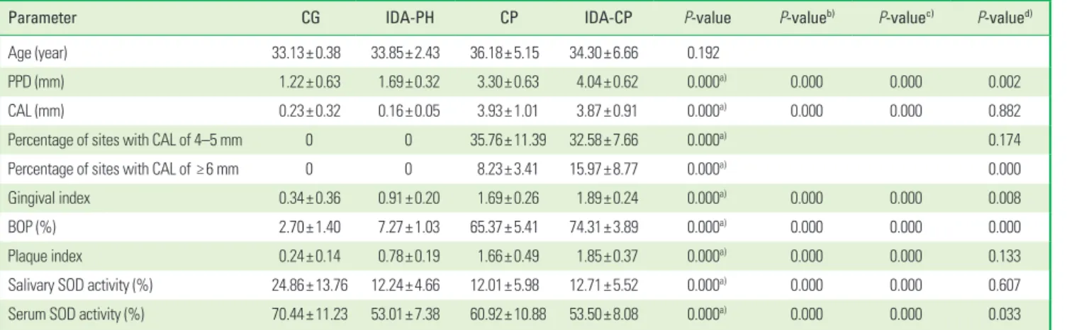
Table 2. Spearman’s rank correlation coefficients among salivary SOD activity (%), serum SOD activity (%), and clinical periodontal parameters in the CG, IDA- PH, and IDA-CP groups.
Parameter Salivay SOD activity (%) Serum SOD activity (%)
r 95% CI P-value r 95% CI P-value
Plaque index –0.384 –0.584 to –0.119 0.002 –0.533 –0.690 to –0.306 0.000
Gingival index –0.372 –0.600 to –0.127 0.003 –0.526 –0.696 to –0.274 0.000
BOP (%) –0.391 –0.593 to –0.135 0.002 –0.574 –0.712 to –0.364 0.000
PPD (mm) –0.511 –0.677 to –0.306 0.000 –0.490 –0.657 to –0.283 0.000
CAL (mm) –0.177 –0.423 to –0.082 0.168 –0.344 –0.547 to –0.131 0.006
Percentage of sites with CAL of 4–5 mm –0.231 –0.440 to –0.010 0.071 –0.308 –0.494 to –0.083 0.015
Percentage of sites with CAL of ≥6 mm –0.340 –0.553 to –0.101 0.007 –0.263 –0.469 to –0.054 0.039
Serum SOD activity (%) 0.400 0.164 to 0.599 0.001
r : Spearman’s rank correlation coefficient, SOD: superoxide dismutase, CG: control group, IDA-PH: periodontally healthy iron deficiency anemia patients, IDA-CP: iron deficiency anemia patients with chronic periodontitis, CI: confidence interval, BOP: bleeding on probing, PPD: probing pocket depth, CAL: clinical attachment loss.
Correlations of SOD activity and clinical periodontal parameters
Spearman’s rank correlation coefficients among salivary, serum SOD activity, and clinical periodontal parameters in iron deficiency anemia patients (pool data of the CG, IDA-PH, and IDA-CP groups)
The data of Table 2 reveal a significant positive correlation be- tween the salivary and the serum SOD activity (P≤0.05). Salivary SOD activity was found to be negatively and significantly correlat- ed with all periodontal parameters (P≤0.05) except CAL and the percentage of sites with CAL of 4–5 mm (P≥0.05). Serum SOD ac- tivity was found to be negatively and significantly correlated with all periodontal parameters including the percentage of sites with CAL of 4–5 and ≥6 mm (P≤0.05).
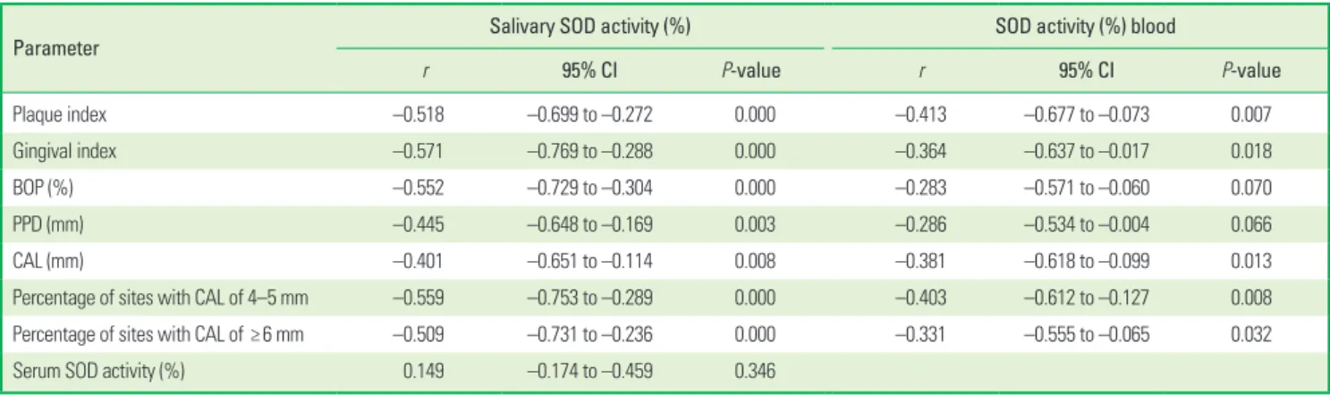
Spearman’s rank correlation coefficients among salivary, serum SOD activity, and clinical periodontal parameters in chronic periodontitis patients (pool data of the CG and CP groups)
Table 3 shows that no correlation was found between the sali- vary and the serum SOD activity (P≥0.05). The salivary SOD activi- ty was found to be negatively and significantly correlated with all the periodontal parameters including the percentage of sites with CAL of 4–5 and ≥6 mm (P≤0.05), whereas the serum SOD activity showed a significant and negative correlation with all periodontal parameters (P≤0.05) except PPD (P≥0.05).
DISCUSSION
Considerable activity of reactive oxygen radicals may lead to the destruction of normal cell functions and the integrity of cell struc- tures. Oxidative stress in biological systems can be induced by the consumption of AOs and/or by an overload of oxidant species, such that AO levels become deficient. Oxidative stress has been deter- mined by the estimation of the products of oxidative damage to lipids, proteins, and DNA or by assaying a single AO compound in
isolation or in groups, or to measure the total AO capacity. By de- tecting a particular AO enzyme activity, we evaluated its impor- tance to a given pathogenic process, for example, the detection of SOD enzyme activity in our study, which represents an important AO defense against excess ROS production in both CP and IDA.
Perusal of the available literature reveals only a single study wherein IDA and CP have both been taken into consideration [19].
To the best of our knowledge, this is the first reported study that evaluated the relationship between chronic periodontitis and iron deficiency anemia with respect to salivary and serum SOD activity levels.
Thus far, numerous studies have been conducted on the associa- tion of the serum and salivary SOD activity levels with periodonti- tis, and the results are equivocal. Kim et al. [20] reported a decrease in the salivary SOD activity in chronic periodontitis patients. Stud- ies by Baltacioglu et al. [21], Canakci et al. [22], and Akalin et al.
[23] have also endorsed the decreased SOD activity in periodontitis.
Conversely, Wei et al. [24] demonstrated a higher activity of SOD in the saliva, serum, and gingival crevicular fluid of chronic periodon- titis patients. In our study, the serum and salivary SOD activity lev- els in the CP patients were significantly lower than those of sys- temically and periodontally healthy individuals. Lower salivary SOD activity could be interpreted as a result of the suppression of SOD production in saliva/PMNLs because of oxidative damage caused by increased ROS/O2- generation [25], while a decrease in the se- rum SOD activity can be attributed to hyperreactive peripheral neutrophils with respect to the production of ROS in response to Fc gamma-receptor stimulation in patients with chronic periodon- titis [26].
A significantly lower value of both salivary and serum SOD ac- tivity was also observed in IDA-PH patients than in the CG at base- line. Studies by Amirkhizi et al. [27], Isler et al. [28], and Kurtoglu et al. [29] have also endorsed the decreased activity of SOD in pa- tients with IDA. In contrast, studies by Acharya et al. [30] and Hafez et al. [31] found increased erythrocytic Cu-Zn SOD activity in IDA Table 3. Spearman’s rank correlation coefficients among salivary SOD activity (%), serum SOD activity (%), and clinical periodontal parameters in the CG and CP groups.
Parameter Salivary SOD activity (%) SOD activity (%) blood
r 95% CI P-value r 95% CI P-value
Plaque index –0.518 –0.699 to –0.272 0.000 –0.413 –0.677 to –0.073 0.007
Gingival index –0.571 –0.769 to –0.288 0.000 –0.364 –0.637 to –0.017 0.018
BOP (%) –0.552 –0.729 to –0.304 0.000 –0.283 –0.571 to –0.060 0.070
PPD (mm) –0.445 –0.648 to –0.169 0.003 –0.286 –0.534 to –0.004 0.066
CAL (mm) –0.401 –0.651 to –0.114 0.008 –0.381 –0.618 to –0.099 0.013
Percentage of sites with CAL of 4–5 mm –0.559 –0.753 to –0.289 0.000 –0.403 –0.612 to –0.127 0.008
Percentage of sites with CAL of ≥6 mm –0.509 –0.731 to –0.236 0.000 –0.331 –0.555 to –0.065 0.032
Serum SOD activity (%) 0.149 –0.174 to –0.459 0.346
SOD: superoxide dismutase, CG: control group, CP: chronic periodontitis, r : Spearman’s rank correlation coefficient, CI: confidence interval, BOP: bleeding on probing, PPD: probing pocket depth, CAL: clinical attachment loss.
patients. In the present study, the lower SOD activity in anemic pa- tients may be linked to increased hemoglobin auto-oxidation ow- ing to hypoxia resulting in the increased RBC production of super- oxide and hydrogen peroxide. These RBC-derived ROS can damage the cell membrane and leak out of the RBC [32]. It is well known that ROS, particularly hydrogen peroxide, inhibit SOD activity [27].
Although there was no significant difference in the salivary and serum SOD activity in the IDA-CP patients and the CP patients, sig- nificantly higher baseline PPD, GI, and BOP% were observed in the IDA-CP group than in the CP group in spite of similar PI values.
Moreover, the present study showed that the IDA-CP patients had worse periodontal conditions, expressed as the percentage of sites with a CAL of ≥6 mm, than the CP group. Higher periodontal de- struction in the IDA-CP group can be attributed to the synergistic effect of both ROS-induced oxidative stress and other mechanisms augmenting inflammation owing to iron deficiency anemia. ROS can directly cause periodontal tissue damage by the degradation of the extracellular matrix components of periodontal tissues or can play an indirect role in potentiating extracellular matrix degrada- tion by matrix metalloproteinase, via the activation of latent en- zymes, such as collagenase and gelatinase [33], and via the inacti- vation of enzyme inhibitors, such as tissue inhibitor of metallopro- teinase and α 1-antiproteinase [34]. In addition, the production of ROS and the subsequent disturbance in the tissue redox status can modulate the expression of a variety of immune and inflammatory molecules via redox-sensitive transcription factors (e.g., nuclear factor κB), thereby causing indirect tissue damage and exacerbat- ing inflammation [35].
Furthermore, IDA has been reported to augment inflammation through a number of mechanisms. Hypersegmented neutrophils found in IDA can cause immune suppression by directly suppressing T-cell responses by the creation of an immunological synapse and the direct delivery of hydrogen peroxide resulting in tissue damage.
Inhibition of the activity of iron-dependent myeloperoxidase, which mediates the bactericidal activity of macrophages [36], may be another associated mechanism. Moreover, alteration in the gin- gival blood flow due to the reduced erythrocyte count might de- crease the oxygen content in the gingival tissues [12]. Hypoxia pro- duced as a result, induced the activation of the transcription factor hypoxia-inducible factor-1, triggering the expression of proinflam- matory genes [37] and increased production of mitochondrial ROS, which activate the endothelial secretion of leukocyte adhesion re- ceptors, thereby promoting the inflammatory response [38].
We also analyzed the relationship between the serum and the salivary SOD activities in iron deficiency anemia patients and found a significant positive correlation, while the CP group did not dem- onstrate any correlation between the salivary and the serum SOD activity. However, a comparison of the test groups (IDA-PH, CP, and IDA-CP) did not show a significant difference in the salivary and serum SOD activity.
The study was carried out with female patients because IDA in males is rare and is generally related to neoplastic diseases [39].
This was the limitation of our study, as we could not evaluate the effects in men.
Being a preliminary study, this study led to the following major conclusions: (1) Higher periodontal inflammation and severe de- struction was observed in the IDA-CP patients than in the CP group. (2) The serum and salivary SOD activities in all test groups were lower than those in the CG. (3) A significant positive correla- tion between the salivary and the serum SOD activity was observed in the iron deficiency anemia group. Furthermore, the serum SOD activity was significantly and negatively correlated with all peri- odontal parameters as well as the percentage of sites with CAL of 4–5 mm and ≥6 mm, while salivary SOD did not show significant correlation with CAL and the percentage of sites with CAL of 4–5 mm. (4) In the CP patients, no correlation was observed between the serum and the salivary SOD activity. The salivary SOD activity was significantly and negatively correlated with all periodontal pa- rameters as well as the percentage of sites with CAL of 4–5 mm and ≥6 mm, while the serum SOD activity was significantly and negatively correlated with PI, GI, and mean CAL as well as the per- centage of sites having CAL of 4–5 and ≥6 mm. (5) IDA influences the serum SOD activity to a greater extent but does not appear to affect the salivary SOD activity in patients with both IDA and CP than in patients having only CP.
The results of our study indicate that IDA patients may be at in- creased risk of periodontal destruction and have lower SOD defense levels. However, studies of larger groups with an interventional na- ture will be required to gain a clearer understanding of the rela- tionship between these two disease entities.
CONFLICT OF INTEREST
No potential conflict of interest relevant to this article was re- ported.
ORCID
Souvik Chakraborty http://orcid.org/0000-0001-5305-1686 Shikha Tewari http://orcid.org/0000-0002-2659-333x
Rajinder Kumar Sharma http://orcid.org/0000-0001-7839-1097 Satish Chander Narula http://orcid.org/0000-0002-1024-2498 Pratap Singh Ghalaut http://orcid.org/0000-0002-7420-1628 Veena Ghalaut http://orcid.org/0000-0002-1716-4277
REFERENCES
1. Sculley DV, Langley-Evans SC. Salivary antioxidants and peri- odontal disease status. Proc Nutr Soc 2002;61:137-43.
2. Canakci CF, Cicek Y, Canakci V. Reactive oxygen species and hu- man inflammatory periodontal diseases. Biochemistry (Mosc) 2005;70:619-28.
3. Ozmeric N. Advances in periodontal disease markers. Clin Chim Acta 2004;343:1-16.
4. Halliwell B, Gutteridge JM. The antioxidants of human extracel- lular fluids. Arch Biochem Biophys 1990;280:1-8.
5. McCord JM. Human disease, free radicals, and the oxidant/anti- oxidant balance. Clin Biochem 1993;26:351-7.
6. Fridovich I. Superoxide anion radical (O2-.), superoxide dismutas- es, and related matters. J Biol Chem 1997;272:18515-7.
7. Mapp PI, Grootveld MC, Blake DR. Hypoxia, oxidative stress and rheumatoid arthritis. Br Med Bull 1995;51:419-36.
8. Macnee W, Rahman I. Oxidants and antioxidants as therapeutic targets in chronic obstructive pulmonary disease. Am J Respir Crit Care Med 1999;160(5 Pt 2):S58-65.
9. Elbim C, Pillet S, Prevost MH, Preira A, Girard PM, Rogine N, et al.
Redox and activation status of monocytes from human immu- nodeficiency virus-infected patients: relationship with viral load.
J Virol 1999;73:4561-6.
10. Halliwell B. The role of oxygen radicals in human disease, with particular reference to the vascular system. Haemostasis 1993;23 Suppl 1:118-26.
11. Toxqui L, De Piero A, Courtois V, Bastida S, Sanchez-Muniz FJ, Vaquero MP. Iron deficiency and overload. Implications in oxida- tive stress and cardiovascular health. Nutr Hosp 2010;25:350-65.
12. Lainson PA, Brady PP, Fraleigh CM. Anemia, a systemic cause of periodontal disease? J Periodontol 1968;39:35-8.
13. Chapple IL, Brock GR, Milward MR, Ling N, Matthews JB. Com- promised GCF total antioxidant capacity in periodontitis: cause or effect? J Clin Periodontol 2007;34:103-10.
14. Page RC, Eke PI. Case definitions for use in population-based sur- veillance of periodontitis. J Periodontol 2007;78(7 Suppl):1387-99.
15. Adamson JW. Iron deficiency and other hypoproliferative anemias.
In: Longo DL, Fauci AS, Kasper DL, Hauser SL, Jameson JL, Loscalzo J. Harrison’s principle of internal medicine. Vol 1. New York : Mc- Graw Hill; 2012. p.844-61.
16. Loe H, Silness J. Periodontal disease in pregnancy. I. Prevalence and severity. Acta Odontol Scand 1963;21:533-51.
17. Silness J, Loe H. Periodontal disease in pregnancy. II. Correlation between oral hygiene and periodontal condtion. Acta Odontol Scand 1964;22:121-35.
18. Tamaki N, Tomofuji T, Ekuni D, Yamanaka R, Yamamoto T, Morita M. Short-term effects of non-surgical periodontal treatment on plasma level of reactive oxygen metabolites in patients with chronic periodontitis. J Periodontol 2009;80:901-6.
19. Enhos S, Duran I, Erdem S, Buyukbas S. Relationship between iron-deficiency anemia and periodontal status in female pa- tients. J Periodontol 2009;80:1750-5.
20. Kim SC, Kim OS, Kim OJ, Kim YJ, Chung HJ. Antioxidant profile of whole saliva after scaling and root planing in periodontal dis- ease. J Periodontal Implant Sci 2010;40:164-71.
21. Baltacioglu E, Akalin FA, Alver A, Balaban F, Unsal M, Karabulut E.
Total antioxidant capacity and superoxide dismutase activity levels in serum and gingival crevicular fluid in post-menopausal women with chronic periodontitis. J Clin Periodontol 2006;33:
385-92.
22. Canakci V, Yildirim A, Canakci CF, Eltas A, Cicek Y, Canakci H. To- tal antioxidant capacity and antioxidant enzymes in serum, sali- va, and gingival crevicular fluid of preeclamptic women with and without periodontal disease. J Periodontol 2007;78:1602-11.
23. Akalin FA, Baltacioglu E, Alver A, Karabulut E. Total antioxidant capacity and superoxide dismutase activity levels in serum and gingival crevicular fluid in pregnant women with chronic peri- odontitis. J Periodontol 2009;80:457-67.
24. Wei D, Zhang XL, Wang YZ, Yang CX, Chen G. Lipid peroxidation levels, total oxidant status and superoxide dismutase in serum, saliva and gingival crevicular fluid in chronic periodontitis pa- tients before and after periodontal therapy. Aust Dent J 2010;
55:70-8.
25. Guarnieri C, Zucchelli G, Bernardi F, Scheda M, Valentini AF, Ca- landriello M. Enhanced superoxide production with no change of the antioxidant activity in gingival fluid of patients with chronic adult periodontitis. Free Radic Res Commun 1991;15:11-6.
26. Matthews JB, Wright HJ, Roberts A, Cooper PR, Chapple IL. Hy- peractivity and reactivity of peripheral blood neutrophils in chronic periodontitis. Clin Exp Immunol 2007;147:255-64.
27. Amirkhizi F, Siassi F, Minaie S, Djalali M, Rahimi A, Chamari M.
Assessment of lipid peroxidation and activities of erythrocyte cytoprotective enzymes in women with iron deficiency anemia. J Res Med Sci 2008; 13:248-54.
28. Isler M, Delibas N, Guclu M, Gultekin F, Sutcu R, Bahceci M, et al.
Superoxide dismutase and glutathione peroxidase in erythro- cytes of patients with iron deficiency anemia: effects of differ- ent treatment modalities. Croat Med J 2002;43:16-9.
29. Kurtoglu E, Ugur A, Baltaci AK, Undar L. Effect of iron supple- mentation on oxidative stress and antioxidant status in iron-de- ficiency anemia. Biol Trace Elem Res 2003;96:117-23.
30. Acharya J, Punchard NA, Taylor JA, Thompson RP, Pearson TC.
Red cell lipid peroxidation and antioxidant enzymes in iron defi- ciency. Eur J Haematol 1991;47:287-91.
31. Hafez FM, Hassab HM, Mourad ZE, Ascalany HE. Red blood cells superoxide dismutase activity in iron deficiency anemia. Alex J Ped 1999; 13:439-42.
32. Rifkind JM, Abugo O. Alterations in erythrocyte deformability under hypoxia: implications for impaired oxygen transport. In:
Hogan MC, Mathieu-Costello O, Poole DC, Wagner PD. Oxygen transport to tissue. Vol 16. New York: Plenum Press; 1994. p.345- 51.
33. Ramamurthy NS, Vernillo AT, Greenwald RA, Lee HM, Sorsa T, Golub LM, et al. Reactive oxygen species activate and tetracy- clines inhibit rat osteoblast collagenase. J Bone Miner Res 1993;
8:1247-53.
34. Shabani F, McNeil J, Tippett L. The oxidative inactivation of tis- sue inhibitor of metalloproteinase-1 (TIMP-1) by hypochlorous acid (HOCI) is suppressed by anti-rheumatic drugs. Free Radic Res 1998;28:115-23.
35. Chapple IL, Matthews JB. The role of reactive oxygen and anti- oxidant species in periodontal tissue destruction. Periodontol
2000 2007;43:160-232.
36. Arredondo M, Nunez MT. Iron and copper metabolism. Mol As- pects Med 2005;26:313-27.
37. Peyssonnaux C, Cejudo-Martin P, Doedens A, Zinkernagel AS, Johnson RS, Nizet V. Cutting edge: Essential role of hypoxia in- ducible factor-1alpha in development of lipopolysaccharide-in- duced sepsis. J Immunol 2007;178:7516-9.
38. Ichimura H, Parthasarathi K, Issekutz AC, Bhattacharya J. Pres- sure-induced leukocyte margination in lung postcapillary ve- nules. Am J Physiol Lung Cell Mol Physiol 2005;289:L407-12.
39. Dallman PR, Yip R, Oski A. Iron deficiency and related nutritional anemias. In: Nathan DG, Oski FA, editors. Hematology of infancy and childhood. 5th ed. Philadelphia: W.B. Saunders; 1998. p.430- 76.
![Figure 1. Comparison of salivary superoxide dismutase (SOD), activity (%) be- be-tween the control group (CG) and the test groups (periodontally healthy iron deficiency anemia patients [IDA-PH], chronic periodontitis patients [CP], iron deficiency anemia](https://thumb-ap.123doks.com/thumbv2/123dokinfo/5316948.165597/4.892.84.420.453.699/comparison-salivary-superoxide-dismutase-periodontally-deficiency-periodontitis-deficiency.webp)