www.jpis.org
pISSN 2093-2278 eISSN 2093-2286 Copyright © 2010 Korean Academy of PeriodontologyThis is an Open Access article distributed under the terms of the Creative Commons Attribution Non-Commercial License (http://creativecommons.org/licenses/by-nc/3.0/).
Clinical evaluation of a collagen matrix to enhance the width of keratinized gingiva around dental implants
Kang-Ho Lee, Byung-Ock Kim, Hyun-Seon Jang* Department of Periodontology, Chosun University School of Dentistry, Gwangju, Korea
Purpose: The purpose of this study was to evaluate the effect of collagen matrix with apically positioned flap (APF) on the width of keratinized gingiva, comparing to the results of APF only and APF combined with free gingival graft (FGG) at the sec- ond implant surgery.
Methods: Nine patients were selected from those who had received treatments at the Department of Periodontics, Chosun University Dental Hospital, Gwangju, Korea. We performed APF, APF combined with FGG, and APF combined with collagen matrix coverage respectively. Clinical evaluation of keratinized gingival was performed by measuring the distance from the gingival crest to the mucogingival junction at the mid-buccal point, using a periodontal probe before and after the surgery.
Results: The ratio of an increase was 0.3, 0.6, and 0.6 for the three subjects in the APF cases, 3, 5, and 7 for the three in the APF combined with FGG case, and 1.5, 0.5, and 3 for the three in the APF combined with collagen matrix coverage case.
Conclusions: This study suggests that the collagen matrix when used as a soft tissue substitute with the aim of increasing the width of keratinized tissue or mucosa, was as effective and predictable as the FGG.
Keywords: Collagen, Dental implantation, Gingiva.
INTRODUCTION
The purposes of soft tissue management around dental implants are successful primary closure, papillary reconstruc- tion, gain of keratinized tissue and preservation of ridge contour. Keratinized tissue is a specialized mucosa covered with keratin or parakeratin which includes the free and at- tached gingiva and extends from the gingival margin to the mucogingival junction. However, The need and significance of keratinized tissue around dental implants is a controver- sial issue. Wennstrom et al. [1] reported that there is no clini- cal difference between teeth with and without adequate ker- atinized tissue and no association between the width of kera- tinized tissue and the presence of bleeding on probing. Ben-
gazi et al. [2] reported that the width of keratinized tissue was a poor predictor for occurrence of soft tissue recession. The recession was primarily the result of a remodeling of the soft tissue for establishing appropriate biological dimensions. Al- brektsson et al. [3] reported that dental implants may have a high survival rate, irrespective of keratinized conditions. Al- though the significance of keratinized tissue is still contro- versial, it is certain that the attached gingiva provides in- creased resistance of the periodontium to external injury, contributes to the stabilization of the gingival margin posi- tion, and aids in the dissipation of physiological forces that are exerted by the muscular fibers of the alveolar mucosa on the gingival tissues [4]. Despite the observation that the lack of keratinized tissue does not influence long term implant
Received: Dec. 31, 2009; Accepted: Mar. 10, 2010
*Correspondence: Hyun-Seon Jang
Department of Periodontology, Chosun University School of Dentistry, 421 Seosuk-dong, Dong-gu, Gwangju 501-825, Korea E mail: [email protected], Tel: +82 -62 -220- 3854, Fax: +82 -62- 224- 4664
techniques to obtain adequate amounts of keratinized tissue around two stage implants, primarily based on the preserva- tion of keratinized tissue over the edentulous ridge. At the time of implant exposure, apically positioned or laterally po- sitioned flaps were proposed to reconstruct keratinized tis- sue of adequate width around implants. When the amount of keratinized tissue over the edentulous ridge was minimal, a free gingival graft (FGG) was suggested. These techniques however are associated with significant patient morbidity due to the need for creating a wound at the palatal donor site. Recently, many of the disadvantages of the classic pro- cedure have been overcome by modification and the use of tissue engineering materials. Wei et al. [6] used an acellular dermal matrix allograft to achieve increased attached gingi- va. Simion [7] used a collagen membrane for soft tissue heal- ing. More recently, a collagen matrix has been proposed as a substitute for the palatal donor tissue to augment gingival tissue dimensions [8]. Here we performed APF only, APF combined with FGG, and APF combined with collagen ma- trix coverage at the second implant surgery and evaluated the width of keratinized tissue around dental implant.
CASE DESCRIPTION
Subjects
Nine patients were selected from those who had received treatments at the Department of Periodontics, Chosun Uni- versity Dental Hospital, Gwangju, Korea. The inclusion crite- ria were as follows:
· The subjects were eriodontally and systemically healthy.
· The subjects presented with at least one site with mini- mal or no keratinized tissue.
· The subjects received a second implant surgery four months after the initial implant surgery.
All patients received the ethics committee approved in- formed consent form. This study protocol was approved by the Chosun University Dental Hospital Institutional Review Board (CDMDIRB-0902-14).
Surgical procedure
We performed APF, APF combined with FGG, and APF combined with collagen matrix coverage (Collatape®, Zim- mer Dental, Carlsbad, USA) at the second implant surgery.
The time point soft tissue management was 4 months after the initial implant surgery. For a proper standardization be- tween baseline and follow up data, periodontal probes were
based on the following criteria:
· If the keratinized tissue had a width greater than three millimeters, the APF technique was performed.
· If the keratinized tissue width was in the two to three- millimeter range, the APF combined with collagen matrix coverage technique was performed.
· If the keratinized tissue was minimal, then the APF com- bined with FGG technique was performed.
At the time of the surgery, local anesthesia was adminis- tered and the surgical procedure was performed. After pa- tient selection was complete, all of the cases underwent a surgical procedure designed to enlarge the area of kerati- nized tissue. The surgical technique used for the groups con- sisted of the following steps:
Case 1 - Apically positioned flap
Using a #15C blade (Ace Surgical Supply Co., Brockton, USA), a mucosal partial-thickness flap was raised. The recipi- ent site was prepared by sharp dissection in order to create a periosteal bed free of any muscle attachment. The resulting flap was sutured at the base of the newly created vestibule with 5-0 non-resorbable nylon sutures (Happylon, Shirakawa Co., Takayama, Japan) (Fig. 1).
Case 2 - APF combined with FGG
FGGs were performed according to the original technique described by Sullivan and Atkins. A recipient bed was pre- pared similar to Case 1 and a free graft was harvested from the palate (Fig. 2).
Case 3 - APF combined with collagen matrix coverage
After preparing the recipient site, collagen matrix was trimmed and shaped to fit the recipient site. Collagen matrix was fixed the same manner as in the FGG method (Fig. 3). All three patient cases were then instructed to rinse twice daily with a chlorhexidine mouth rinse (0.12%) for 2 weeks. Anti- inflammatory therapy (Amoxicillin 625 mg) was prescribed, and the patients were given instructions to take this drug for three days. Sutures were removed after 10 days. The collagen matrices used in this clinical evaluation were Collatape® (Zimmer Dental, Carlsbad, USA) in sterile, individual bubble packs. These matrices are fabricated using collagen obtained from bovine deep flexor (Achilles) tendon, and function to control bleeding, stabilize blood clots, protect wound beds, and provide a matrix for tissue ingrowth, and they absorb in approximately 10-14 days. However, if exposed directly to the
oral environment, they will be absorbed more rapidly. To test the toxicity of Collatape® (Zimmer Dental, Carlsbad, USA), Luitaud et al. [9] seeded epithelial cells onto the CollaTape, and epithelium formation was analyzed at set times. The epi- thelial cells adhered, proliferated, and began to stratify as early as 2 days post-seeding. Advanced stratification was ob- served at 6 days post-seeding. This result confirms that Col- latape® (Zimmer Dental, Carlsbad, USA) material is nontoxic and capable of supporting fibroblast adhesion and growth.
Clinical measurements
Clinical evaluation of keratinized gingival was performed by measuring the distance from the gingival crest to the mucogingival junction at the mid-buccal point, using a peri- odontal probe (PCP 10®, Hu-Friedy, Chicago, USA). It was re- corded pre-operatively and after soft tissue healing.
The subject consisted of 9 patients; 3 subjects were allocat- ed in the APF case, 3 subjects were allocated in the APF com- bined with FGG, and 3 subjects were allocated in the APF combined with collagen matrix coverage. No patients in any Figure 1. Case 1 - Apically positioned flap. (A) Pre-surgical image of #36 implant site. Note the presence of approximately 3 mm width of ker- atinized tissue exists. (B) Split partial thickness flap was elevated and sutured at the base of the newly created vestibule with 5-0 non-resorb- able nylon sutures. (C) Five months after prosthetic setting, the keratinized tissue was well maintained. However, the vertical incision at the time of surgery produced a frenum at #35 distal area.
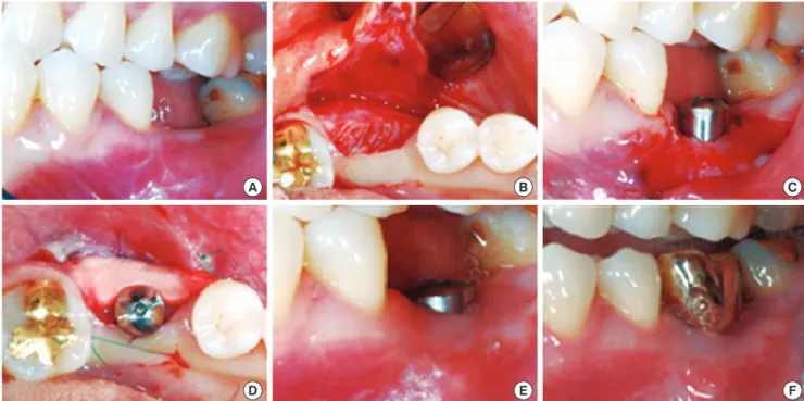
Figure 2. Case 2 - Apically positioned flap combined with free gingival grafts (FGG). (A) Pre-surgical image of #36 and 37 implant sites. Note the minimal amount of keratinized tissue on the edentulous ridge. (B) Split partial-thickness flap was elevated and sutured at the base of the newly created vestibule with 5-0 non-resorbable nylon sutures. (C) Dimension of the FGG retrieved from the patient’s palate. (D) FGG su- tured on the recipient bed. (E) Healing of the FGG after three weeks post surgery. Note the presence of 3-4 mm of keratinized tissue around the healing abutment. (F) Some shrinkage has taken place after 3 months.
A B C
D A
E B
F C
of the cases developed any significant complications. The change of keratinized tissue width is shown in Table 1. The increased ratio was 0.3, 0.6, and 0.6 for the three subjects in the APF cases 3, 5, and 7 for the three subjects in the APF combined with FGG cases, and 1.5, 0.5, and 3 for the three subjects in the APF combined with collagen matrix coverage cases. These results showed greatest increase in the kerati-
nized tissue for the FGG cases, and a mild to moderate in- crease for the APF and APF combined with collagen matrix coverage case. At baseline, the width of the keratinized tissue in the FGG cases were minimal and the other cases showed a similar width, which ranged from 1 to 3 mm. The amount of the keratinized tissue increased over the 3 to 4-week post- surgical period in all three cases. The APF combined with collagen matrix coverage cases show a similar or greater in- crease of keratinized tissue increase as in APF only areas.
Similarly to the APF combined with FGG areas, the APF com- bined with collagen matrix coverage cases showed more fa- vorable physiologic morphology then the APF only cases.
DISCUSSION
The controversy regarding the need for an ‘adequate’ width of keratinized tissue around teeth in order to preserve peri- odontal health still exists. There are clinical situations where the presence of a certain width of keratinized tissue may be important in maintaining periodontal health and preventing soft tissue recession, such as in the areas around fixed pros- thetic restorations [10]. In spite of the observation that the lack of keratinized tissue may not influence implant survival, the careful management of soft tissue around implants is Figure 3. Case 3 - Apically positioned flap combined with collagen matrix coverage. (A) Pre-surgical image of #36 implant site.
2-3 mm of keratinized tissue is present. Note the adjacent teeth buccal-gingival line. Keratinized tissue loss was detected after tooth loss. (B) Split partial-thickness flap was elevated (C) Flap was sutured at the base of the newly created vestibule with 5-0 non-resorbable nylon sutures. (D) Collagen matrix covered the recipient bed. (E) One months later, 2 mm of keratinized tissue gain was observed vertically and horizontally. (F) Well maintained keratinized tissue after 6 months.
Table 1. Dimensional change of keratinized gingiva before and after surgery.
Patient
No. Cases Site
Width of keratinized tissue Baseline
(mm) Post-surgery (mm) Increase
(ratio)
1 APF #47 3 4 0.3
2 APF #36 3 5 0.6
3 APF #35 3 5 0.6
4 APF + FGG #34, 35, 36 0.5 2 3
5 APF + FGG #36, 37 0.5 3 5
6 APF + FGG #14, 15, 16 0.5 4 7
7 APF + CM #47 1 2.5 1.5
8 APF + CM #14 2 3 0.5
9 APF + CM #36 1 4 3
APF: apically positioned flap, FGG: free gingival graft, CM: collagen matrix.
D A
E B
F C
atinized tissue has been achieved traditionally using the FGG [16]. Augmentation of keratinized tissue width and vestibular deepening with autogenous FGGs has been reported to be a predictable and effective method [17-19].
Although the incidence of complications is very low, dis- comfort and pain at the donor site are frequently observed.
This technique causes other wounds at the palatal site, and increases the morbidity of the patient.
To avoid this morbidity, a substitute for palatal donor tissue has been studied. For example, acellular dermal matrix al- lograft, collagen membrane, and collagen matrix have been used, instead of palatal tissue. The acellular dermal matrix al- lograft showed good results at soft tissue augmentation, be- cause this material is derived from human cadavers, howev- er, it is associated with ethical concerns and the possible risk of disease transmission [20-22].
Collagen membrane has also been shown to have a posi- tive effect on soft tissue augmentation and healing, but colla- gen matrix has a more porous layer meaning greater kerati- nized tissue can be achieved because of the space creating effect and blood clot formation [23]. Thus, the collagen matrix can be expected to be more effective at augmenting kerati- nized tissue. The main objective of this clinical study was to evaluate the changes in width of keratinized tissue following three surgical techniques, APF, APF combined with FGG, and APF combined with collagen matrix coverage. The result from this clinical study indicated that after 3-4 weeks, all of the cases achieved keratinized tissue. Furthermore, after 3-4 weeks, the three cases also achieved proliferation and matu- ration. The FGG cases showed a greater increase of kerati- nized tissue, and the APF combined with collagen matrix coverage cases showed more keratinized tissue increase than the APF only cases. The keratinized tissue after surgery in the APF combined with collagen matrix coverage cases were characterized by more physiologic and favorable morpholo- gy than the APF only cases. It is believed that the collagen matrix acts as a scaffold to prevent the mucosal relapse, and protection of the recipient bed.
Evidence of the advantages of collagen matrix is insuffi- cient. Therefore, further studies are necessary to determine the influence of collagen matrix on the recipient bed, and on how to prolong the short absorption period of collagen ma- trix (10-14 days).
CONFLICT OF INTEREST
No potential conflict of interest relevant to this article was reported.
Wennstrom JL, Bengazi F, Lekholm U. The influence of 1.
the masticatory mucosa on the peri-implant soft tissue condition. Clin Oral Implants Res 1994;5:1-8.
Bengazi F, Wennstrom JL, Lekholm U. Recession of the 2.
soft tissue margin at oral implants. A 2-year longitudinal prospective study. Clin Oral Implants Res 1996;7:303-10.
Albrektsson T, Zarb G, Worthington P, Eriksson AR. The 3.
long-term efficacy of currently used dental implants: a re- view and proposed criteria of success. Int J Oral Maxillofac Implants 1986;1:11-25.
Lang NP, Loe H. The relationship between the width of 4.
keratinized gingiva and gingival health. J Periodontol 1972;43:623-7.
Langer B, Sullivan DY. Osseointegration: its impact on the 5.
interrelationship of periodontics and restorative dentist- ry: Part I. Int J Periodontics Restorative Dent 1989;9:84- 105.
Wei PC, Laurell L, Geivelis M, Lingen MW, Maddalozzo D.
6.
Acellular dermal matrix allografts to achieve increased at- tached gingiva. Part 1. A clinical study. J Periodontol 2000;
71:1297-305.
Simion M. Soft tissue healing on application of a natural 7.
collagen matrix. 6th Congress of the European Federation of Periodontology; 2009 June 4-6; Stockholm, Sweden.
Sanz M, Lorenzo R, Aranda JJ, Martin C, Orsini M. Clinical 8.
evaluation of a new collagen matrix (Mucograft prototype) to enhance the width of keratinized tissue in patients with fixed prosthetic restorations: a randomized prospective clinical trial. J Clin Periodontol 2009;36:868-76.
Luitaud C, Laflamme C, Semlali A, Saidi S, Grenier G, 9.
Zakrzewski A, et al. Development of an engineering au- tologous palatal mucosa-like tissue for potential clinical applications. J Biomed Mater Res B Appl Biomater 2007;
83:554-61.
Barone R, Clauser C, Grassi R, Merli M, Prato GP. A proto- 10.
col for maintaining or increasing the width of masticatory mucosa around submerged implants: a 1-year prospective study on 53 patients. Int J Periodontics Restorative Dent 1998;18:377-87.
Prato GP, Clauser C, Cortellini P. Periodontal plastic and 11.
mucogingival surgery. Periodontol 2000 1995;9:90-105.
Dorfman HS, Kennedy JE, Bird WC. Longitudinal evalua- 12.
tion of free autogenous gingival grafts. J Clin Periodontol 1980;7:316-24.
Wennstrom J, Lindhe J, Nyman S. Role of keratinized gin- 13.
giva for gingival health. Clinical and histologic study of normal and regenerated gingival tissue in dogs. J Clin Periodontol 1981;8:311-28.
Kennedy JE, Bird WC, Palcanis KG, Dorfman HS. A longi- 15.
tudinal evaluation of varying widths of attached gingiva. J Clin Periodontol 1985;12:667-75.
Langer B, Langer L. Subepithelial connective tissue graft 16.
technique for root coverage. J Periodontol 1985;56:715-20.
Bohannan HM. Studies in the alteration of vestibular 17.
depth I. Complete denudation. J Periodontol 1962;33:120-7.
Egli U, Vollmer WH, Rateitschak KH. Follow-up studies of 18.
free gingival grafts. J Clin Periodontol 1975;2:98-104.
Han TJ, Takei HH, Carranza FA. The strip gingival au- 19.
tograft technique. Int J Periodontics Restorative Dent 1993;
13:180-7.
lar dermal graft and palatal autograft in the reconstruc- tion of keratinized gingiva around dental implants: a case report. Int J Periodontics Restorative Dent 2006;26:287-92.
Imberman M. Gingival augmentation with an acellular 22.
dermal matrix revisited: surgical technique for gingival grafting. Pract Proced Aesthet Dent 2007;19:123-8.
Hammerle CH, Jung RE, Feloutzis A. A systematic review 23.
of the survival of implants in bone sites augmented with barrier membranes (guided bone regeneration) in partial- ly edentulous patients. J Clin Periodontol 2002;29 Suppl 3:226-31.