Introduction
The attached gingiva (AG) consists of keratinized epithelium, dense connective tissue and periosteum, and plays an important role in protecting the periodontal structures [1]. For many years, the presence of an ‘adequate’ amount of keratinized gingiva was considered to be a keystone for the maintenance of periodontal health [2,3].
The presence of site-related conditions, e.g., gingival
recession, thin periodontium and root prominence, combined with a reduced or missing amount of AG, may indicate a gingival augmentation procedure. Serino et al. [4] reported that sites with gingival recession should be considered susceptible to additional apical displacement of the soft tissue margin.
The American Academy of Periodontology [5] suggested the following indications for gingival augmentation procedures:
to prevent soft tissue damage in the presence of alveolar bone dehiscence during natural or orthodontic tooth eruption; to halt progressive marginal gingival recession; to improve plaque control and patient comfort around the teeth and implants;
and to increase the insufficient dimensions of the gingiva in conjunction with fixed or removable prosthetic dentistry.
Since Friedman introduced the term ‘mucogingival surgery’
in the 1950’s, a range of procedures have been used to correct
The increase of keratinized and attached gingiva using collagen wound dressing in dogs: A clinical and histomorphometric comparative study
Sang-Joun Yu
1, In-Chae Na
1, Han-Wook Jang
1, Moon-Jin Jung
2, Byung-Ock Kim
1,*
Departments of
1Periodontology and
2Oral Histology, School of Dentistry, Chosun University, Gwangju, Korea
ABSTRACT
Purpose: This study examined the width of keratinized gingiva and attached gingiva after an apically repositioned flap (APF group), APF combined with free gingival grafts (FGG group), and APF combined with collagen wound dressing (Collatape®
group), both clinically and histomorphometrically.
Materials and Methods: The right and left maxillary canine areas of eight mongrel dogs were used (16 surgical sites). In the maxilla, the canine areas were used as experimental sites. Three different surgical techniques were performed on the sixteen canine areas(apically repositioned flap; APF group, APF combined with free gingival grafts; FGG group, and APF combined with collagen wound dressing; Collatape® group). After 6 weeks, clinical and histomorphometric evaluation were done.
Results: The FGG group showed more attached gingiva and a more favorable physiological appearance than the other groups.
However, there was no significant difference in keratinized gingiva and attached gingiva among the three groups. According to the results of the histomorphometric examination, keratinized gingiva were formed in all groups. The FGG group showed thicker epithelium and connective tissue than the APF group. Collatape® group showed thicker connective tissue than the APF group.
Conclusion: The clinical and histomorphometric results suggested that APF combined with collagen wound dressing promotes more favorable healing of the keratinized and attached gingiva.
Key Words: Animal experimentation, Free tissue flaps, Membranes, Wound healing
Received Sep 5, 2014; Revised version received Mar 16, 2015 Accepted Mar 16, 2015
Corresponding author: Byung-Ock Kim
Department of Periodontology, School of Dentistry, Chosun University, 303 Pilmun-daero, Dong-gu, Gwangju 501-825, Korea Tel: 82-62-220-3856, Fax: 82-62-224-4664
E-mail: [email protected]
problems associated with the lack of AG [6]. One of the first surgical techniques designed to correct such problems was an apically repositioned flap (APF) [7]. This technique allowed surgeons to increase or preserve the area of AG by moving the tissue apically and exposing a variable band of crestal bone depending on how much AG was required [7]. Another techniques to increase AG was a free gingival graft (FGG) [8]
and free connective tissue graft [9,10]. Two of the advantages of these techniques are the availability of adequate donor tissue and ability to treat multiple teeth. The disadvantages include technical difficulty, postoperative discomfort, and poor continuity with adjacent tissue in color or shape.
Recently, many of the disadvantages of the classic procedure have been overcome by a modification of the procedure and the use of tissue engineering materials [7,11]. Collagen wound dressings have been used to stabilize blood clots, and protect the wound bed. Furthermore, they absorb blood and wound exudates and promote hemostasis, thus aiding wound healing while concurrently enhancing patient’s comfort [12].
The purpose of this study was to evaluate the width of keratinized gingiva and AG after an APF, APF combined with FGG, and APF combined with collagen wound dressing, clinically and histomor phometrically in dogs.
Materials and Methods
Surgical procedure
Eight mongrel dogs, approximately 1-year-old and weighing 17 to 19 kg each, were used in this experiment. The study protocol was approved by the Chosun University Dental
Hospital Institutional Review Board (#CDMDIRB-0902-A29).
Supragingival scaling was performed on all dogs prior to surgery. General anesthesia was induced by an injection of tiletamine-zolazepam (Zoletil 50
®; Virbac, Carros, France; 5 to 10 mg/kg, intramuscular) and xyalzine HCL (Rompun
®; Bayer, Seoul, Korea; 0.15 mL/kg, intramuscular).
In both quadrants of the maxilla, the canine areas were used as experimental sites. Three different surgical techniques were performed on the sixteen canine areas (Table 1).
First, only an APF was performed (APF group). APF was performed according to the modified technique described by Carnio and Miller [7]. Before making the incisions, the level of crestal bone was probed to detect the presence of bone dehiscence. A periodontal probe or anesthetic needle can be used via the gingival sulcus. A horizontal beveled incision was made in the attached portion of the keratinized gingiva slightly apical to the alveolar crest. Notch was formed 3 mm under gingival margin with round bur. Mesial and distal extensions of the initial horizontal incision were then made (15 mm).
Two vertical incisions were made on the mesial and distal ends connecting the horizontal incision (15 mm). These incisions extended beyond the mucogingival junction (MGJ). A split- thickness flap was elevated, moved apically, positioned at the desired level, and fixed with a periosteal horizontal suture using resorbable suture materials (Monosyn
®5-0; B. Braun Melsungen AG, Bethlehem, PA, USA). The size of the exposed periosteal bed was 15×15 mm (Fig. 1A).
Second, APF combined with a FGG was performed (FGG group). The APF procedure was performed in the same manner described for the first technique. The FGG was harvested from the palate, trimmed and shaped to fit the recipient site (Fig.
1B). The graft thickness was approximately 1.5 mm. The graft was fixed to the periosteal bed using a horizontal key suture.
Pressure was applied to the recipient site for 3 minutes after suturing to ensure hemostasis and tissue adaptation.
Third, APF combined with collagen wound dressing (Collatape
®; Zimmer Dental, Carlsbad, CA, USA) was performed (Collatape
®group). Twofold Collatape
®was used for the same thickness as the FGG. After preparing the recipient site, Collatape
®was trimmed and shaped to fit the recipient site (Fig. 1C). Collatape
®was fixed in a similar manner as described for the FGG method. Pressure was also applied for 3 minutes.
Table 1. Surgical Areas in the Eight Dogs
Dog No. Maxillary canine
Right Left
Dog 1 Dog 2 Dog 3 Dog 4 Dog 5 Dog 6 Dog 7 Dog 8
APF group FGG group Collatape® group APF group FGG group Collatape® group FGG group APF group
FGG group
Collatape® group
APF group
FGG group
Collatape® group
APF group
Collatape® group
Collatape® group
APF group: apically repositioned flap only, FGG group: apically
repositioned flap+free gingival graft, Collatape® group: apically re-
positioned flap+Collatape®.
Postsurgical care
After surgery, each dog was injected with an antibacterial agent (Gentamicin 0.1mL/kg; Daesung, Gwangju, Korea) for seven days. Tooth cleaning with 0.2% chlorohexidine digluconate was performed three times per week for 4 weeks.
The sutures were removed two weeks after surgery.
Clinical measurements
The index was marked on the mid-buccal surface of the canine, 3 mm from the gingival margin. The probing depth (PD) was measured at three points (mesio-buccal, mid-buccal, and disto-buccal) to the nearest millimeter with a force-controlled probe (tip diameter: 0.45 mm; probing force: 20 g/pressure).
The PDs at the three points were averaged for the purpose of analysis. The width of keratinized gingival (KG) at the mid- buccal point was measured from the MGJ to the free gingival margin. The width and thickness of the AG was calculated by subtracting the PD at the mid-buccal point from the width of
the KG to the nearest millimeter (Fig. 1A, F).
Histologic and histomorphometric evaluation
Six weeks after APF, a soft tissue biopsy was harvested from
A B C
D E F
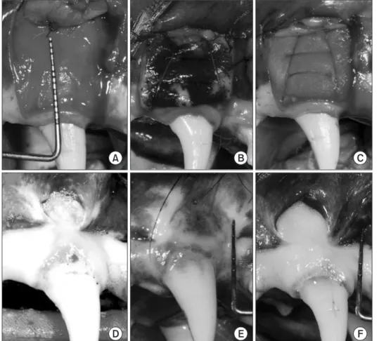
Fig. 1. Clinical procedure. (A) Apically repositioned flap (APF) was done.
Recipient bed preparation was per- formed (15×15 mm) (APF group). (B) Free gingival graft (FGG) harvested from the palate (15×15×1.5 mm).
Horizontal key suture to fix the graft on the recipient bed (FGG group).
(C) Two fold collagen wound dress- ing (Collatape®) was similar thick- ness to the gingival graft (15×15×1.5 mm). Horizontal key suture to fix the Collatape® on the recipient bed (col- latape® group). (D-F) Six weeks heal- ing after surgery. Smooth and physi- ological morphology of the attached gingiva was observed (D: APF group, E: FGG group, F: Collatape® group).
Epi
CT
Fig. 2. Histomorphometric evaluation (H&E, ×40).
Epi: thickness of epithelium, CT: thickness of connective tissue.
each dog under local anesthesia. The biopsy was performed in these areas. All the specimens were fixed in a 10% neutral buffered formalin solution for further descriptive histological analyses. After they were dehydrated in a graded ethanol series, the specimens were embedded in paraffin and serially sectioned in 5 µm thick sections. Each section was stained individually with H&E stain. The thickness of epithelium and connective tissue in coronal area of all sections was observed by using a optical microscope (LEICA DM750; Leica Microsystems, Wetzlar, Germany) equipped with a digital camera (LEICA ICC50 camera; Leica Microsystems) with ×40 and ×100
magnification and histomorphometric measurements was attained by calculating the average length of each section by measuring the length of the sections 10 times each using the i-SOLUTION Lite
®processing and analysis program (IMT i-Solution Inc., Daejeon, Korea) on a personal computer (Fig. 2).
The thickness of the section’s epithelium and connective tissue was measured perpendicularly to the surface of the tissue that attached to root.
Statistical analysis
A statistical software program (SPSS 16.0; SPSS Inc., Chicago, A
APF group 2.5
2.0
1.5
1.0
0.5
Probingdepth(mm)
0
FGG group APF group
25
20
15
10
5
Widthofkeratinizedgingiva(mm)
0
FGG group
Collatape group
RCollatape group
RB
* * *
Baseline Post-surgery
*
APF group 25
20
15
10
5
Widthofattachedgingiva(mm)
0
FGG group Collatape group
RAPF group 35
30 25 20 15 10
5
Shrinkageofkeratinizedgingiva(%)
0
FGG group Collatape group
RC D
* *
Baseline Post-surgery
Baseline Post-surgery
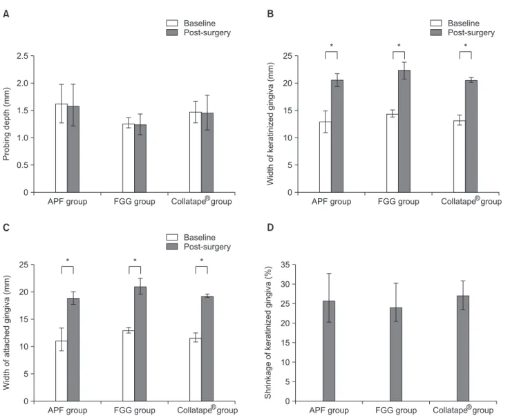
Fig. 3. Clinical parameters at the baseline and post-surgery. (A) Probing depth after surgery. (B) Width of keratinized gingiva after surgery. (C) Width of attached gingiva after surgery. (D) Shrinkage of keratinized gingiva after surgery.
APF group: apically repositioned flap only, FGG group: apically repositioned flap+free gingival graft, Collatape® group: apically repositioned flap+Collatape®.
*Statistically significant differences (p<0.05) by paired t-test.
IL, USA) was used for all statistical analyses. A paired t-test was performed to analyze the differences between the baseline and six weeks after surgery. An analysis of the variance (ANOVA)
was performed to examine the difference in the continuous clinical parameters between the three surgical procedures. In addition, histomorphometric measurement was evaluated by
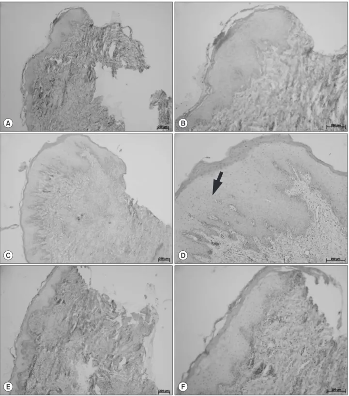
Fig. 4. Histological view. (A, B) Apically repositioned flap (APF) only group. (C, D) APF combined with free gingival graft group (black arrow:
gingival epithelium). (E, F) APF combined with Collatape® coverage (H&E; A, C, E: ×40, B, D, F: ×100).
A B
C D
E F
ANOVA and post-mortem of Games-Howel. A p-value <0.05 was considered significant.
Results
Clinical findings
Healing of all groups was uneventful. The soft tissue grafts were fully integrated without any signs of necrosis (Fig. 1D-F).
Fig. 3 lists the preoperative and postoperative clinical measurements. Treatment with the three surgical procedures resulted in significant augmentation of the apico-coronal dimensions of the keratinized gingiva and AG (p<0.05) (Fig. 1).
In the APF group, the mean apico-coronal dimension of the keratinized gingiva was 12.85 mm (range, 7.2 to 16.2 mm) preoperatively and 20.40 mm (range, 17.0 to 21.9 mm) postoperatively. The mean apico-coronal dimension of the AG was 11.23 mm (range, 5.4 to 14.2 mm) preoperatively and 18.80 mm (range, 15.5 to 20.4 mm) postoperatively (Fig. 3B, C).
In the FGG group, the mean apico-coronal dimension of the keratinized gingiva was 14.30 mm (range, 13.1 to 15.3 mm) preoperatively and 22.23 mm (range, 19.7 to 26.5 mm) postoperatively. The mean apico-coronal dimension of the AG was 13.03 mm (range, 11.9 to 14.0 mm) preoperatively and 20.98 mm (range, 18.2 to 25.2 mm) postoperatively (Fig. 3B, C).
In the Collatape
®group, the mean apico-coronal dimension of the keratinized gingiva was 13.13 mm (range, 10.8 to 15.1 mm) preoperatively and 20.43 mm (range, 19.8 to 21.5 mm)
postoperatively. The mean apico-coronal dimension of the AG was 11.65 mm (range, 9.1 to 13.5 mm) preoperatively and 19.15 mm (range, 18.3 to 19.9 mm) postoperatively (Fig. 3B, C).
There was no significant difference in PD detected pre- and postoperatively in each procedure (Fig. 3A).
The average shrinkage of apico-coronal dimension in keratinized gingiva marked 25.5% (range, 4.95% to 38.63%) in the APF group, 23.9% (reange, 5.69% to 33.33%) in the FGG group and 27.03% (range, 16.67% to 34.22%) in the Collatape
®group, respectively (Fig. 3D).
Histologic and histomorphometric findings
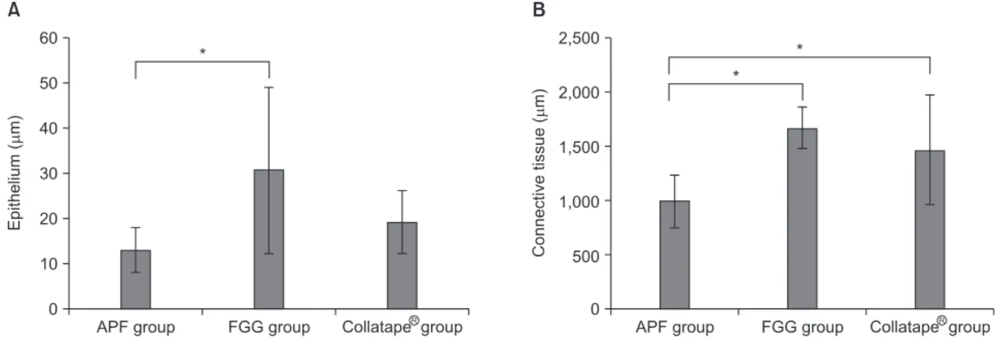
According to the results of the histological examination, keratinized gingiva was formed in all groups (Fig. 4). The FGG group showed the thickest epithelium, and the APF group showed the thinnest epithelium and connective tissue in coronal area (Fig. 5).
The thickness of the keratinized gingiva was measured using the telescopic histology images. In the APF group, the average thickness of epithelium and entire connective tissue was 13.2 µm (range, 9.3 to 24.0 µm) and 1,002.7 µm (range, 706.7 to 1,440.0 µm), respectively. In the FGG group, the average thickness of epithelium and connective tissue was 30.8 µm (range, 12.0 to 49.3 µm) and 1,677.3 µm (range, 1,413.3 to 2,000.0 µm), respectively. In the Collatape
®group, the average thickness of whole epithelium and keratinized layer was 19.2 µm (range, 10.7 to 30.7 µm) and 1,476 µm (range, 600.0
APF group 60
50 40 30 20 10
Epithelium(m)
0
FGG group Collatape group
RA
APF group 2,500
2,000
1,500
1,000
Connectivetissue(m)