ORIGINAL ARTICLE
위 신생물의 기저 등급에 따른 내시경 절제술 후 재발 양상
정고은1, 정수진1, 양종인1, 진은효1, 박민정2, 김상균3, 김주성1,3
서울대학교병원 강남센터 헬스케어연구소 내과1, 셰이크 칼리파 전문병원 내과2, 서울대학교 의과대학 내과학교실 및 간연구소3
Development of Metachronous Tumors after Endoscopic Resection for Gastric Neoplasm according to the Baseline Tumor Grade at a Health Checkup Center
Goh Eun Chung1, Su Jin Chung1, Jong In Yang1, Eun Hyo Jin1, Min Jung Park2, Sang Gyun Kim3 and Joo Sung Kim1,3
Department of Internal Medicine, Healthcare Research Institute, Healthcare System Gangnam Center, Seoul National University Hospital1, Seoul, Korea, Department of Internal Medicine, Sheikh Khalifa Specialty Hospital2, Ras AlKhaimah, UAE, Department of Internal Medicine and Liver Research Institute, Seoul National University College of Medicine3, Seoul, Korea
Background/Aims: Endoscopic resection (ER) procedure has been performed widely to treat gastric neoplasms. Here, we compared the long-term prognosis based on the clinical features of three types of recurred gastric neoplasms after ER, including low-grade dys- plasia (LGD), high-grade dysplasia (HGD), and early gastric carcinoma (EGC).
Methods: Between 2003 and 2014, subjects who were diagnosed with gastric neoplasm during screening endoscopy were included.
The baseline clinicopathologic and tumor recurrence were analyzed.
Results: Of the 316 patients enrolled, 170 patients (53.8%) were categorized into the LGD group, 34 patients (10.8%) into the HGD group, and 112 patients (35.4%) into the EGC group. The median follow-up duration was 4.2 years. Among the total, 14 patients experi- enced a development of metachronous gastric cancer; 4 patients (2.3%) in the LGD group, 3 patients (8.3%) in the HGD group, and 7 patients (6.1%) in the EGC group. Metachronous gastric neoplasm had developed in 17 LGD patients (10.0%), 5 HGD patients (14.7%), and 14 EGC patients (12.5%). There was no significant difference in the incidence of metachronous gastric cancer and neo- plasm among the three groups (p=0.15 and p=0.72, respectively).
Conclusions: We identified that the incidence rates of gastric neoplasm and cancer after endoscopic treatment were not significantly different between the LGD, HGD, and EGC groups. (Korean J Gastroenterol 2017;70:223-231)
Key Words: Gastric neoplasm; Recurrence; Adenoma
Received June 14, 2017. Revised August 15, 2017. Accepted August 22, 2017.
CC This is an open access article distributed under the terms of the Creative Commons Attribution Non-Commercial License (http://creativecommons.org/licenses/
by-nc/4.0) which permits unrestricted non-commercial use, distribution, and reproduction in any medium, provided the original work is properly cited.
Copyright © 2017. Korean Society of Gastroenterology.
교신저자: 정수진, 06236, 서울시 강남구 테헤란로 152, 서울대학교병원 강남센터 헬스케어연구소 내과
Correspondence to: Su Jin Chung, Department of Internal Medicine, Healthcare Research Institute, Healthcare System Gangnam Center, Seoul National University Hospital, 152 Teheran-ro, Gangnam-gu, Seoul 06236, Korea. Tel: +82-2-2112-5751, Fax: +82-2-2112-5635, E-mail: medjsj7@hanmail.net
Financial support: This study was supported by grant 04-2015-0760 from the Seoul National University Hospital Research Fund.
Conflict of interest: None.
INTRODUCTION
Stomach cancer is a common malignancy.1 Detection at an early stage reduces morbidity and improves survival. If there is no metastasis in the lymph nodes, the clinical outcome of early stage gastric cancer is very good, with a 5-year survival rate of 99%.2 Gastric dysplasia is considered as a premalig-
nant lesion, suggesting an increased risk of gastric cancer ac- cording to the grade of the dysplasia.3-6
Endoscopic resection (ER) has been widely performed to treat certain cases of gastric neoplasm. However, metachro- nous neoplasm can develop at another site in the stomach because the remnant mucosa may still be affected by pre- cancerous lesions, such as, intestinal metaplasia or gastric
atrophy, thus resulting in increased risk for recurrence.7 In fact, the incidence rate of metachronous or synchronous ne- oplasm after ER has been reported to be 3.0-20.9%.8-10 Previous studies have identified multiple predictors for meta- chronous neoplasm of the stomach, including Helicobactoer pylori (H. pylori) infection, intestinal metaplasia or gastric atrophy, histologic findings, and grade of dysplasia.11,12 Therefore, scheduled endoscopic surveillance is important, especially for high risk patients who have undergone pre- vious ER.
Clear guidelines regarding clinical management of gastric neoplasm after ER are lacking. Moreover, the recommended interval and period of follow-up exams vary widely. Thus, the purpose of this study was to investigate the long-term prog- nosis based on clinical features of three types of recurred gastric neoplasm after ER, including low grade dysplasia (LGD), high grade dysplasia (HGD), and early gastric cancer (EGC). The results of this study may guide individualized sur- veillance strategies based on the primary disease.
SUBJECTS AND METHODS
1. Patients
Between October 2003 and December 2014, subjects who were diagnosed with gastric neoplasm (LGD, HGD, and EGC) during a screening endoscopy at Seoul National University Hospital Healthcare System Gangnam Center were enrolled. The microscopic criteria of gastric neoplasm were defined in accordance with the Vienna classification;
LGD in category 3, HGD in category 4, and invasive carcinoma in category 5.13
Among the 406 cases of ER, 82 cases with less than 12 month of follow-up, 5 cases with additional operation imme- diately after ER, and 3 cases without ER due to old age were all excluded. This study was approved by the Institutional Review Board of Seoul National University; requirement for written consent was waived.
2. Endoscopy procedure and follow-up
In almost all cases, ER was performed with the endoscopic submucosal dissection (ESD) technique. Indications for ER were as follows: low- or high-grade dysplasia lesion regard- less of tumor size and shape; if the lesion was EGC, histologi-
cally-confirmed adenocarcinoma with well or moderate-dif- ferentiation, with the depth of invasion confined to mucosa, a lesion less than 2 cm without evidence of distant or lymph node metastasis. Patients with LGD received several treat- ment modalities, including ESD, endoscopic ablation with ar- gon plasma coagulation, endoscopic mucosal resection, or simple excisional biopsy, and regular surveillance endoscopy with re-biopsy when the size was less than 5 mm,14 based on the size of the lesion, endoscopist’s recommendation, as well as pa- tient preference. After ER for gastric neoplasm, a follow-up en- doscopy to detect any mucosal abnormalities was scheduled at 3, 6, and 12 months after ER, and annually thereafter. We de- fined local recurrence as a lesion found on the ER scar during a surveillance endoscopy.15 Metachronous’ lesion was de- fined as a lesion that had newly developed more than 1 year after the treatment of the primary lesion.
3. Clinicopathological evaluations
H. pylori infection was considered positive based on either positive findings on histological examination or a rapid ure- ase test. Mucosal atrophy or intestinal metaplasia was con- sidered to be present based on pathological examination.
Endoscopic severity of gastric atrophy was assessed based on the location of the atrophic border via endoscopy, by dis- tinctive differences in the visible capillary network, height of the gastric mucosa, and color, as reported by Kimura and Takemoto16 and Liu et al.17 The extent of EAG was categorized into six grades (C1 to O3) in accordance with the Kimura-Takemoto classification. We grouped the six categories into two types:
antral dominant and corpus dominant types.16
4. Statistics
Chi square test or Student’s t-test was performed to com- pare the baseline characteristics in accordance with the pathological type. Kaplan-Meier method with a log-rank test was performed to identify factors affecting tumor recurrence.
Cox proportional hazard model was used to investigate the in- dependent factors associated with increased risk of meta- chronous gastric neoplasm. All statistical analyses were per- formed using SPSS statistics for windows version 22.0 (SPSS Inc., Chicago, IL, USA). p-values of less than 0.05 were consid- ered statistically significant.
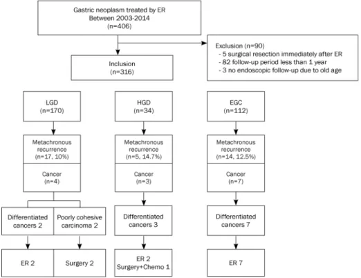
Fig. 1. Enrollment process, patterns of metachronous neoplasia, and treatment modalities in each group. ER, endoscopic resection; LGD, low grade dysplasia; HGD, high grade dysplasia; EGC, early gastric cancer.
RESULTS
1. Baseline characteristics of study population
During the study period, a total of 406 patients underwent ER for primary treatment of gastric neoplasm. Of these, 5 pa- tients underwent additional gastrectomy due to incomplete ER. Moreover, we excluded 82 patients with less than 12 months of follow-up and 3 patients who refused endoscopic follow-up due to old age. The remaining 316 patients were en- rolled for final analysis. Overall, the study included 249 males (79.1%) and 67 females (20.9%), with a mean age of 58.6±
8.8 years (median 58 years, range 36-88 years). In total, 170 patients (53.8%) were categorized into the LGD group, 34 pa- tients (10.8%) into the HGD group, and 112 patients (35.4%) into the EGC group (Fig. 1).
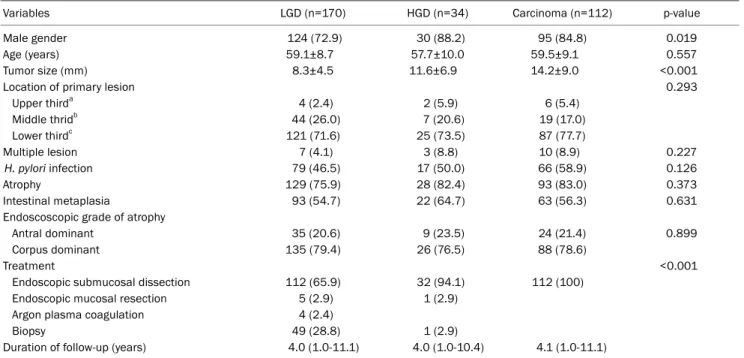
The baseline characteristics of the three groups are shown in Table 1. In the LGD group, the proportion of males was low- er (72.9% vs. 88.2% and 84.8%; p=0.019) and the tumor size was smaller (8.3±4.5 mm vs. 11.6±6.9 mm and 14.2±9.0 mm; p<0.001). There were no significant differences among the three groups with respect to age, location of the primary tumor, lesion multiplicity, H. pylori infection, atrophy and in- testinal metaplasia, follow-up duration, as well as the num- ber of follow-up endoscopies. In total, 49 patients (28.8%) in the LGD group and one patient (2.9%) in the HGD group were
treated using simple biopsy.
2. Development of metachronous neoplasm
The overall mean follow-up period was 4.5±2.4 years and ranged from 1.0 to 11.1 years (median 4.2 years). The lon- gest interval between the initial ER and metachronous tumor recurrence was 6.3 years (range 1.1-6.3, median 3.2 years).
During the follow-up period, a total of 14 metachronous gas- tric cancers were newly developed: 4 patients (2.3%) in the LGD group, 3 patients (8.3%) in the HGD group, and 7 pa- tients (6.1%) in EGC group. The incidence rate of gastric can- cer after endoscopy treatment was 15.2 cases per 1,000 per- son-years in the LGD group, 18.5 cases per 1,000 per- son-years in the HGD group, and 20.9 cases per 1,000 per- son-years in the EGC group. Metachronous gastric neoplasm developed in 17 LGD patients (10.0%), 5 HGD patients (14.7%), and 14 EGC patients (12.5%). The incidence of metachronous neoplasm was 28.6, 45.7, and 39.7 cases per 1,000 person-years in the LGD, HGD, and EGC groups, respectively.
The cumulative incidence of metachronous gastric cancer and neoplasm had no significant difference among the three groups via Kaplan-Meier analysis (p=0.15 and p=0.72, re- spectively, Fig. 2). When we combined the HGD and EGC groups, the cumulative incidence of metachronous gastric
Table 1. Clinical Characteristics according to the Pathologic Type of Initial Gastric Neoplasm
Variables LGD (n=170) HGD (n=34) Carcinoma (n=112) p-value
Male gender 124 (72.9) 30 (88.2) 95 (84.8) 0.019
Age (years) 59.1±8.7 57.7±10.0 59.5±9.1 0.557
Tumor size (mm) 8.3±4.5 11.6±6.9 14.2±9.0 <0.001
Location of primary lesion 0.293
Upper thirda 4 (2.4) 2 (5.9) 6 (5.4)
Middle thridb 44 (26.0) 7 (20.6) 19 (17.0)
Lower thirdc 121 (71.6) 25 (73.5) 87 (77.7)
Multiple lesion 7 (4.1) 3 (8.8) 10 (8.9) 0.227
H. pylori infection 79 (46.5) 17 (50.0) 66 (58.9) 0.126
Atrophy 129 (75.9) 28 (82.4) 93 (83.0) 0.373
Intestinal metaplasia 93 (54.7) 22 (64.7) 63 (56.3) 0.631
Endoscoscopic grade of atrophy
Antral dominant 35 (20.6) 9 (23.5) 24 (21.4) 0.899
Corpus dominant 135 (79.4) 26 (76.5) 88 (78.6)
Treatment <0.001
Endoscopic submucosal dissection 112 (65.9) 32 (94.1) 112 (100)
Endoscopic mucosal resection 5 (2.9) 1 (2.9)
Argon plasma coagulation 4 (2.4)
Biopsy 49 (28.8) 1 (2.9)
Duration of follow-up (years) 4.0 (1.0-11.1) 4.0 (1.0-10.4) 4.1 (1.0-11.1)
Values are presented as n (%), mean±standard deviation or median (range). The status of H. pylori infection was considered positive if the result of 1 or both tests (histologic examination or rapid urease test) was positive.
LGD, low grade dysplasia; HGD, high grade dysplasia; H. pylori, Helicobacter pylori.
aCardia, fundus or high body; bMid body or lower body; cAntrum or angle.
Fig. 2. Development of metachronous neoplasm according to the baseline tumor grade. (A) Kaplan-Meier analysis of the cumulative incidence of gastric cancer recurrence with respect to the pathologic type of the initial neoplasm. (B) Kaplan-Meier analysis of cumulative incidence of meta- chronous neoplasm with respect to the pathologic type of initial neoplasm. HGD, high-grade dysplasia; EGC, early gastric cancer; LGD, low-grade dysplasia.
cancer was higher in the HGD+EGC group than in the LGD group, with a borderline statistical significance (p=0.06, Fig. 3A).
However, the incidence rate of the overall gastric neoplasm after ER did not differ between the two groups. (p=0.43 ac- cording to the log-rank test, Fig. 3B).
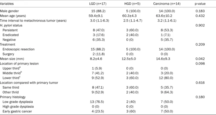
Table 2 compares the clinical features of 36 patients with metachronous gastric neoplasm according to the baseline
tumor grade. There was no significant difference among the three groups in gender, age at metachronous recurrence, treatment modality, and interval to metachronous tumor.
Most patients with metachronous neoplasm (34 of 36, 94.4%) were successfully treated by ER. Metachronous tu- mor size was greater in the HGD and EGC groups than in the LGD group (12.5±5.0 mm and 14.6±9.3 mm vs. 8.2±4.6 mm,
A B
Fig. 3. Comparison of the development of metachronous neoplasm between two groups. (A) Kaplan-Meier analysis of the cumulative incidence of gastric cancer recurrence between two groups. (B) Kaplan-Meier analysis of the cumulative incidence of metachronous neoplasm between the two groups. HGD, high-grade dysplasia; EGC, early gastric cancer; LGD, low-grade dysplasia.
Table 2. Characteristics of Patients with Metachronous Gastric Neoplasm and Their Metachronous Lesions according to the Baseline Tumor Grade
Variables LGD (n=17) HGD (n=5) Carcinoma (n=14) p-value
Male gender 15 (88.2) 5 (100.0) 14 (100.0) 0.183
Mean age (years) 59.4±9.1 60.3±4.3 63.6±10.2 0.432
Time interval to metachronous tumor (years) 3.0 (1.1-6.3) 2.5 (1.1-4.7) 3.2 (1.1-6.1)
H. pylori status 0.902
Persistent 8 (47.0) 3 (60.0) 8 (53.3)
Eradicated 3 (17.6) 2 (40.0) 1 (7.1)
Negative 6 (35.3) 0 (0) 5 (35.7)
Treatment 0.209
Endoscopic resection 15 (88.2) 5 (100.0) 14 (100.0)
Surgery 2 (11.8) 0 (0) 0 (0)
Mean size (mm) 8.2±4.6 12.5±5.0 14.6±9.3 0.042
Location of primary lesion 0.098
Upper thirda 1 (5.9) 0 (0) 0 (0)
Middle thridb 7 (41.2) 2 (40.0) 3 (20.0)
Lower thirdc 9 (52.9) 3 (60.0) 12 (80.0)
Location compared with primary tumor 0.616
Same third 8 (47.1) 3 (60.0) 5 (35.7)
Other thrid 9 (52.9) 2 (40.0) 9 (64.3)
Primary histology 0.180
Low grade dysplasia 13 (76.5) 2 (40) 7 (50.0)
High grade dysplasia 0 (0) 0 (0) 0 (0)
Early gastric cancer 4 (23.5) 3 (60) 7 (50.0)
Values are presented as n (%), mean±standard deviation or median (range).
LGD, low grade dysplasia; HGD, high grade dysplasia; H. pylori, Helicobacter pylori.
aCardia, fundus or high body; bMid body or lower body; cAntrum or angle.
p=0.042). No significant difference was found in the location and histology of metachronous neoplasm among the three groups. Metachronous tumors were in the same third loca- tion as the primary lesion in half of the total (47.1% in the LGD group, 60.0% in the HGD group, and 35.7% in the EGC group).
Fig. 1 shows the patterns of metachronous cancer and treat-
ment modalities in each group. Among the 14 cases of meta- chronous cancer, two EGC cases underwent gastrectomy due to undifferentiated histology. Advanced cancer was identi- fied in one case in the HGD group and was treated with gas- trectomy with additional chemotherapy due to combined liv- er metastasis.
A B
Table 3. Comparison of Clinical Characteristics of Patients with Low Grade Dysplasia between the Simple Excisional Biopsy-removed Group and ESD-treated Group
Variables Simple excisional biopsy-removed
group (n=49)
ESD-treated group
(n=112) p-value
Age (years) 56.7±9.1 59.0±8.5 0.161
Male 35 (71.4) 82 (73.2) 0.847
Tumor size (mm) 5.7±2.1 9.2±4.2 <0.001
Follow-up duration (years) 4.0±2.2 4.6±2.3 0.148
Metachronous neoplasm 7 (14.3) 9 (8.0) 0.473
Local recurrence 3 (6.1) 2 (1.8) 0.382
Values are presented as n (%) or mean±standard deviation.
ESD, endoscopic submucosal dissection.
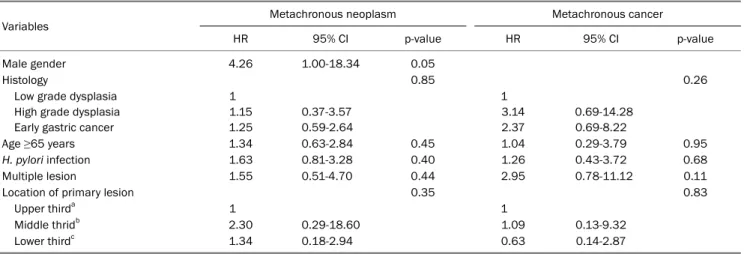
Table 4. Multivariate Cox Regression Analysis for the Cumulative Incidence of Metachronous Gastric Cancer and Neoplasm
Variables Metachronous neoplasm Metachronous cancer
HR 95% CI p-value HR 95% CI p-value
Male gender 4.26 1.00-18.34 0.05
Histology 0.85 0.26
Low grade dysplasia High grade dysplasia Early gastric cancer
1 1.15 1.25
0.37-3.57 0.59-2.64
1 3.14 2.37
0.69-14.28 0.69-8.22
Age ≥65 years 1.34 0.63-2.84 0.45 1.04 0.29-3.79 0.95
H. pylori infection 1.63 0.81-3.28 0.40 1.26 0.43-3.72 0.68
Multiple lesion 1.55 0.51-4.70 0.44 2.95 0.78-11.12 0.11
Location of primary lesion 0.35 0.83
Upper thirda 1 1
Middle thridb 2.30 0.29-18.60 1.09 0.13-9.32
Lower thirdc 1.34 0.18-2.94 0.63 0.14-2.87
HR, hazard ratio; CI, confidence interval; H. pylori, Helicobacter pylori.
aCardia, fundus or high body; bMid body or lower body; cAntrum or angle.
Table 3 provides the clinical characteristics and outcomes of patients with LGD comparing two sub-groups according to the treatment modality; the simple excisional biopsy-removed group vs. the ESD-treated group. Tumor size was significantly smaller in the simple excisional biopsy-removed group com- pared with the ESD-treated group (5.7±2.1 vs. 9.2±4.2, p<0.001). The simple excisional biopsy-removed group had higher rates of metachronous gastric neoplasm and local re- currence compared with the ESD-treated group; however, there was no statistical significance.
3. Risk factors of tumor recurrence after endoscopic treatment
We performed a multivariate Cox proportional hazard anal- ysis to determine the risk of metachronous gastric cancer or neoplasm. Of the variables, only male gender increased the risk for metachronous gastric neoplasm (hazard ratio, 4.26;
95% confidence interval, 1.00-18.34, Table 4). And all of the
metachronous gastric cancer had developed in male. The baseline histological grade of gastric neoplasia was not a sig- nificant predictor for the development of metachronous neoplasm. Older age, lesion multiplicity, H. pylori infection, and background precursor lesions (atrophy or intestinal met- aplasia) were not associated with the risk of metachronous neoplasm or cancer. Among patients who were treated with ESD, en-block resection or submucosal invasion were asso- ciated with increased risk of metachronous neoplasm; how- ever, without statistical significance.
DISCUSSION
We aimed to determine the long-term prognosis based on the clinical features of gastric neoplasm after ER with respect to the grade of dysplasia, including LGD, HGD, and EGC in a primary screening center, not in a tertiary referral center. The incidence rates of gastric neoplasm and cancer after endos-
copy treatment were not significantly different among the three groups. Consistent with our findings, a recent retro- spective study has reported that histological features of gas- tric neoplasm did not affect the development of metachro- nous gastric neoplasm.18 However, in the following points, our study differed from this previous study. First, our study participants were relatively healthy who participated in a health-checkup program, while the previous study was con- ducted at a referral center in an outpatient clinic. Thus, the generalizability is greater in this study. Second, our study had a relatively long-term follow-up period with a median of 50 months, whereas in the previous study, the median follow-up period was 28 months; therefore, this study shows long-term outcomes with greater precision. Finally, in our study, we showed a comparison among three subgroups based on the primary tumor histology, separating the HGD group from in- vasive neoplasia, which shows long-term outcomes of three separate subgroups; in the previous study, however, only two subgroups were used.
The incidence rate of metachronous gastric neoplasm was comparable with the previous Korean study,19 while relatively higher compared with another previous report, as 3.4%, which were evaluated in patients with H. pylori eradication;20 this difference may be attributable to the different H. pylori eradication rate after ER. The preventive effect of H. pylori eradication for metachronous gastric cancer has not been confirmed yet. A prospective study performed in Korea has shown that H. pylori eradication after endoscopy treatment did not significantly decrease the incidence of metachronous cancer.21 Contrastingly, in a randomized controlled study, it was shown that the risk of metachronous carcinoma was re- duced in patients with prophylactic H. pylori eradication.22 In fact, H. pylori eradication has been shown to prevent meta- chronous gastric cancer after ER; and as a result, it has been incorporated in a recent treatment guideline.23 However, H. py- lori eradication after ER, at the moment, is not covered by the National Health Insurance system in Korea.24,25
Despite the removal of gastric neoplasm with the sur- rounding mucosa by ER, most of the gastric tissues with atro- phy or intestinal metaplasia may still remain. Thus, secon- dary lesions can frequently develop in the preserved stom- ach mucosa. In this study, the precursor lesions, such as in- testinal metaplasia, gastric atrophy, or H. pylori infection, did not differ among the three groups; therefore, there may have
been no significant differences in the incidences of meta- chronous gastric neoplasm. This finding may indicate the concept of field for cancerization, i.e., the exposure of a whole field of tissue to continuous carcinogen resulting the occur- rence of cancers in a predisposed filed during multistep process.26 However, when we combined the two groups to- gether (HGD and EGC), the incidence of metachronous gas- tric cancer was higher in the combined group than in the LGD group with borderline statistical significance. This suggests that there is a difference in the premalignant potential be- tween LGD and HGD. In fact, a large cohort study with 10 years of follow-up reported that the annual incidence of gas- tric cancer increased according to the severity of premalig- nant lesion at the initial diagnosis; 0.6% in mild-to-moderate vs. 6% in severe dysplasia.27 This confirms our results. In con- trast to our results, the cumulative incidence of gastric can- cer was not significantly different between the LGD group and the HGD+EGC group in a previous study.17 This difference may be attributable to the difference in the follow-up periods and discrepancies between the two study populations.
Our study demonstrated the long-term outcomes of sub- jects with LGD treated with simple excisional biopsy without additional ESD compared with subjects treated with ESD.
There was no significant difference in the incidences of meta- chronous neoplasm and local recurrence between the sim- ple excisional biopsy-removed group and the ESD-treated group. In this context, these results suggest that ‘simple exci- sional biopsy and follow-up’ can be cautiously applied for small LGD in selected settings, such as old age or the pres- ence of comorbidity.
There have been no clinical guidelines for the surveillance interval and duration after endoscopic treatment. In a pre- vious study, patients received follow-up at 2, 6, and 12 months and annually thereafter.18 In the present study, meta- chronous gastric cancer developed in a patient with EGC 5.7 years after ER and was retreated using ESD, suggesting the importance of long-term regular surveillance. In one case in the HGD group, advanced gastric cancer was found and treat- ed with gastrectomy with additional chemotherapy due to combined liver metastasis. This indicates that careful sur- veillance via endoscopy should be encouraged for the dyspla- sia group as well as for the EGC group with the same levels of attention.
Several factors should be considered when interpreting
the results of this study. First, given the retrospective design of the study, the follow-up duration varied widely and there was a problem with the judgement of H. pylori infection status. A prospective study with a large number of partic- ipants is warranted to verify our results. Second, we did not evaluate the histologic severity of intestinal metaplasia or gastric atrophy. Further study based on the histological evalu- ation for the extent of intestinal metaplasia or gastric atrophy is needed to elaborate the results of this study. Third, the sample size may be too small, particularly considering that there were three different categories compared. Fourth, be- cause the specimen obtained using a biopsy material was not representative of the entire lesion, pathological diagnosis based on forcep biopsy may have been inaccurate.28 Thus, patients with LGD treated with simple excisional biopsy in this study may have confounding effects for the interpretation of long-term follow-up prognosis of ER. Finally, because there is no widely accepted universal interval for a follow-up endos- copy in patients with adenoma,29 the period of follow-up en- doscopy differed between patients with adenoma and those with carcinoma in this study, which may have influenced the quality of the study.
In conclusion, our study has demonstrated the clinical out- come and long-term prognosis of gastric neoplasm treated with ER with respect to the grade of dysplasia. The incidence rates of gastric neoplasm and cancer after endoscopy treat- ment were not significantly different among the three groups –LGD, HGD, and EGC. Long-term careful endoscopic surveil- lance should be considered in patients with dysplasia as well as in those with EGC who underwent previous endoscopy treatment.
REFERENCES
1. Torre LA, Bray F, Siegel RL, Ferlay J, Lortet-Tieulent J, Jemal A.
Global cancer statistics, 2012. CA Cancer J Clin 2015;65:87-108.
2. Japanese Gastric Cancer Association Registration Committee, Maruyama K, Kaminishi M, et al. Gastric cancer treated in 1991 in Japan: data analysis of nationwide registry. Gastric Cancer 2006;9:51-66.
3. Orlowska J, Jarosz D, Pachlewski J, Butruk E. Malignant trans- formation of benign epithelial gastric polyps. Am J Gastroenterol 1995;90:2152-2159.
4. Fujiwara Y, Arakawa T, Fukuda T, et al. Diagnosis of borderline ad- enomas of the stomach by endoscopic mucosal resection. Endoscopy 1996;28:425-430.
5. Kato M, Nishida T, Tsutsui S, et al. Endoscopic submucosal dis-
section as a treatment for gastric noninvasive neoplasia: a multi- center study by Osaka University ESD study group. J Gastroenterol 2011;46:325-331.
6. Yamada H, Ikegami M, Shimoda T, Takagi N, Maruyama M. Long-term follow-up study of gastric adenoma/dysplasia. Endoscopy 2004;36:
390-396.
7. Arima N, Adachi K, Katsube T, et al. Predictive factors for meta- chronous recurrence of early gastric cancer after endoscopic treatment. J Clin Gastroenterol 1999;29:44-47.
8. Kato M, Nishida T, Yamamoto K, et al. Scheduled endoscopic sur- veillance controls secondary cancer after curative endoscopic resection for early gastric cancer: a multicentre retrospective co- hort study by Osaka University ESD study group. Gut 2013;62:
1425-1432.
9. Nasu J, Doi T, Endo H, Nishina T, Hirasaki S, Hyodo I. Character- istics of metachronous multiple early gastric cancers after endo- scopic mucosal resection. Endoscopy 2005;37:990-993.
10. Lim JH, Kim SG, Choi J, Im JP, Kim JS, Jung HC. Risk factors for synchronous or metachronous tumor development after endo- scopic resection of gastric neoplasms. Gastric Cancer 2015;18:
817-823.
11. Seo JH, Park JC, Kim YJ, Shin SK, Lee YC, Lee SK. Undifferentiated histology after endoscopic resection may predict synchronous and metachronous occurrence of early gastric cancer. Digestion 2010;81:35-42.
12. Maehata Y, Nakamura S, Fujisawa K, et al. Long-term effect of Helicobacter pylori eradication on the development of metachro- nous gastric cancer after endoscopic resection of early gastric cancer. Gastrointest Endosc 2012;75:39-46.
13. Schlemper RJ, Riddell RH, Kato Y, et al. The Vienna classification of gastrointestinal epithelial neoplasia. Gut 2000;47:251-255.
14. Srivastava A, Lauwers GY. Gastric epithelial dysplasia: the Western perspective. Dig Liver Dis 2008;40:641-649.
15. Lee JY, Choi IJ, Cho SJ, et al. Routine follow-up biopsies after complete endoscopic resection for early gastric cancer may be unnecessary.
J Gastric Cancer 2012;12:88-98.
16. Kimura K, Takemoto T. An endoscopic recognition of the atrophic border and its significance in chronic gastritis. Endoscopy 1969;
3:87-97.
17. Liu Y, Uemura N, Xiao SD, Tytgat GN, Kate FJ. Agreement between endoscopic and histological gastric atrophy scores. J Gastroenterol 2005;40:123-127.
18. Yoon SB, Park JM, Lim CH, et al. Incidence of gastric cancer after endoscopic resection of gastric adenoma. Gastrointest Endosc 2016;83:1176-1183.
19. Yoon H, Kim N, Shin CM, et al. Risk factors for metachronous gas- tric neoplasms in patients who underwent endoscopic resection of a gastric neoplasm. Gut Liver 2016;10:228-236.
20. Jung S, Park CH, Kim EH, et al. Preventing metachronous gastric lesions after endoscopic submucosal dissection through Helico- bacter pylori eradication. J Gastroenterol Hepatol 2015;30:75-81.
21. Choi J, Kim SG, Yoon H, et al. Eradication of Helicobacter pylori after endoscopic resection of gastric tumors does not reduce in- cidence of metachronous gastric carcinoma. Clin Gastroenterol Hepatol 2014;12:793-800.e1.
22. Fukase K, Kato M, Kikuchi S, et al. Effect of eradication of
Helicobacter pylori on incidence of metachronous gastric carci- noma after endoscopic resection of early gastric cancer: an open- label, randomised controlled trial. Lancet 2008;372:392-397.
23. Kim SG, Jung HK, Lee HL, et al. Guidelines for the diagnosis and treatment of Helicobacter pylori infection in Korea, 2013 revised edition. J Gastroenterol Hepatol 2014;29:1371-1386.
24. Lee SY. New guidelines for Helicobacter pylori treatment: com- parisons between Korea and Japan. Korean J Gastroenterol 2014;63:151-157.
25. Jung DH, Kim JH, Lee YC, et al. Helicobacter pylori eradication re- duces the metachronous recurrence of gastric neoplasms by at- tenuating the precancerous process. J Gastric Cancer 2015;15:
246-255.
26. Papadimitrakopoulou VA, Shin DM, Hong WK. Molecular and cel-
lular biomarkers for field cancerization and multistep process in head and neck tumorigenesis. Cancer Metastasis Rev 1996;15:
53-76.
27. de Vries AC, van Grieken NC, Looman CW, et al. Gastric cancer risk in patients with premalignant gastric lesions: a nationwide cohort study in the Netherlands. Gastroenterology 2008;134:
945-952.
28. Park DI, Rhee PL, Kim JE, et al. Risk factors suggesting malignant transformation of gastric adenoma: univariate and multivariate analysis. Endoscopy 2001;33;501-506.
29. Lage J, Uedo N, Dinis-Ribeiro M, Yao K. Surveillance of patients with gastric precancerous conditions. Best Pract Res Clin Gastroenterol 2016;30:913-922.