Dental malocclusions coupled with skeletal discrepan- cies usually require a combination of surgical and ortho- dontic interventions. Most common orthognathic proce- dures involved in facial, functional, and esthetic correc- tions are Le Fort-I osteotomy, mandibular bilateral sagittal split osteotomy (BSSO), and genioplasty.1
The recent introduction of cone-beam computed tomo- graphy (CBCT) into dentistry has given the surgeon the ability to perform real-time, virtual surgical planning.2It also helps in the diagnosis and dento-facial evaluation of pre-surgical orthognathic cases.2Coupled with a relative- ly low radiation dose and a high spatial resolution, CBCT is rapidly becoming the imaging modality of choice in the pre-surgical evaluation of orthognathic surgical procedures.
Most common incidental findings that appeared during
the interpretation of CBCT volumes are associated with the sinonasal complex, pharyngeal wall and airway, tem- poromandibular joint (TMJ), and the jaws.3
This report is the first to describe a rare case of an oste- olytic lesion found in the clivus of a patient with an other- wise unremarkable history, discovered during the pre-sur- gical interpretation of a CBCT volume.
Case Report
A 27-year-old male patient visited the Department of Orthodontics in the University of Connecticut, School of Dental Medicine with the chief complaint of an “asym- metric face.” The consulting orthodontist performed a facial and dental evaluation and arrived at a working diag- nosis of facial asymmetry associated with skeletal mal- occlusion. An initial panoramic radiograph was taken to evaluate the maxillofacial complex. The panoramic image and posterior-anterior (PA view) showed an asymmetry of the mandible with deviation to the left side and slightly
Clival lesion incidentally discovered on cone-beam computed tomography: A case report and review of the literature
Aniket B. Jadhav1,*, Aditya Tadinada1, Kandasamy Rengasamy1, Douglas Fellows2, Alan G. Lurie1
1Department of Oral and Maxillofacial Radiology, University of Connecticut School of Dental Medicine, Farmington, CT, USA
2Division of Diagnostic Sciences and Therapeutics, University of Connecticut School of Medicine, Farmington, CT, USA
ABSTRACT
An osteolytic lesion with a small central area of mineralization and sclerotic borders was discovered incidentally in the clivus on the cone-beam computed tomography (CBCT) of a 27-year-old male patient. This benign appearance indicated a primary differential diagnosis of non-aggressive lesions such as fibro-osseous lesions and arrested pneu- matization. Further, on magnetic resonance imaging (MRI), the lesion showed a homogenously low T1 signal inten- sity with mild internal enhancement after post-gadolinium and a heterogeneous T2 signal intensity. These signal characteristics might be attributed to the fibrous tissues, chondroid matrix, calcific material, or cystic component of the lesion; thus, chondroblastoma and chondromyxoid fibroma were added to the differential diagnosis. Although this report was limited by the lack of final diagnosis and the patient lost to follow-up, the incidental skull base find- ing would be important for interpreting the entire volume of CBCT by a qualified oral and maxillofacial radiologist.
(Imaging Sci Dent 2014; 44: 165-9)
KEY WORDS: Cranial Fossa, Posterior; Cone-Beam Computed Tomography; Skull Base Neoplasms; Incidental Findings
Received July 8, 2013; Revised December 11, 2013; Accepted December 15, 2013
*Correspondence to : Dr. Aniket B. Jadhav
Department of Diagnostic and Biomedical Sciences, University of Texas School of Dentistry at Houston, 7500 Cambridge St, Suite 5371, Houston, TX 77054, USA Tel) 1-713-486-4134, Fax) 1-713-486-4416, E-mail) [email protected]
Copyright ⓒ 2014 by Korean Academy of Oral and Maxillofacial Radiology
This is an Open Access article distributed under the terms of the Creative Commons Attribution Non-Commercial License (http://creativecommons.org/licenses/by-nc/3.0) which permits unrestricted non-commercial use, distribution, and reproduction in any medium, provided the original work is properly cited.
Imaging Science in Dentistry∙pISSN 2233-7822 eISSN 2233-7830
increased dimension of the right side of the jaw (Fig. 1).
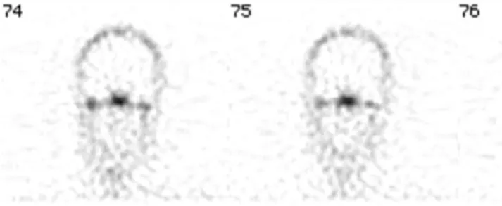
The oral and maxillofacial (OMF) surgeon suspected con- dylar hyperplasia, and to further evaluate the growth of the condyle, a single-photon emission computed tomog- raphy (SPECT) study of the mandible was requested. The SPECT study showed a mild increase in the tracer uptake on the right side, but it was not significant enough to sug- gest active growth (Fig. 2). To correct the facial asymmetry and the skeletal malocclusion, which was the patient’s chief complaint, an orthognathic surgery was planned.
The patient was at this point referred to the Advanced Oral and Maxillofacial Imaging Center at the University of Connecticut School of Dental Medicine for a pre-surgical evaluation using CBCT.
The CBCT image was acquired with a Hitachi CB Mercu- Ray CBCT unit (Hitachi Medical Corporation, Tokyo, Japan) with 12-inch×12-inch field of view (FOV) at 120 kVp and 15 mA, as per the departmental imaging proto- col for a pre-orthognathic surgical evaluation. During the radiographic interpretation of the acquired scan volume, an ellipsoidal, well demarcated, osteolytic lesion with a
small central area of mineralization was incidentally dis- covered on the clivus/basisphenoid (Fig. 3). The lesion extended laterally from the midline to the left margin of the clivus and sagittally to the posterior border of the sphenoid sinus. The borders of this lesion were hyperostotic, and the lesion had the overall dimensions of 12 mm×8.5 mm
×8.5 mm at its greatest right angle diameters. No mass effect was noted on the adjacent bony structures, most importantly, the carotid canals. Given its location and radiographic appearance of a non-aggressive nature, a primary differential diagnosis of a benign fibro-osseous lesion and arrested pneumatization was made.
Since CBCT inherently lacks the differential attenuation for soft tissue, this case was discussed with a neuroradiol- ogist who recommended magnetic resonance imaging (MRI) to evaluate the internal contents and possible soft tissue involvement. An MRI of the brain was performed with sagittal and axial T1 pre- and post-gadolinium, T2, fluid attenuation inversion recovery (FLAIR), diffusion weighted images (DWI), apparent diffusion coefficient (ADC), and gradient-echo (GRE) sequences. On the T1 and FLAIR sequences (Figs. 4A and B), the lesion showed hypointensity, while on the T2 weighted sequence (Fig.
4C), it had a heterogeneous signal intensity. Mild internal enhancement was noted on the post-gadolinium T1 images (Fig. 4D). These MRI findings might be attributed to the possible fibrous, chondroid, or cystic component of the lesion, and a differential diagnosis of chondromyxoid fib- roma and chondroblastoma was made.
The DWI, ADC, and GRE sequences showed marked magnetic susceptibility secondary to orthodontic braces and were not of diagnostic value.
Clival lesion incidentally discovered on cone-beam computed tomography: A case report and review of the literature
Fig. 1.A. A panoramic radiograph of the patient shows increased ramal height on right side. B. A posterio-anterior skull view shows facial asymmetry and deviated jaw.
A B
Fig. 2.SPECT study shows very mild uptake in the right mandibu- lar condyle.
Discussion
The posterior portion of a sphenoid body along with the basiocciput forms the clivus. The spheno-occipital syn- chondrosis, which is one of the last sutures in the body to fuse, is primarily responsible for the growth of the skull base postnatally.4 Margins of the clivus include a petro-
occipital fissure anterior-laterally while basiocciput and the exo-occipital bone form the posterior-lateral margin.
Anteriorly, it blends into the posterior body of the sphe- noid sinus, and posteriorly, it continues as the anterior aspect of the foramen magnum.
Anatomically, the clivus can be found well on the sagit- tal sections on CBCT, CT, and MRI. The clivus consists
Fig. 3.A. Axial CBCT shows osteolytic lesion on clivus with hyperostotic borders and small area of mineralization internally. B. Coronal image shows lateral extent of the lesion and sclerotic rim. C. Sagittal image shows anterio-posterior extension of the lesion.
A B C
Fig. 4.A and B. Coronal T1 weighted (A) and axial FLAIR MR images show low signal intensity. C. Coronal T2 weighted image shows heterogeneous intensity inter- nally. D. Coronal T1 post gadolinium image shows very mild internal enhancement.
A B
C D
of a central cancellous bone and a peripheral cortical bone.
On the MRI, the signal intensity depends upon the nature and the amount of marrow in the cancellous clivus. In young individuals, the hematopoietic component predom- inates, which results in a low T1 signal. As the fatty trans- formation of the marrow occurs with advancing age, the T1 signal becomes brighter. Inflammation, tumors, vascular lesions, and various other abnormalities can cause altered signal intensity due to the replacement of the fatty marrow.5 The apparent site of the lesion is often an indicator of its embryological origin, and most tumors develop according to the tissue present in a particular region. Arrested pneu- matization can be confused with a variety of skull base lesions; on CT images, this could appear as an osteolytic lesion with sclerotic margins and sometimes internal cur- vilinear calcifications. MRI shows a high T1 signal, which corresponds to the fat, while T2 appears from a low-to- high signal intensity.6 In our case, all of the above MRI characteristics were met except that there was no fat signal on the T1 acquisitions.
Tumors in the clivus are uncommon and can be inciden- tal findings on the CT and the MRI. Primary bone neo- plasms, including chordoma, chondrosarcoma, plasma- cytoma, sellar lesions, benign fatty lesions, and metastatic lesions are known to involve the clivus.7-10
Chordoma is an osteolytic lesion that usually has sharp borders with bony fragments internally, as opposed to ring- like calcifications often seen in chondrosarcomas. Both of these lesions have a tendency to expand and cause destruc- tion of the adjacent structures. In our case, the lesion was non-aggressive appearing with sclerotic margins, which represented a typical bone reaction to a slow growth pro- cess. Solitary plasmacytoma is rare in the clivus and usu- ally progresses to multiple myeloma. These osteolytic lesions appear hyperdense on CT and have an intermediate- to-high signal intensity on both T1 and T2 MRI.11
Chondroid tumors, including chondromyxoid fibroma, originate in the cartilaginous remnants of the skull base synchondrosis.12Petroclival synchondrosis is known to be involved most commonly. Chondroblastomas are epiphy- seal tumors of long bones but can occur in the skull base, although cases have been reported involving the temporal bone and rarely, the sphenoid bone.13-15 These osteolytic lesions with sclerotic margins tend to expand and show internal calcifications. On the MRI, they are usually hypo- intense on T1 and heterogeneously intense on T2 with vari- able contrast enhancement following gadolinium. These imaging characteristics are consistent with our case except that the lesion was not expansile.
Sellar lesions, such as pituitary macroadenomas, can invade and expand the sella turcica laterally into the cav- ernous sinuses and inferiorly into the clivus.8 Neoplastic lesion and infections of the nasopharynx can invade from below into the skull base, notably against the clivus and the foramen lacerum.16
Several studies have evaluated the prevalence of inciden- tal findings on CBCT.3,17A majority of such incidental find- ings are reported in the sinonasal complex, C-spine, and the soft tissues. Incidental findings, which are also known as incidentaloma, may be defined as “an incidentally dis- covered mass or lesion detected by CT, MRI, or other imaging modality performed for an unrelated reason.”18 A majority of such incidental findings are benign; how- ever, as some of them may be serious, it is essential to evaluate and report such lesions.
Due to the utilization of variable FOVs on CBCT, it is not uncommon to see the skull base on larger FOV scans, and thus, it has become important to understand the patho- logies and their imaging characteristics on CT and MRI.
The relevance of this case is to present a rare but potential- ly significant incidental finding in the skull base. It is often important to have the entire volume of a large FOV CBCT interpreted by a qualified OMF radiologist. Although this case report is limited by the lack of final diagnosis and the patient was lost to follow-up, the incidental skull base finding is important and instructive for all dentists using large FOV CBCT imaging in caring for their patients.
References
1. Alhadidi A, Cevidanes LH, Paniagua B, Cook R, Festy F, Tyndall D. 3D quantification of mandibular asymmetry using the SPHARM-PDM tool box. Int J Comput Assist Radiol Surg 2012; 7: 265-71.
2. Swennen GR, Mollemans W, Schutyser F. Three-dimensional treatment planning of orthognathic surgery in the era of virtual imaging. J Oral Maxillofac Surg 2009; 67; 2080-92.
3. Price JB, Thaw KL, Tyndall DA, Ludlow JB, Padilla RJ. Inci- dental findings from cone beam computed tomography of the maxillofacial region: a descriptive retrospective study. Clin Oral Implants Res 2012; 23: 1261-8.
4. Laine FJ, Nadel L, Braun IF. CT and MR imaging of the cen- tral skull base. Part 1: Techniques, embryologic development, and anatomy. Radiographics 1990; 10: 591-602.
5. Chaljub G, Van Fleet R, Guinto FC Jr, Crow WN, Martinez L, Kumar R. MR imaging of clival and paraclival lesions. AJR Am J Roentgenol 1992; 159: 1069-74.
6. Welker KM, DeLone DR, Lane JI, Gilbertson JR. Arrested pneumatization of the skull base: imaging characteristic. AJR Am J Roentgenol 2008; 190: 1691-6.
7. Bloch OG, Jian BJ, Yang I, Han SJ, Aranda D, Ahn BJ, et al.
Cranial chondrosarcoma and recurrence. Skull Base 2010; 20:
Clival lesion incidentally discovered on cone-beam computed tomography: A case report and review of the literature
149-56.
8. Borges A. Skull base tumours: Part II. Central skull base tum- ours and intrinsic tumours of the bony skull base. Eur J Radiol 2008; 66: 348-62.
9. Douis H, Saifuddin A. The imaging of cartilaginous bone tumours. II. Chondrosarcoma. Skeletal Radiol 2013; 42: 611- 26.
10. Géhanne C, Delpierre I, Damry N, Devroede B, Brihaye P, Christophe C. Skull base chordoma: CT and MRI features.
JBR-BTR 2005; 88: 325-7.
11. Wein RO, Popat SR, Doerr TD, Dutcher PO. Plasma cell tum- ors of the skull base: four case reports and literature review.
Skull Base 2002; 12: 77-86.
12. Borges A. Imaging of the central skull base. Neuroimaging Clin N Am 2009; 19: 669-96.
13. Ben Salem D, Allaoui M, Dumousset E, Ponnelle T, Justrabo E, Martin D, et al. Chondroblastoma of the temporal bone
associated with a persistent hypoglossal artery. Acta Neurochir (Wien) 2002; 144: 1315-8.
14. Hatano M, De Donato G, Falcioni M, Sanna M. Chondroblas- toma of the temporal bone. Acta Otolaryngol 2011; 131: 890-5.
15. Dran G, Niesar E, Vandenbos F, Noel G, Paquis P, Lonjon M.
Chondroblastoma of the apex portion of petrousal bone. Childs Nerv Syst 2007; 23: 231-5.
16. Weber AL. Imaging of the skull base. Eur J Radiol 1996; 22:
68-81.
17. Scarfe WC, Li Z, Aboelmaaty W, Scott SA, Farman AG.
Maxillofacial cone beam computed tomography: essence, ele- ments and steps to interpretation. Aust Dent J 2012; 57 Suppl 1: 46-60.
18. Berland LL, Silverman SG, Gore RM, Mayo-Smith WW, Megibow AJ, Yee J, et al. Managing incidental findings on abdominal CT: white paper of the ACR incidental findings committee. J Am Coll Radiol 2010; 7: 754-73.