Received on March 27, 2009. Accepted on April 3, 2009.
*Corresponding Author. Tel: 82-2-3277-3248; Fax: 82-2-3277-3760; E-mail: [email protected]
Keywords: TRAP (transmembrane adaptor protein), LAT (Linker for activation of T cells), LIME (Lck-interacting trans- membrane adaptor protein), lymphocyte
Transmembrane Adaptor Proteins Positively Regulating the Activation of Lymphocytes
Inyoung Park and Yungdae Yun*
Department of Life Science, Ewha Womans' University, Seoul 120-750, Korea
Engagement of the immunoreceptors initiates signaling cas- cades resulting in lymphocyte activation and differentiation to effector cells, which are essential for the elimination of pathogens from the body. For the transduction of these im- munoreceptor-mediated signals, several linker proteins term- ed transmembrane adaptor proteins (TRAPs) were shown to be required. TRAPs serve as platforms for the assembly and membrane targeting of the specific signaling proteins. Amo- ng seven TRAPs identified so far, LAT and LIME were shown to act as a positive regulator in TCR-mediated signaling pathways. In this review, we will discuss the functions of LAT and LIME in modulating T cell development, activation and differentiation.
[Immune Network 2009;9(2):53-57]
INTRODUCTION
Lymphocytes respond to various extracellular stimuli medi- ated by surface molecules such as immunoreceptors (e.g., the T cell receptors; TCR, the B cell receptors; BCR, or the Fc receptors; FcR) and additional coreceptors (e.g., CD28 for T cells and CD19 for B cells). As the earliest immunor- eceptor-mediated biochemical events, Src-family protein ty- rosine kinases (PTKs) become activated. In turn, activated Src PTKs phosphorylate immunoreceptor tyrosine-based activa- tion motifs (ITAMs) within the cytoplasmic tails of immunor- eceptor-associated signaling molecules (e.g., CD3 complexes in T cells and Igα and Igβ in B cells). Subsequently, Syk-family PTKs are recruited through their SH2 domains and activated by phosphorylation by Src-family PTKs (1). Once
activated, Syk family PTKs in combination with Src-family PTKs phosphorylate a variety of downstream effector mole- cules, including transmembrane adaptor proteins (TRAPs).
TRAPs contain up to 10 tyrosine-based signaling motifs (TBSM) in the cytoplasmic region. Upon antigen receptor en- gagement, these tyrosine sites are phosphorylated by Src- and/or Syk-family PTKs and serve as the binding motifs for signaling molecules possessing either SH2 or phosphotyr- osine-binding (PTB) domains. In this manner, TRAPs serve as platforms to enhance signaling efficiency by assembling and concentrating signaling components to the plasma mem- brane proximal sites.
The transmembrane adaptor protein family includes seven members named LAT (The linker for activation of T cells) (2), LIME (Lck-interacting transmembrane adaptor protein) (3,4), LAX (linker for activation if X cells) (5), NTAL/LAB (non T-cell activation liner/linker fir activation of B cells) (6), PAG/Cbp (phosphoprotein associated with glycosphingoli- pid-enriched domain/Csk-binding protein) (7), SIT (SHP2-in- teracting TRAP) (8), and TRIM (TCR-interacting molecule) (9).
TRAPs have common structural features by possessing a short extracellular domain, a single transmembrane domain, and a long cytoplasmic region with several potential tyrosine phos- phorylation sites. In the juxtamembrane portion of their cyto- plasmic region, LAT, LIME, NTAL/LAB, and PAG/Cbp possess dicystein motif CXXC (C; Cystein, X; any amino acid) that serves as palmitoylation sites. Palmitoylation of this motif al- lows the targeting of the transmembrane adaptor proteins to lipid rafts, a specialized region of plasma membrane enriched with other signaling molecules, such as Src-family PTKs (10).
On the other hand, LAX, SIT and TRIM, which lack palmitoy-
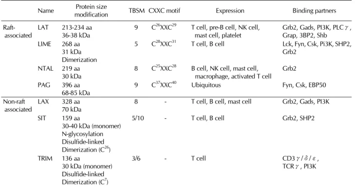
Table I. Characteristics of TRAPs Protein size
Name TBSM CXXC motif Expression Binding partners
modification Raft-
associated LAT 213-234 aa
36-38 kDa 9 C26XXC29 T cell, pre-B cell, NK cell,
mast cell, platelet Grb2, Gads, PI3K, PLCγ, Grap, 3BP2, Shb
LIME 268 aa 31 kDa Dimerization
5 C28XXC31 T cell, B cell Lck, Fyn, Csk, Pi3K, SHP2, Grb2
NTAL 219 aa
30 kDa 8 C25XXC28 B cell, NK cell, mast cell,
macrophage, activated T cell Grb2
PAG 396 aa
68-85 kDa 9 C37XXC40 Ubiquitous Fyn, Csk, EBP50
Non-raft
associated LAX 328 aa
70 kDa 8 - T cell, B cell, mast cell Grb2, Gads, PI3K
SIT 159 aa
30-40 kDa (monomer) N-glycosylation Disulfide-linked Dimerization (C26)
5/10 - T cell, B cell Grb2, SHP2
TRIM 136 aa
30 kDa (monomer) Disulfide-linked Dimerization (C7)
3/6 - T cell CD3γ/δ/ε,
TCRγ, PI3K
lation sites, are mainly localized in non-raft region and seem to be involved in signaling cascades in different cellular compartments. In spite of their similar structural features, the functions of TRAPs are relatively specialized depending on their expression patterns and binding partners (Table I).
Through the previous studies using cell lines and primary immune cells isolated from genetically engineered mice, TRAPs were shown to integrate immunoreceptor-mediated signals in either positive or negative manner. Particularly in this review, we will focus on LAT and LIME, which exert pos- itive regulatory functions in T lymphocytes. For the readers of this review, the characteristics of each TRAPs are summar- ized in Table I.
LINKER FOR ACTIVATION OF T CELLS (LAT)
LAT was initially identified as a phosphoprotein which is rap- idly phosphorylated following TCR ligation (2). The expres- sion of LAT is limited to thymic and peripheral T cells, NK cells, mast cells, megakaryocytes, platelet, and bone-mar- row-derived pre-B cells, but not mature B cells and mono- cytes (2). LAT possesses nine TBSMs in cytoplasmic tail, and becomes phosphorylated at least five tyrosine residues by
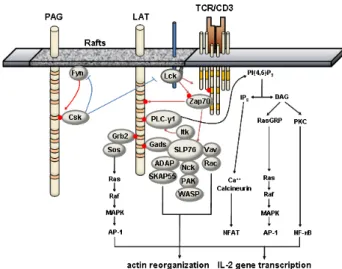
ZAP-70 or Syk kinases upon immunoreceptor engagement. In addition, LAT is phosphorylated by Itk upon CD28 engage- ment (11). After phosphorylation, LAT recruits Grb2, Gads, Grap, PLCγ1, p85 PI3K, Vav, 3BP2, and Shb directly via their SH2-domain, and mediates activation of Ras/MAPK, intra- cellular calcium influx, and cytoskeleton reorganization (12,13). Four distal tyrosines primarily responsible for LAT function are Y132 for PLCγ1, Y171 for PI3K, Y171 and Y191 for Gads, Y191 and Y226 for Vav, Y171, Y191, and Y226 for Grb2, respectively (13). Through the constitutive interaction with Gads, SLP-76 is recruited to LAT upon TCR engagement and in turn, serves as a platform for several signaling mole- cules such as Vav, NCK, ITK and ADAP. The LAT-SLP-76-PLC γ1 complex formation is required for phosphorylation of PLC γ1, allowing optimal calcium mobilization following TCR ligation. On the other hand, the recruitment of Vav, NCK and ADAP through SLP-76 seems to propagate actin polymer- ization and integrin activation. In addition, the recruitment of Grb2 is essential in the activation of Ras/MAPK signaling pathway (Fig. 1).
In LAT-deficient Jurkat T cell line, optimal tyrosine phos- phorylation of PLCγ1, Vav, and SLP-76 are impaired, result- ing in the diminished TCR-mediated calcium mobilization and
Figure 1. Signaling pathways mediated by LAT. Upon TCR engage- ment, Src-family kinase, Lck, is activated and phosphorylates the tyrosine residues within ITAM motifs on CD3γ/ε/ζ chains.
Subsequently, Syk-family kinase, ZAP-70, is recruited through its SH2 domain and become activated by phosphorylation by Lck. Activated ZAP-70 phosphorylates the tyrosine residues within TBSMs of raft-associated LAT, allowing the recruitment of other signaling molecules possessing SH2 domains or phosphotyrosine-binding (PTB) domains including Gads, Grb2, PLCγ1 at the membrane proximal sites. Through the constitutive interaction with Gads, SLP-76 are recruited to LAT and become phosphorylated by ZAP-70. The recruitment of Itk via SLP-76 is required for full activation of PLCγ1.
Activated PLCγ1 hydrolyze the phosphatidylinositol 4, 5 bisphos- phate (PIP2) to inositol 3, 4, 5-triohosphate (IP3) and diacylglycerol (DAG), leading to the calcium mobilization and NF-κB activation.
Tyrosine-phosphorylated SLP-76 also provides docking sites for Vav, Nck and ADAP, forming signaling complexes for actin reorganization.
Together with Grb2/Sos-mediated Ras/MAPK activation, these signal- ing pathways cooperatively activate the IL-2 poduction and T cell proliferation. These positive signaling pathways can be negatively regulated by the inhibition of Lck activity by PAG-associated Csk kinase.
MAPK activation (14,15). When SLP-76 is targeted con- stitutively to plasma membrane in LAT-deficient Jurkat T cells, the signaling defects are restored, suggesting that major signal transduction downstream of LAT is mediated by SLP-76 re- cruited to lipid rafts (16).
Through the extensive studies using genetically engineered mice, it was revealed that LAT is critical in thymocyte devel- opment as well as mature T cell activation. In LAT-deficient mice, B cells NK cells, and platelets appear to develop nor- mally, but crucial defects are found in thymocyte develop- ment at double-negative stage, resulting in lack of mature pe- ripheral T cell population (17). This suggests that LAT is es- sential for the pre-TCR signal transduction. Moreover, func- tional restoration studies using the LAT knock-in mice harbor- ing individual mutations of these tyrosine residues support
the importance of LAT in T cell functions. Similar to LAT-defi- cient mice, mutant mice with point mutations in all distal four tyrosine residues have defects in thymocyte development (18). LAT Y132F mice which possess a mutation in PLCγ1 binding site also have partial defects in thymocyte develop- ment in DN stage (19,20). Unexpectedly, however, Y132F mice develop autoimmune phenotype with augmented Th2- type cytokines and B cell proliferation. These results suggest that LAT may also function as a negative modulator in lymphocytes.
As previously indicated, LAT is palmitoylated at two cystein residues, Cys26 and Cys29, near the transmembrane domain in cytoplasmic region, and palmitoylated LAT is targeted con- stitutively to lipid rafts. Inhibition of palmitoylation on these cystein residues abrogates lipid raft targeting of LAT (12).
However, it seems that LAT localization to lipid rafts is not essential during normal T cell activation and development.
The reconstitution of LAT-deficient cell line with the LAX-LAT chimeric protein consisting of the cytoplasmic region of LAT fused with the transmembrane region of non-raft TRAP, LAX restored MAPK activation, calcium flux, and NFAT activation in LAT-deficient cells (21). Furthermore, the defects in thymo- cyte development and peripheral T cell responses in LAT-de- ficient mice are rescued by the reconstitution with the chi- meric LAX-LAT protein (21). Therefore, it seems that the lo- calization to lipid rafts may not directly affect the function of LAT in normal T cell activation and development. On the oth- er hand, the raft localization of LAT may differentially affect its function in anergic T cells. It has been recently shown that palmitoylation of LAT is defective in anergic T cells (22). The early TCR signaling events such as CD3ξ-chain phosphor- ylation or Zap-70 phosphorylation are intact in these cells, however, tyrosine phosphorylation of LAT and calcium mobi- lization by PLCγ1 activation are significantly diminished.
Interestingly, the palmitoylation defect in anergic T cells is likely specific to LAT, yet the mechanism is still unclear.
LCK-INTERACTING TRANSMEMBRANE ADAPTOR PROTEIN (LIME)
Another raft-associated TRAP, LIME was identified as a pos- itive regulator of immunoreceptor signaling (3,4). Originally, LIME was identified as a binding partner of Lck by yeast two hybrid screening (3). In Jurkat T cells, unlike LAT or NTAL, which are phosphorylated by Syk-family PTKs, LIME interact with and is phosphorylated by Lck upon TCR stimulation.
Subsequently, LIME recruits the cytoplasmic proteins such as Lck, Fyn, Vav, p85 PI3K, Grb2, Gads, Shp-2, and Csk (3,4) to membrane proximal sites. In addition, LIME was shown to be phosphorylated by cross-linking of CD4 or CD8 cor- eceptors (4). LIME also possesses a dicystein palmitoylation motif and localize in the lipid rafts.
The binding partners of LIME are, in part, common with those of LAT, which include Gads, p85 PI3K, and Grb2 (3,13,14). When ectopically overexpressed in Jurkat T cells, LIME promotes TCR-mediated signaling pathways to drive cal- cium mobilization, MAPK activation, and IL-2 production (3).
In contrast to the cell line studies, however, LIME-deficient mice show no significant alteration in the development of thy- mocytes, peripheral T cells, as well as other immune cells such as B cells, mast cells, and macrophages (Unpublished data, ref (23)). In addition, TCR-mediated responses such as T cell proliferation and cytokine production are normal in LIME-deficient mice, suggesting that LIME is dispensible in the development and activation of naïve T cells. The reason for the discrepancy between the results from cell line and knock-out mice studies is not clear at this stage. Unlike LAT, LIME expression is barely detectable in thymocytes and rest- ing T cells but largely upregulated upon T cell activation (3).
Thus, it is likely that LAT acts as a main signal transducer of pre-TCR and TCR signaling for the development of thymo- cytes and the activation of naïve peripheral T cells, whereas LIME may function in the later stage of T cell activation. It will be interesting to test whether the function of LIME is re- dundant to that of LAT in activated/effector T cells.
LIME is also expressed in B cells. In B cell lines, after its phosphorylation by Lyn, LIME augments BCR-mediated sig- naling pathways leading to activation of NFAT and NF-κB pathways (24). However, the experiments using LIME-defi- cient mice show that LIME is dispensable in the development and the function of B cells (23). Furthermore, the absence of LIME has no further effect on the autoimmune syndrome of NTAL-deficient mice (23). Interestingly, histological analy- sis reveals that LIME is expressed in plasma cells, as well as myeloma/plasmacytoma (25). Further studies are required to elucidate the functions of LIME especially in the effector T cells or plasma B cells.
CONCLUSION
So far, both LAT and LIME were found to positively regulate TCR-mediated signaling events. However, unlike LAT-defi-
cient mice, LIME-deficient mice are normal in the thymocyte development and peripheral T cell activation. Although the binding partners are partially overlapping, LAT and LIME also have several distinct characteristics. First, LAT is phosphory- lated by Syk-family kinases, while LIME is phosphorylated by Lck. Second, the expression patterns of LAT and LIME are different. LAT is expressed starting from double negative stage of thymocytes and, in peripheral T cells, the level is constant regardless of the activation status. On the other hand, LIME expression is barely detectable in thymocytes and resting peripheral T cells and upregulated upon T cell activa- tion and differentiation to effector T cells. In addition, LIME but not LAT is also expressed in B cells. Therefore, it is ex- pected that the biological function of LAT and LIME are different. As LIME is induciblely expressed upon TCR engage- ment, it will be interesting to study the physiological sig- nificance of LIME in effector/memory T cells.
ACKNOWLEDGEMENTS
This work was supported by a grant from Ewha Womans University. I. Park was supported by BK21 project.
CONFLICTS OF INTEREST
The authors have no financial conflict of interest.
REFERENCES
1. Latour S, Veillette A: Proximal protein tyrosine kinases in immunoreceptor signaling. Curr Opin Immunol 13;299-306, 2001
2. Zhang W, Sloan-Lancaster J, Kitchen J, Trible RP, Samelson LE: LAT: the ZAP-70 tyrosine kinase substrate that links T cell receptor to cellular activation. Cell 92;83-92, 1998 3. Hur EM, Son M, Lee OH, Choi YB, Park C, Lee H, Yun
Y: LIME, a novel transmembrane adaptor protein, associates with p56lck and mediates T cell activation. J Exp Med 198;1463-1473, 2003
4. Brdicková N, Brdicka T, Angelisová P, Horváth O, Spicka J, Hilgert I, Paces J, Simeoni L, Kliche S, Merten C, Schraven B, Horejsí V: LIME: a new membrane Raft-associated adap- tor protein involved in CD4 and CD8 coreceptor signaling.
J Exp Med 198;1453-1462, 2003
5. Zhu M, Janssen E, Leung K, Zhang W: Molecular cloning of a novel gene encoding a membrane-associated adaptor protein (LAX) in lymphocyte signaling. J Biol Chem 277;46151-46158, 2002
6. Brdicka T, Imrich M, Angelisová P, Brdicková N, Horváth O, Spicka J, Hilgert I, Lusková P, Dráber P, Novák P,
Engels N, Wienands J, Simeoni L, Osterreicher J, Aguado E, Malissen M, Schraven B, Horejsí V: Non-T cell activation linker (NTAL): a transmembrane adaptor protein involved in immunoreceptor signaling. J Exp Med 196;1617-1626, 2002
7. Brdicka T, Pavlistoví D, Leo A, Bruyns E, Korinek V, Angelisová P, Scherer J, Shevchenko A, Hilgert I, Cerný J, Drbal K, Kuramitsu Y, Kornacker B, Horejsí V, Schraven B: Phosphoprotein associated with glycosphingolipid-en- riched microdomains (PAG), a novel ubiquitously ex- pressed transmembrane adaptor protein, binds the protein tyrosine kinase csk and is involved in regulation of T cell activation. J Exp Med 191;1591-1604, 2000
8. Pfrepper KI, Marie-Cardine A, Simeoni L, Kuramitsu Y, Leo A, Spicka J, Hilgert I, Scherer J, Schraven B: Structural and functional dissection of the cytoplasmic domain of the transmembrane adaptor protein SIT (SHP2-interacting trans- membrane adaptor protein). Eur J Immunol 31;1825-1836, 2001
9. Bruyns E, Marie-Cardine A, Kirchgessner H, Sagolla K, Shevchenko A, Mann M, Autschbach F, Bensussan A, Meuer S, Schraven B: T cell receptor (TCR) interacting molecule (TRIM), a novel disulfide-linked dimer associated with the TCR-CD3-zeta complex, recruits intracellular signaling pro- teins to the plasma membrane. J Exp Med 188;561-575, 1998
10. Resh MD: Palmitoylation of ligands, receptors, and intra- cellular signaling molecules. Sci STKE 359;re14, 2006 11. Michel F, Attal-Bonnefoy G, Mangino G, Mise-Omata S,
Acuto O: CD28 as a molecular amplifier extending TCR liga- tion and signaling capabilities. Immunity 15;935-945, 2001 12. Zhang W, Trible RP, Samelson LE: LAT palmitoylation: its
essential role in membrane microdomain targeting and ty- rosine phosphorylation during T cell activation. Immunity 9;239-246, 1998
13. Zhang W, Trible RP, Zhu M, Liu SK, McGlade CJ, Samelson LE: Association of Grb2, Gads, and phospholipase C-gam- ma 1 with phosphorylated LAT tyrosine residues. Effect of LAT tyrosine mutations on T cell angigen receptor-mediated signaling. J Biol Chem 275;23355-23361, 2000
14. Finco TS, Kadlecek T, Zhang W, Samelson LE, Weiss A:
LAT is required for TCR-mediated activation of PLCgamma1 and the Ras pathway. Immunity 9;617-626, 1998 15. Zhang W, Irvin BJ, Trible RP, Abraham RT, Samelson LE:
Functional analysis of LAT in TCR-mediated signaling path- ways using a LAT-deficient Jurkat cell line. Int Immunol
11;943-950, 1999
16. Boerth NJ, Sadler JJ, Bauer DE, Clements JL, Gheith SM, Koretzky GA: Recruitment of SLP-76 to the membrane and glycolipid-enriched membrane microdomains replaces the requirement for linker for activation of T cells in T cell re- ceptor signaling. J Exp Med 192;1047-1058, 2000
17. Zhang W, Sommers CL, Burshtyn DN, Stebbins CC, DeJarnette JB, Trible RP, Grinberg A, Tsay HC, Jacobs HM, Kessler CM, Long EO, Love PE, Samelson LE: Essential role of LAT in T cell development. Immunity 10;323-332, 1999 18. Sommers CL, Menon RK, Grinberg A, Zhang W, Samelson
LE, Love PE: Knock-in mutation of the distal four tyrosines of linker for activation of T cells blocks murine T cell development. J Exp Med 194;135-142, 2001
19. Aguado E, Richelme S, Nuñez-Cruz S, Miazek A, Mura AM, Richelme M, Guo XJ, Sainty D, He HT, Malissen B, Malissen M: Induction of T helper type 2 immunity by a point muta- tion in the LAT adaptor. Science 296;2036-2040, 2002 20. Sommers CL, Park CS, Lee J, Feng C, Fuller CL, Grinberg
A, Hildebrand JA, Lacaná E, Menon RK, Shores EW, Samelson LE, Love PE: A LAT mutation that inhibits T cell development yet induces lymphoproliferation. Science 296;
2040-2043, 2002
21. Zhu M, Shen S, Liu Y, Granillo O, Zhang W: Cutting Edge:
localization of linker for activation of T cells to lipid rafts is not essential in T cell activation and development. J Immunol 174;31-35, 2005
22. Hundt M, Tabata H, Jeon MS, Hayashi K, Tanaka Y, Krishna R, De Giorgio L, Liu YC, Fukata M, Altman A: Impaired acti- vation and localization of LAT in anergic T cells as a con- sequence of a selective palmitoylation defect. Immunity 24;513-522, 2006
23. Grégoire C, Simova S, Wang Y, Sansoni A, Richelme S, Schmidt-Giese A, Simeoni L, Angelisova P, Reinhold D, Schraven B, Horejsi V, Malissen B, Malissen M: Deletion of the LIME adaptor protein minimally affects T and B cell de- velopment and function. Eur J Immunol 37;3259-3269, 2007 24. Ahn E, Lee H, Yun Y: LIME acts as a transmembrane adapt- er mediating BCR-dependent B-cell activation. Blood 107;1521-1527, 2006
25. Tedoldi S, Paterson JC, Hansmann ML, Natkunam Y, Rüdiger T, Angelisova P, Du MQ, Roberton H, Roncador G, Sanchez L, Pozzobon M, Masir N, Barry R, Pileri S, Mason DY, Marafioti T, Horejsi V: Transmembrane adaptor molecules: a new category of lymphoid-cell markers. Blood 107;213-221, 2006