Received on October 17, 2011. Revised on November 7, 2011. Accepted on November 11, 2011.
CC This is an open access article distributed under the terms of the Creative Commons Attribution Non-Commercial License (http://creativecommons.org/licenses/by-nc/3.0) which permits unrestricted non-commercial use, distribu- tion, and reproduction in any medium, provided the original work is properly cited.
*Corresponding Author. Tel: 82-10-2315-2227; Fax: 82-2-745-9528; E-mail: [email protected] Keywords: Dendritic cells, Maturation, Experimental Autoimmune Uveoretinitis (EAU)
Maturation-Resistant Dendritic Cells Ameliorate Experimental Autoimmune Uveoretinitis
Keunhee Oh1,2, Yon Su Kim2,3 and Dong-Sup Lee1,2*
1Laboratory of Immunology, Transplantation Research Institute, Departments of 2Biomedical Sciences and 3Internal Medicine, Seoul National University College of Medicine, Seoul 110-799, Korea
Background: Endogenous uveitis is a chronic inflammatory eye disease of human, which frequently leads to blindness.
Experimental autoimmune uveoretinitis (EAU) is an animal disease model of human endogenous uveitis and can be in- duced in susceptible animals by immunization with retinal antigens. EAU resembles the key immunological character- istics of human disease in that both are CD4+ T-cell medi- ated diseases. Dendritic cells (DCs) are specialized anti- gen-presenting cells that are uniquely capable of activating naïve T cells. Regulation of immune responses through mod- ulation of DCs has thus been tried extensively. Recently our group reported that donor strain-derived immature DC pre- treatment successfully controlled the adverse immune re- sponse during allogeneic transplantation. Methods: EAU was induced by immunization with human interphotoreceptor reti- noid-binding protein (IRBP) peptide1-20. Dendritic cells were differentiated from bone marrow in the presence of recombi- nant GM-CSF. Results: In this study, we used paraformalde- hyde-fixed bone marrow-derived DCs to maintain them in an immature state. Pretreatment with fixed immature DCs, but not fixed mature DCs, ameliorated the disease progression of EAU by inhibiting uveitogenic CD4+ T cell activation and differentiation. Conclusion: Application of iBMDC prepared according to the protocol of this study would provide an im- portant treatment modality for the autoimmune diseases and transplantation rejection.
[Immune Network 2011;11(6):399-405]
INTRODUCTION
Endogenous uveitis is a chronic inflammatory eye disease,
leading to blindness not infrequently (1). Even though, uveitis often appears in association with systemic diseases such as Vogt-Koyanagi-Harada disease or Behcet’s disease, the ma- jority of uveitis is still of an idiopathic origin (2). Experimental autoimmune uveoretinitis (EAU) is a disease model of human endogenous uveitis and can be induced in susceptible animals by immunization with retinal antigens (3). Uveitogenic anti- gens which can induce EAU are retinal soluble antigen, inter- photoreceptor retinoid-binding protein (IRBP), rhodopsin, op- sin, recoverin, and phosducin (4). EAU is a CD4+ T cell-medi- ated disease. Uveitogenic effector CD4+ T cells infiltrate into the eyes and are responsible for the pathogenesis (5).
Dendritic cells (DCs) are specialized antigen-presenting cells that are uniquely capable of activating naïve T cells (6).
When pathogens invade the tissue, tissue-resident DCs endo- cytose antigens and process them for presentation on the MHC molecules. With appropriate inflammatory signals pro- voked by pathogens, DCs upregulated the expression of sur- face MHC molecules containing antigenic peptides and co-stimulatory molecules on their surface (7). High level sur- face expression of MHC class II and co-stimulatory molecules are whole-mark of activated dendritic cells and are pre- requisite for effective activation of naïve CD4+ T cells (8).
In contrast to activated DCs, immature DCs with low levels of MHC and co-stimulatory molecules have been implicated in the regulation of immune responses through diverse effec- tor mechanisms (9). Particularly, immature DCs are able to induce a state of hyporesponsiveness in T cells, the phenom- enon has been used for the control or suppression of the im-
and differentiation.
MATERIALS AND METHODS Animals and induction of EAU
C57BL/6 (B6) female mice (Jackson, Bar Harbor, MA), 8-10 weeks of age, were immunized s.c. in the both footpads and tail-base with 250μg of human interphotoreceptor retinoid- binding protein (IRBP) peptide1-20 (GPTHLFQPSLVLDMAKV- LLD) (Peptron, Daejeon, Korea) in 100μl of emulsion in com- plete Freund’s adjuvant (Sigma-Aldrich, St. Louis, MO) su- pplemented with Mycobacterium tuberculosis (strain H37 Ra;
Difco, Detroit, MI) to 1.5 mg/ml (14). Mice were received 1.0μg of pertussis toxin (Sigma-Aldrich), intraperitoneally at the time of the immunization. All mice were bred and main- tained in specific pathogen-free conditions at the animal fa- cility of Seoul National University College of Medicine. All an- imal experiments were performed with the approval of the Institutional Animal Care and Use Committee (IACUC, SNU 050502003) at Seoul National University.
Histopathological scoring
Eyes were removed from mice sacrificed 21 days after the immunization with human IRBP peptide, and then fixed in 4% buffered paraformaldehyde (PFA), embedded in paraffin, sectioned, and stained with hematoxylin and eosin for histo- pathological analysis. The severity of the disease was de- termined for each eye and scored on a scale of 0∼4 in half- point increments according to a semiquantitative system (15).
Generation of bone marrow-derived dendritic cells Bone marrow cells from the both femurs and tibias were ob- tained from 6∼8-week-old mice. Cells were passed through a nylon mesh and red blood cells were lysed. Following washing, granulocytes, B cells and red blood cell precursors
1μg/ml lipopolysaccharide (Salmonella abortus equi; Sigma- Aldrich) was added to the cell culture for 18 hrs before harvest. To render DCs maturation-resistant, they were fixed by resuspension in 2% (weight/volume) paraformaldehyde for 10 min, then washed three times. Fixed immature or fixed mature cells were used for the following experiments.
IRBP-specific CD4+ T cell proliferation
CD4+ T cells from draining inguinal and popliteal lymph no- des of mice were purified using magnetic beads on day 7 after immunization with human IRBP peptide, then co-cul- tured (2×10⁵ cells/well) with γ-irradiated, syngeneic spleno- cytes (4×10⁵ cells/well) with or without 30μM of IRBP pep- tide in round-bottom 96 well plate. Culture was incubated for 72 hrs at 37oC in a 5% CO2 atmosphere, pulsed [3H]- thymi- dine (1μCi/well) during the last 12 hrs, then incorporated radioactivity was counted.
Migration of bone marrow-derived dendritic cells In vitro generated DCs from bone marrow of GFP transgenic mice (Jackson, Bar Harbor, MA) were fixed in 2% PFA and injected intravenously with 5×10⁶ cells into B6 mice. On 14 days after injection with DCs, spleens and inguinal lymph no- des were frozen in OCT compound, sectioned (4μm), and examined under fluorescence microscopy.
Intracellular cytokine staining and flowcytometric analysis
After restimulation, the cells were fixed with 4% paraf- ormaldehyde and permeabilized with 0.5% Triton X-100.
Cells were stained with anti-CD4, anti-CD62L, and anti-IFN-γ (all from BD-Pharmingen, San Diego, CA). Flow cytometric analysis was conducted using a FACSCalibur (BD Bioscience, San Jose, CA).
Figure 1. Mature-resistant DC ameliorates EAU in mice. (A) Histology of eyes. C57BL/6 (B6) mice were intravenously injected with 5×10⁶ BM-derived fixed immature DCs (iBMDC), fixed mature DCs (mBMDC), or media. Two days after the BMDC transfer, mice were immunized with human IRBP peptide1-20, Both eyes were removed, fixed in 4% paraformaldehyde, and embedded in paraffin. Sections (4μm) were prepared and stained with hematoxylin and eosin (original magnification, ×100). Midsagittal section for each eyes were used. V, vitreous; GL, ganglion layer; INL, inner nuclear layer; ONL, outer nuclear layer; RPE, retinal pigmented epithelium (RPE); Ch, choroid. Retina folding and infiltrations (arrows). (B) Histopathological score of EAU. Data are presented as the mean±SEM of n=10 mice for each group from two independent experiments. (C) IRBP-specific proliferation of CD4+ T cells. CD4+ T cells from draining lymph nodes were purified with magnetic beads on the 7th day after immunization, then co-cultured (2×10⁵ cells/well) with irradiated, syngeneic splenocytes (4×10⁵ cells/well) with 30 mM of IRBP peptide. The cultures were incubated for 3 days and pulsed with [3H]-thymidine (1 mCi/well) for the last 12 hrs. Data represent means±SD of two independent determinations with draining lymph nodes from n=3 mice/group.
RESULTS
Maturation-resistant DCs ameliorate EAU in mice Female C57BL/6 (B6) mice were immunized with an uveito- genic human IRBP 1∼20 peptide. Two days before immuni- zation, mice were intravenously injected with 5×10⁶ BM-de- rived fixed immature DCs (iBMDC), fixed mature DCs (mBMDC), or media. B6 mice pretreated with mBMDC devel- oped more severe uveoretinitis compared with the moderate disease in media-pretreated mice, whereas mice pretreated with iBMDC revealed improved disease phenotype compared with media-pretreated control. Extensive tissue damage, in- cluding retinal folding, heavy inflammatory cell infiltration in- to the vitreous humor, and choroidal granuloma formation were noted in the eyes of mBMDC-pretreated mice (Fig. 1A).
In contrast to this, mice pretreated with iBMDC exhibited very
mild inflammatory cell infiltration and local retinal destruction (Fig. 1A). The median disease scores were 1.8 (mBMDC-pre- treated), 0.6 (iBMDC-pretreated), and 1.4 (media-pretreated) (Fig. 1B). IRBP-specific CD4+ T lymphocyte proliferation pre- pared from the draining lymph nodes was greatly reduced in iBMDC-pretreated mice compared to the media-pretreated control (Fig. 1C).
Pretreatment with immature DCs reduced the activation and differentiation of CD4+ T cells
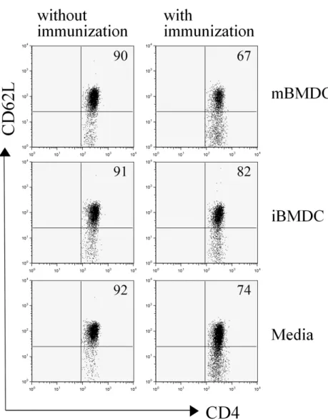
To evaluate the initial uveitogenic CD4+ T cell responses fol- lowing immunization in the mBMDC or iBMDC-pretreated mice, lymphocytes prepared from draining lymph nodes were analyzed on the 7th day after immunization for the activation marker. CD62L-negative activated T cells among CD4+ T cells were increased following immunization with IRBP peptide
Figure 2. iBMDC-pretreatment reduced the acti- vation of CD4+ T cells. Draining lymph node cells from the mice pretreated with mBMDC, iBMDC, or media and immunized with human IRBP peptide were harvested on the 7th day after immunization. T-cell activation was ana- lyzed using flowcytometer after staining with anti-CD4 and anti-CD62L antibodies. The data are representative of three independent deter- minations.
while iBMDC pretreatment decreased the percentage of acti- vated CD4+ T cells compared to the media-pretreated control (Fig. 2).
Uveitogenic CD4+ T cell differentiation in the draining lymph nodes were also analyzed on the 7th day after immunization. IFN-γ-expressing CD4+ T cells among uveito- genic T cells were decreased in the iBMDC-pretreated mice and increased in the mBMDC-pretreated mice compared with media-pretreated control mice (Fig. 3).
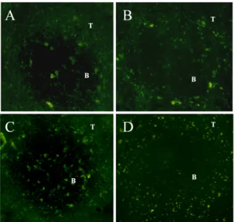
Both immature and mature DCs migrate into the T-cell zone of the secondary lymphoid organs To evaluate the site of immunoregulation on the activation and differentiation of uveitogenic CD4+ T cells implemented
by pretreated iBMDC, B6 mice were received iBMDC and mBMDC prepared from syngeneic GFP transgenic mice. On 14 days after injection with DCs, spleens and inguinal lymph nodes were examined under fluorescence microscopy. Both the fixed immature DCs and fixed mature DCs migrated into the T-cell zones of secondary lymphoid organs such as spleen and inguinal lymph nodes and stayed there at least for 14 days following injection (Fig. 4).
DISCUSSION
In this study, we demonstrated that pretreatment with fixed immature bone marrow-derived dendritic cells (iBMDC) ame- liorated experimental autoimmune uveoretinitis (EAU) by in-
Figure 3. Intracellular cytokine expression of uveitogenic CD4+ T cells. Draining lymph node cells were harvested from the mice received mBMDC, iBMDC, or media on the 7th day after immunization with IRBP peptide and were restimulated in vitro with PMA and ionomycin as described in Materials and Methods. Cells were analyzed using flowcytometer after staining with anti-CD4 and anti-IFN-γ antibodies. The data are representative of two independent determinations.
Figure 4. Both mBMDC and iBMDC migrate into T-cell zone of the spleen and lymph nodes. B6 mice were received mBMDC and iBMDC prepared from GFP transgenic mice. On 14 days after injection with DCs, spleens and inguinal lymph nodes were frozen in OCT compound. Sections (4μm) were examined under fluorescence microscopy (original magnification, ×200). Spleen (A) and inguinal lymph node (B) from the mice received iBMDC. Spleen (C) and inguinal lymph node (D) from the mice received mBMDC. T cell zone (T) and B cell zone (B) were indicated.
hibiting uveitogenic CD4+ T cell activation and differen- tiation. Pretreated fixed immature dendritic cells migrated into the T-cell zone of the secondary lymphoid organs and af- fected the uveitogenic CD4+ T cell immune responses.
EAU is an animal disease model of human endogenous uveitis and resembles the key immunological characteristics of human disease in that both are CD4+ T-cell mediated dis- eases (5). In EAU pathogenesis, Th1 immune responses have been suggested to be essential factors. Susceptibility to dis- ease paralleled Th1 responsiveness among the different mouse or rat strains (16). In this study we found that pre- treated iBMDC effectively inhibited uveitogenic Th1 differ- entiation in the draining lymph nodes. As the transferred fixed dendritic cells migrated and survived in the secondary lymphoid organs for more than 2 weeks, we assumed that iBMDC directly inhibited initial uveitogenic CD4+ T cell acti- vation and subsequent differentiation in the T-cell zone of these organs. Recently, Th17 cells have also been implicated in the disease progression of autoimmune eye diseases of hu- man and animal models, including uveitis. Th17 cells among peripheral blood lymphocytes were increased in active uveitis patients and treatment with blocking anti-IL-17 antibody miti- gated EAU in animal models (17). IFN-γ and IL-17 were sug- gested to have distinct pathogenic roles in different animal models of autoimmune uveoretinitis uveiretinitis (18,19).
Thus, evaluation of the effect of iBMDC on the Th17 differ- entiation of uveitogenic CD4+ T cells would be very im- portant in the following study.
As the immature DCs are important in maintaining self tol- erance, many researchers have tried to regulate autoimmune response using immature DCs (8). However, previous at- tempts have not obtained consistent results, possibly because the immaturity of transferred DCs have not sustained in vivo
ability of DCs to function as antigen presenting cells and/or affected the survival of DCs in the recipient. Paraformalde- hyde fixation, however, did not eliminate the potential of DCs in supporting allogeneic T-cell proliferation (13). Also as both the fixed immature DCs and fixed mature DCs survived over 2 weeks in the secondary lymphoid organs of the recipients, the concern of second possibility was ruled out. Thus, appli- cation of iBMDC prepared according to the protocol of this study would provide an important treatment modality for the autoimmune diseases and transplantation rejection.
ACKNOWLEDGEMENTS
This work was supported by grants from MarineBio Technol- ogy Project, Ministry of Land, Transport and Maritime Affairs (D-S.L.).
CONFLICTS OF INTEREST
The authors have no financial conflict of interest.
REFERENCES
1. Ahn JK, Chung H, Lee DS, Yu YS, Yu HG: CD8brightCD56+
T cells are cytotoxic effectors in patients with active Behcet's uveitis. J Immunol 175;6133-6142, 2005.
2. Yu HG, Lee DS, Seo JM, Ahn JK, Yu YS, Lee WJ, Chung H:
The number of CD8+ T cells and NKT cells increases in the aqueous humor of patients with Behçet's uveitis. Clin Exp Immunol 137;437-443, 2004.
3. Caspi RR, Chan CC, Wiggert B, Chader GJ: The mouse as a model of experimental autoimmune uveoretinitis (EAU).
Curr Eye Res 9(Suppl);169-174, 1990.
4. Nussenblatt RB, Caspi RR, Mahdi R, Chan CC, Roberge F, Lider O, Weiner HL: Inhibition of S-antigen induced ex- perimental autoimmune uveoretinitis by oral induction of tol- erance with S-antigen. J Immunol 144;1689-1695, 1990.
9. Wakkach A, Fournier N, Brun V, Breittmayer JP, Cottrez F, Groux H: Characterization of dendritic cells that induce toler- ance and T regulatory 1 cell differentiation in vivo. Immunity 18;605-617, 2003.
10. Adorini L: Tolerogenic dendritic cells induced by vitamin D receptor ligands enhance regulatory T cells inhibiting auto- immune diabetes. Ann N Y Acad Sci 987;258-261, 2003.
11. Roelen DL, Schuurhuis DH, van den Boogaardt DE, Koekkoek K, van Miert PP, van Schip JJ, Laban S, Rea D, Melief CJ, Offringa R, Ossendorp F, Claas FH: Prolongation of skin graft survival by modulation of the alloimmune re- sponse with alternatively activated dendritic cells. Transplan- tation 76;1608-1615, 2003.
12. Raimondi G, Thomson AW: Dendritic cells, tolerance and therapy of organ allograft rejection. Contrib Nephrol 146;105-120, 2005.
13. Kang HG, Lee JE, Yang SH, Lee SH, Gao W, Strom TB, Oh K, Lee DS, Kim YS: Donor-strain-derived immature dendritic cell pre-treatment induced hyporesponsiveness against alloge- neic antigens. Immunology 129;567-577, 2010.
14. Oh K, Byoun OJ, Ham DI, Kim YS, Lee DS: Invariant NKT cells regulate experimental autoimmune uveitis through in- hibition of Th17 differentiation. Eur J Immunol 41;392-402, 2011.
15. Chan CC, Caspi RR, Ni M, Leake WC, Wiggert B, Chader GJ, Nussenblatt RB: Pathology of experimental autoimmune uve- oretinitis in mice. J Autoimmun 3;247-255, 1990.
16. Sun B, Rizzo LV, Sun SH, Chan CC, Wiggert B, Wilder RL, Caspi RR: Genetic susceptibility to experimental autoimmune uveitis involves more than a predisposition to generate a T helper-1-like or a T helper-2-like response. J Immunol 159;
1004-1011, 1997.
17. Amadi-Obi A, Yu CR, Liu X, Mahdi RM, Clarke GL, Nussenblatt RB, Gery I, Lee YS, Egwuagu CE: TH17 cells con- tribute to uveitis and scleritis and are expanded by IL-2 and inhibited by IL-27/STAT1. Nat Med 13;711-718, 2007.
18. Tang J, Zhu W, Silver PB, Su SB, Chan CC, Caspi RR: Autoim- mune uveitis elicited with antigen-pulsed dendritic cells has a distinct clinical signature and is driven by unique effector mechanisms: initial encounter with autoantigen defines dis- ease phenotype. J Immunol 178;5578-5587, 2007.
19. Luger D, Silver PB, Tang J, Cua D, Chen Z, Iwakura Y, Bowman EP, Sgambellone NM, Chan CC, Caspi RR: Either a Th17 or a Th1 effector response can drive autoimmunity: con- ditions of disease induction affect dominant effector category.
J Exp Med 205;799-810, 2008.
20. Gregori S, Casorati M, Amuchastegui S, Smiroldo S, Davalli AM, Adorini L: Regulatory T cells induced by 1 alpha,25-dihy- droxyvitamin D3 and mycophenolate mofetil treatment medi- ate transplantation tolerance. J Immunol 167;1945-1953, 2001.
21. Hayamizu K, Huie P, Sibley RK, Strober S: Monocyte-derived dendritic cell precursors facilitate tolerance to heart allografts after total lymphoid irradiation. Transplantation 66;1285-1291, 1998.