Received on July 7, 2010. Revised on July 20, 2010. Accepted on July 23, 2010.
CC This is an open access article distributed under the terms of the Creative Commons Attribution Non-Commercial License (http://creativecommons.org/licenses/by-nc/3.0) which permits unrestricted non-commercial use, distribu- tion, and reproduction in any medium, provided the original work is properly cited.
*Corresponding Author. Tel: 82-31-219-5071; Fax: 82-31-219-5079; E-mail: [email protected] Keywords: Hepadnaviridae, T-lymphocytes, Cytotoxic, Viral proteins, Apoptosis, Interferon-γ
Expression of Hepatitis B Virus X Protein in Hepatocytes Suppresses CD8 + T Cell Activity
Mi Jin Lee1, Young-hee Jin1, Kyongmin Kim1, Yangkyu Choi2, Hyoung-Chin Kim3 and Sun Park1*
1Department of Microbiology and Immunology, Ajou University School of Medicine, Suwon 442-721, 2Department of Laboratory Animal Medicine, College of Veterinary Medicine, Konkuk University, Seoul 143-701, 3Biomedical Mouse Resource Center, Korea Research Institute of Bioscience and Biotechnology, Ochang 363-883, Korea
Background: CD8+ T cells contribute to the clearance of Hepatitis B virus (HBV) infection and an insufficient CD8+ T cell response may be one of the major factors leading to chronic HBV infection. Since the HBx antigen of HBV can up-regulate cellular expression of several immunomodulatory molecules, we hypothesized that HBx expression in hep- atocytes might affect CD8+ T cell activity. Methods: We ana- lyzed the activation and apoptosis of CD8+ T cells co-cul- tured with primary hepatocytes rendered capable of express- ing HBx by recombinant baculovirus infection. Results:
Expression of HBx in hepatocytes induced low production of interferon-γ and apoptosis of CD8+ T cells, with no effect on CD8 T cell proliferation. However, transcriptional levels of H-2K, ICAM-1 and PD-1 ligand did not correlate with HBx expression in hepatocytes. Conclusion: Our results suggest that HBx may inhibit CD8+ T cell response by regulation of interferon-γ production and apoptosis.
[Immune Network 2010;10(4):126-134]
INTRODUCTION
Chronic infections caused by hepatitis B virus (HBV) afflict some 400 million people globally and kill over 500,000 peo- ple annually. Death is due mainly to complications of cir- rhosis and hepatocellular carcinoma (1). Although factors that appear to have an impact on the progression to chronic hep- atitis B are not fully understood, an insufficient the immune response to HBV is regarded as an important factor based on the higher probability of developing chronic hepatitis B
in individuals infected perinatally (90%) or during childhood (20∼30%), situations when the immune system is thought to be immature (2).
Evidence supporting a critical role of a CD8+ T cell re- sponse in HBV infection has accumulated. A chimpanzee model of HBV infection revealed that CD8+ T cells are the main effector cells responsible for viral clearance and disease pathogenesis during acute HBV infection (3). HBV-specific CD8 T cells contribute to viral clearance by cytolysis of in- fected hepatocytes as well as by a noncytolytic process in- volving suppression of the hepatocellular HBV gene ex- pression via production of interferon-gamma (IFN-γ) and tu- mor necrosis factor-alpha (TNF-α) (4,5). Strong and multi- specific CD8 T cell responses to HBV have been demon- strated in self-limited acute hepatitis B patients, while weak CD8 T cell responses are displayed in chronically infected pa- tients (6-8). Recently, an exhausted phenotype of HBV-specif- ic CD8 T cells was demonstrated in chronic HBV infection (9), however, the underlying mechanisms for the weak CD8 T cell immune responses in chronic hepatitis B patients re- main unclear.
The CD8 T cell response in the liver has unique features.
The liver is believed to be the site for the priming of naive CD8+ T cells as well as for accumulation and apoptosis of activated CD8+ T cells. Intrahepatic activation of CD8+ T cells has been demonstrated in a liver transplantation model with- out liver-derived antigen-presenting cells (10,11). Further- more, the liver induces full CD8+ T cell activation and differ-
entiation, while activated CD8 T cells are trapped in the liver partly due to the high expression of intercellular adhesion molecule-1 (ICAM-1) and vascular cell adhesion molecule-1 (VCAM-1) on hepatic sinusoidal endothelium (12). CD8 T cell apoptosis in the liver is related with several molecules such as TNF-α, Fas ligand, and programmed death-1 ligand (PD-L1;B7-H1) (13-15). It has been suggested that these unique characteristics of the liver may predispose this organ to the persistence of infections.
X protein of HBV (HBx) is implicated in inflammation and immunomodulation. HBx in human hepatoma cell lines in- duces transcription of inflammatory cytokines such as TNF-α interkeukin (IL)-18, and IL-8 (16-18). Also, HBx increases the expression of molecules that are important in the immune re- sponse such as major histocompatibility complex (MHC) mol- ecules, ICAM-I and Fas ligand (19-22). Since these molecules have been implicated in intrahepatic activation, trapping, and apoptosis of CD8 T cells, we investigated whether HBx ex- pression in hepatocytes could modulate CD8 T cell activation and apoptosis. We report that HBx expression in hepatocytes does not affect CD8+ T cell proliferation but suppresses IFN-γ production as well as the survival of CD8+ T cells.
MATERIALS AND METHODS
Construction of baculoviral vectors and production of recombinant baculoviruses
To facilitate the introduction of the HBx gene into primary hepatocytes, a recombinant baculoviral vector was con- structed using pAcSG2-CMV, which contains the eukaryotic gene expression cassette derived from pIRES-EGFP (Clontech, Mountain View, CA) (23). The gene sequences for the en- hanced green fluorescent protein and internal ribosome entry site were removed using BamHI and NotI. The DNA fragment coding HBx was amplified using the primers 5’-CTAGCT- AGCATGGCTGCTCGGGTGTG-3’ and 5’-AACTGCAGTTAGG- CAGAGGTGAAAAAGTTGC-3’, and using pGEX-4T-HBx (24) as a template. The PCR product was introduced into pAcSG2-CMV downstream of the cytomegalovirus promoter and was confirmed by sequencing. Recombinant baculovi- ruses were produced in Sf9 cells (BD Biosciences Pharmin- gen, San Diego, CA) cotransfected with baculoviral Gold DNA (BD Biosciences) and baculoviral transfer vectors. Recombi- nant baculoviruses were amplified in Sf9 cells and con- centrated by centrifugation of the culture supernatant at 6,000 rpm for 16 h at 4oC. The virus pellet was resuspended in
DMEM-F12 medium (GibcoBRL, Carlsbad, CA) and the viral titer was determined using a plaque forming unit assay.
Baculovirus infection of primary hepatocytes Isolation of hepatocytes was done as described previously (23). Briefly, the livers of male C57BL/6 (B6) mice (Deahan Biolink, Seoul, Korea) were perfused with liver perfusion me- dium (GibcoBRL) via the portal vein. Released hepatocytes were washed, seeded into 48-well plates at a density of 2×10⁴ hepatocytes/well and allowed to adhere overnight.
Recombinant baculoviruses were added to the adhered hep- atocyte culture and 1 h later the culture medium was replaced.
Purification of CD8+ T cells
Lymph nodes and spleen cells were isolated from MataHari TCR transgenic mice (25) that were generously provided by Dr. Matzinger, United States National Institutes of Health.
These mice have CD8 T cells expressing TCR specific to the H-Ypb peptide bound to H-2Db. CD8+ T cells were purified using magnetic beads by incubation with beads coupled to anti-mouse CD8 antibody (Miltenyi Biotec, Auburn, CA) for 30 min at 4oC prior to passage twice through a MACS mag- netic cell separation column (Miltenyi Biotec). CD8+ cell pu- rity was ascertained by labeling cells with PE-conjugated an- ti-CD8 antibody (BD Biosciences) and flow cytometry using the BD Vantage system (BD Biosciences). The purity was consistently 90∼98%.
Proliferation and apoptosis of CD8+ T cells co-cultured with primary hepatocytes
Purified MataHari CD8+ T cells (2×10⁵ cells/well) were added to hepatocyte cultures pre-infected with baculoviruses. To an- alyze the proliferation of CD8 T cells, the cells were labeled with CFSE (Molecular Probes, Eugene, OR) prior to addition to the hepatocyte-culture. After 2 days, CD8 T cells were har- vested in phosphate-buffered saline containing 1 mM EDTA, stained with antibodies against CD8 and CD45 (BD Biosciences) and analyzed using flow cytometry (Becton Dickinson, Sunnyvale, CA). To enumerate apoptotic cells, un- labeled CD8 T cells were co-cultured, harvested and stained with antibodies against CD8 and CD45, and then a terminal deoxynucleotidyltransferase-mediated dUTP-biotin nick end-la- beling (TUNEL) assay was conducted (Roche Applied Science, Indianapolis, IN).
Figure 1. Baculovirus-mediated HBx expression in hepatocytes. (A) HEK293 cells were transfected with a baculoviral transfer vector containing the HBx gene (pAcSG2-CMV- HBx) or with an empty control vector (pAcSG2-CMV). Western blotting was performed 48 h later on cell lysates using rabbit sera specific to HBx (upper panel) and anti-tubulin antibody (lower panel). CON denotes untransfected cells. (B) Huh7 cells were infected with recombinant baculoviruses containing HBx gene (HBx) or with control baculoviruses (CMV). Purified baculoviruses at a multiplicity of infection (M.O.I) of 1000 were added into Huh7 cell culture and 1 h later the culture medium was replaced. Cell lysates obtained 48 h later were subjected to Western blotting to detect HBx. (C) Primary murine hepatocytes were isolated and infected with recombinant baculoviruses at a M.O.I. of 1000 for 1 h. At the indicated time points after infection, total RNA was prepared from cells and DNase I treated RNA was reverse transcribed. PCR for the amplification of HBx transcript (upper and middle panels) was performed using cDNA or RNA as template.
RT-PCR for beta-actin was performed (lower panel). HBx denotes hepatocytes infected with baculoviruses containing HBx gene, CMV denotes hepatocytes infected with control baculoviruses and CON denotes uninfected hepatocytes.
Cytokine production by CD8+ T cells co-cultured with primary hepatocytes
For the production of IFN-γ and IL-10 in co-culture of CD8 T cells and primary hepatocytes, the supernatant obtained from a 48 h culture was used for an enzyme-linked im- munosorbant assay (ELISA) using an IFN-γ ELISA kit (Pierce Endogen, Rockford, IL) and an IL-10 ELISA kit (Biosource, Carlsbad, CA).
RT-PCR and Western blot
For detection of HBx transcript in baculovirus infected hep- atocytes, total RNA of hepatocytes infected with recombinant baculoviruses was isolated at the indicated time points using RNAzol-B (Tel-Test, Friendswood, TX). To remove con- taminated vector DNA, the RNA samples were treated with 1 U/μl DNase I (Invitrogen) for 15 min at room temperature and were reverse transcribed using oligo-dT primers. PCR was performed using cDNA or RNA as a template and a pri- mer set for HBx.
For the analysis of transcriptional levels of H-2K, ICAM-1 or PD-L1, total RNA of hepatocytes from male C57BL/6 or HBx transgenic mice (26) (kindly provided by Dr. Dae-Yul
Yu, KRIBB, Korea) was isolated. The cDNA was synthesized using the Superscript II first-strand cDNA synthesis system (Invitrogen) according to the manufacturer’s instructions.
Semi-quantitative RT-PCR was performed using serially di- luted cDNA and primer sets for H-2k, ICAM-1 or PD-L1: H-2k forward, 5’-CTGCAGGGGATGGAACCTTC, H-2k reverse, 5’- CTTCACGCTAGAGAATGAGG; ICAM-1 forward, 5’-TGGAAC- TGCACGTGCTGTAT, ICAM-1 reverse, 5’-ACCATTCTGTTCA- AAAGCAG, PD-L1 forward, 5’-GGAATTGTCTCAGAATGGTC, PD-L1 reverse, 5’-GTAGTTGCTTCTAGGAAGGAG. For nor- malization of PCR product, RT-PCR for beta-actin was con- ducted using forward primer, 5’-CAGGTCATCACCATTGGC- AATGAG and reverse primer, 5’-CAGCACTGTGTTGGCGT- ACAGGTC. PCR products were separated using a 1.5% agar- ose gel and band intensities were analyzed with the Molecular Imager Gel Doc XR System (Bio-Rad Laboratories, Hercules, CA). For detection of HBx protein, HEK293 cells transfected with baculovirus transfer vector and Huh7 cells in- fected with recombinant baculoviruses were lysed in RIPA buffer containing 1 mM 4-(2-aminoethyl)-benzenesulfonyl flu- oride, 0.3μM aprotinin and 1μM leupeptin. Proteins were separated by 15% sodium dodecyl sulfate-polyacrylamide gel
Figure 2. HBx expression in hepatocytes enhances apoptosis of antigen-stimulated CD8+ T cells. Primary hepatocytes isolated from C57BL/6 male mice were infected with baculoviruses at a M.O.I. of 1000 for 1 h and then the culture medium was replaced. Twenty-four hours later, purified Matahari CD8 T cells were added to cultures of primary hepatocytes. After 48 h of co-culture, CD8 T cells were harvested and the frequency of apoptotic CD8+ T cells was analyzed by TUNEL and flow cytometry. The TUNEL histograms that are displayed are gated on CD8+ cells. Culture of uninfected hepatocytes (CON), culture of hepatocytes infected with baculoviruses containing HBx gene (HBx) or with control baculoviruses (CMV) Upper and lower panels depict separate independent experiments.
electrophoresis and transferred to a polyvinylidene difluoride membrane. HBx protein was visualized by Western blot anal- ysis using rabbit sera specific to HBx (24) and the enhanced chemiluminescent system (GE Healthcare, Buckinghamshire, UK).
RESULTS
HBx is expressed in primary hepatocytes following baculovirus-mediated HBx gene transduction For efficient introduction of the HBx gene into primary hep- atocytes, recombinant baculoviruses containing the gene in the eukaryotic gene expression cassette were produced and utilized. HBx gene expression in mammalian cells using the baculoviral vector system was demonstrated (Fig. 1). Using an antibody specific to HBx, a protein with an approximate molecular weight of 17 kDa was detected in Western blots
of HEK293 cells transfected with the baculoviral transfer vec- tor harboring the HBx gene (pAcSG2-CMV-HBx) (Fig. 1A).
Western blots of HEK293 cells transfected with the control empty vector pAcSG2-CMV did not detect the 17 kDa band (Fig. 1A). HBx was also detected in Huh7 human hepatoma cells infected with recombinant baculoviruses containing the HBx gene, but not in Huh7 cells infected with control baculo- viruses lacking the HBx gene (Fig. 1B). HBx expression was also evident in primary murine hepatocytes infected with re- combinant baculoviruses using reverse transcription-polymer- ase chain reaction (RT-PCR) (Fig. 1C). HBx gene transcription was detected in hepatocytes infected with baculoviruses con- taining the HBx gene for 3 days post-infection but not with control baculoviruses. The possibility of amplification of HBx gene from transduced baculoviral DNA was excluded by the absence of the gene in PCR using RNA samples as templates.
TUNEL
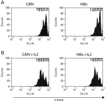
Figure 3. HBx expression in hepatocytes does not modulate proliferation of antigen-stimulated CD8+ T cells. Primary hepatocytes isolated from C57BL/6 male mice were infected with baculoviruses at a M.O.I. of 1000 for 1 h and then the culture medium was replaced.
Twenty-four hours later purified Matahari CD8 T cells labeled with CFSE were added without IL-2 (A) or with IL-2 (B) to cultures of primary hepatocytes. After 48 h of co-culture, CD8 T cells were harvested and analyzed by flow cytometry. CFSE histograms are gated on CD8+ cells. Culture of hepatocytes infected with baculoviruses containing HBx gene (HBx) or with control baculoviruses (CMV)
Figure 4. HBx expression in hepatocytes reduces IFN-γ production in co-culture with antigen-stimulated CD8+ T cells. Primary hepa- tocytes isolated from C57BL/6 male mice were infected with baculoviruses at a M.O.I. of 1000 for 1 h and then the culture medium was replaced. Twenty-four hours later purified Matahari CD8 T cells were added and 48 h later the supernatant was obtained from the co-culture, culture of hepatocytes (hepatocyte only) and culture of purified Matahari CD8+ T cells (Lympho). The IFN-γ concentration was determined using ELISA. The data is mean of two independent experiments. HBx denotes hepatocytes infected with baculoviruses containing HBx gene, CMV denotes hepatocytes infected with control baculoviruses and CON denotes uninfected hepatocytes.
HBx expression in hepatocytes increases apoptosis of antigeng specific CD8+ T cells
The influence of HBx expression in hepatocytes on apoptosis of CD8 T cells recognizing hepatocyte surface antigens was assessed next. For this, apoptosis of CD8+ T cells cultured with hepatocytes that did and did not express HBx was determined. CD8 T cells were isolated from female MataHari mice, which express a transgenic T-cell receptor (TCR) specif- ic to the male antigenic peptide H-Ypb presented by H-2Db. Hepatocytes were isolated from male C57BL/6 (H-2b) mice and infected with baculoviruses containing the HBx gene pri- or to co-culture with CD8 T cells. As a result, hepatocytes presented antigen to MataHari CD8 T cells in co-culture and expressed HBx when these cells encountered CD8 T cells.
After 48 h of co-culture, the percentage of apoptotic CD8 T cells in the presence of expressed HBx was higher than that of the two controls (cultures of non-infected hepatocytes and hepatocytes infected with control baculoviruses), while the controls were similar to one another (Fig. 2). These results
suggest that HBx expression may render hepatocytes capable of promoting apoptosis of CD8 T cells that recognize hep- atocyte surface antigen.
HBx expression in hepatocytes does not affect proliferation of antigen specific CD8+ T cells
To assess whether the increased frequency of CD8 T cell apoptosis was due to aberrant activation of CD8 T cells by HBx-expressing hepatocytes, CD8 T cell proliferation in co-culture was analyzed. CD8+ T cell division was visualized by monitoring 5,6-carboxy-fluorescein diacetate succinimidyl ester (CFSE) dilution after 48 h of co-culture. The division frequency of MataHari CD8 T cells in the presence of ex- pressed HBx was similar to that of control CD8 T cells.
Addition of IL-2 into the co-culture increased the number of dividing CD8 T cells regardless of HBx expression (Fig. 3).
These results imply that HBx expression in hepatoctyes may not influence CD8+ T cell proliferation driven by hepatocyte surface antigen.
HBx expression in hepatocytes reduces IFN-γ pro- duction in co-culture with Antigen specific CD8+ T cells
IFN-γ is involved in suppression of HBV transcription level and activation of immune cells important for antiviral im- munity such as T cells and NK cells. We analyzed whether IFN-γ production was influenced by HBx expression of hep- atocytes in co-culture with CD8 T cells. Compared to IFN-γ production in co-cultures of CD8 T cells with normal hep- atocytes, IFN-γ production was increased in co-cultures of hepatocytes infected with baculoviruses, regardless whether HBx was expressed or not. However, IFN-γ production in co-cultures where HBx was expressed was lower than that in co-cultures with hepatocytes infected with control bacu- loviruses. IFN-γ production was barely detected in cultures of lymphocytes alone or hepatocytes alone (Fig. 4). These results imply that IFN-γ production may be suppressed when CD8 T cells are interacted with antigen on hepatocytes expressing HBx. IL-10 production was assessed in this co-cul- ture system; however, it was always below detectable limits (data not shown).
Baculovirus infection rather than HBx expression upregulates transcription of H-2K, ICAM-1 and PD-L1 in primary hepatocytes
MHC and ICAM-1 expression are elevated in human hep- atoma cell lines through transfection of either the HBV or HBx genes (19-22). We reasoned that upregulation of these genes by HBx may not have occurred in our system based on the results of CD8 T cell proliferation and IFN-γ pro- duction. Appropriately, we evaluated transcriptional levels of H-2K and ICAM-1 in hepatocytes infected with baculoviruses that did and did not express HBx. As expected, enhanced H-2K transcription in hepatocytes was observed solely upon baculovirus infection; the expression of HBx did not further enhance H-2K transcription. Similarly, a transient increase of ICAM-1 transcription was detected in hepatocytes infected with baculoviruses regardless of whether HBx was expressed (Fig. 5). Finally, to clarify the nature of the enhanced apopto- sis of CD8 T cells by HBx expression in hepatocytes, we ex- amined hepatocytic transcription of PD-L1, a molecule im- plicated in apoptosis of CD8 T cells in the liver (3,15). Like H-2K and ICAM-1, PD-L1 transcription was upregulated by baculovirus infection but not further upregulated by HBx ex- pression (Fig. 6A). To exclude the possibility that baculovirus infection may have masked the effect of HBx on PD-L1 in-
duction, we compared PD-L1 mRNA levels in hepatocytes from HBx transgenic mice with those from normal mice. No differences were evident (Fig. 6B). These observations in- dicate that regulation of PD-L1 expression may not account for the increase of CD8 T cell apoptosis by HBx expression in hepatocytes.
DISCUSSION
HBV-specific CD8 T cell responses are critical in viral clear- ance and immune-mediated liver injury (3). CD8 T cell re- sponses are stronger in acute HBV infection than in chronic HBV infection, and unregulated CD8 T cell response to HBV may lead to fulminant hepatitis (6-8,27). Despite the associa- tions between strength of CD8 T cell responses and outcome of HBV infection, it has been unclear if HBV proteins affect CD8 T cell response. In this study, we found that expression in primary hepatocytes of HBx, one of four HBV proteins, limits CD8 T cell activity via enhanced apoptosis of CD8+ T cells and reduces the production of IFN-γ an important cyto- kine for suppression of HBV replication in hepatocytes.
Several viral proteins have been reported to suppress CD8 T cell function. Expression of structural proteins of hepatitis C virus (HCV) in hepatocytes increases apoptosis of CD8 T cells in mice (28). Dendritic cells infected with human im- munodeficiency virus (HIV) that contain viral protein R (Vpr) dysregulate CD8 T cell proliferation and induce apoptosis (29). Our results indicate a role of HBx in immune evasion of HBV through suppression of CD8 T cell function.
Recently, CD8 T cell exhaustion has been observed in sev- eral chronic viral infections such as those caused by HIV, HCV, and HBV (9,30,31). The phenotype of exhausted CD8 T cells is characterized by impaired proliferation and IFN-γ production, and by increased apoptosis. Presently, CD8 T cells cultured under HBx expressing conditions showed an increased frequency of apoptosis and reduced production of IFN-γ but displayed no difference in division number com- pared to CD8 T cells cultured with hepatocytes infected with control viruses. Also, production of IFN-γ in the presence of expressed HBx was higher than upon culture with unin- fected hepatocytes. Thus, in our co-culture system, CD8 T cells seem not to have been the exhausted phenotype.
However, surface expression of PD-1, CD127, CTLA4 or CD57 on T cells needs to be investigated to confirm this suggestion.
PD-L1 and PD-1 interaction suppresses IFN-γ production by CD8 T cells (32). Furthermore, a human hepatoma cell
Figure 6. Baculoviral infection rather than HBx expression increases the PD-L1 mRNA levels in hepatocytes. Primary hepatocytes were isolated from C57BL/6 male mice (A and normal in B) and from HBx transgenic mice (transgenic in B). Hepatocytes were cultured for the indicated time. Relative mRNA levels of PD-L1 were analyzed using semi-quantitative RT-PCR. Band intensity of PCR product is normalized to that of β-actin in each sample. Data represents the mean±standard deviation of three independent experiments. In A HBx denotes hepatocytes infected with baculoviruses containing HBx gene, CMV denotes hepatocytes infected with control baculoviruses and CON denotes uninfected hepatocytes.
Figure 5. Baculoviral infection rather than HBx expression increases the mRNA levels of H-2K and ICAM-1 in hepatocytes. Primary hepatocytes isolated from C57BL/6 male mice were cultured for the indicated time as uninfected (CON) or infected either with baculoviruses containing HBx gene (HBx) or with control baculoviruses (CMV) at a M.O.I. of 1000 for 1 h and then the culture medium was replaced. After culture for the indicated time relative mRNA levels of H-2K (A) and ICAM-1 (B) were analyzed using semi-quantitative RT-PCR. Band intensity of PCR product is normalized to that of β-actin in each sample.
line upregulates apoptosis of co-cultured Jurkat T cells when its PD-L1 transcription is induced by IFN-α or -γ treatment (33). PD-L1 transcription is enhanced in primary human hep- atocytes infected with adenoviruses and in HepG2.2.15, a
hepatocellular carcinoma cell line in which HBV replication occurs (33). Thus, it has been speculated that HBV-induced intrahepatic PD-L1 expression may lead to apoptosis of HBV-specific CD8 T cells expressing high levels of PD-1.
However, HBx expression alone did not enhance PD-L1 tran- scription in HBx transgenic murine hepatocytes and, regard- less of HBx expression, upregulation of PD-L1 transcription by baculovirus-infection of hepatocytes was observed. Thus, induction of PD-L1 expression seems not involved in HBx-mediated suppression of CD8 T cell activity.
Contrary to previous reports, baculovirus mediated HBx ex- pression in hepatocytes did not presently transactivate H-2K and ICAM-1. Previous reports showed that HBx expression induces transcription of MHC and ICAM-1 in human hep- atoma cell lines compared to transfection of empty vector (19-21). The discrepancy may be from the method for HBx gene introduction into cells; baculovirus infection may mask the effect of HBx on induction of these genes in our system.
Additionally, primary murine hepatocytes may differ from hu- man hepatoma cell lines in response to HBx.
In conclusion, the present study reveals that HBx down- regulates CD8 T cell activity through the modulation of IFN-γ production and apoptosis, and indicates the irrelevance of PD-L1, ICAM-1 and H-2K to HBx-mediated CD8 T cell suppression. Exploration of molecules involved in HBx-in- duced CD8 T cell apoptosis and evaluation of the suppressive role of HBx in CD8 T cell immune response in vivo should be undertaken.
ACKNOWLEDGEMENTS
This work was supported by the Basic Research Program of the Korea Science & Engineering Foundation (KOSEF) (R04-2002-000-20101-0) and by KOSEF through the Chronic Inflammatory Disease Research Center, Ajou University (Grant R13-2003-019).
CONFLICTS OF INTEREST
The authors have no financial conflict of interest.
REFERENCES
1. Fattovich G, Bortolotti F, Donato F: Natural history of chronic hepatitis B: special emphasis on disease pro- gression and prognostic factors. J Hepatol 48;335-352, 2008 2. Tovo PA, Lazier L, Versace A: Hepatitis B virus and hep-
atitis C virus infections in children. Curr Opin Infect Dis 18;261-266, 2005
3. Thimme R, Wieland S, Steiger C, Ghrayeb J, Reimann KA, Purcell RH, Chisari FV: CD8(+) T cells mediate viral clear-
ance and disease pathogenesis during acute hepatitis B vi- rus infection. J Virol 77;68-76, 2003
4. Guidotti LG, Ando K, Hobbs MV, Ishikawa T, Runkel L, Schreiber RD, Chisari FV: Cytotoxic T lymphocytes inhibit hepatitis B virus gene expression by a noncytolytic mecha- nism in transgenic mice. Proc Natl Acad Sci USA 91;3764- 3768, 1994
5. Guidotti LG, Rochford R, Chung J, Shapiro M, Purcell R, Chisari FV: Viral clearance without destruction of infected cells during acute HBV infection. Science 284;825-829, 1999 6. Rehermann B, Fowler P, Sidney J, Person J, Redeker A,
Brown M, Moss B, Sette A, Chisari FV: The cytotoxic T lym- phocyte response to multiple hepatitis B virus polymerase epitopes during and after acute viral hepatitis. J Exp Med 181;1047-1058, 1995
7. Webster GJ, Reignat S, Maini MK, Whalley SA, Ogg GS, King A, Brown D, Amlot PL, Williams R, Vergani D, Dusheiko GM, Bertoletti A: Incubation phase of acute hep- atitis B in man: dynamic of cellular immune mechanisms.
Hepatology 32;1117-1124, 2000
8. Löhr HF, Krug S, Herr W, Weyer S, Schlaak J, Wölfel T, Gerken G, Meyer zum Büschenfelde KH: Quantitative and functional analysis of core-specific T-helper cell and CTL activities in acute and chronic hepatitis B. Liver 18;405-413, 1998
9. Peng G, Li S, Wu W, Tan X, Chen Y, Chen Z: PD-1 upregu- lation is associated with HBV-specific T cell dysfunction in chronic hepatitis B patients. Mol Immunol 45;963-970, 2008 10. Wuensch SA, Pierce RH, Crispe IN: Local intrahepatic CD8+
T cell activation by a non-self-antigen results in full func- tional differentiation. J Immunol 177;1689-1697, 2006 11. Klein I, Crispe IN: Complete differentiation of CD8+ T cells
activated locally within the transplanted liver. J Exp Med 203;437-447, 2006
12. John B, Crispe IN: Passive and active mechanisms trap acti- vated CD8+ T cells in the liver. J Immunol 172;5222-5229, 2004
13. Murray DA, Crispe IN: TNF-alpha controls intrahepatic T cell apoptosis and peripheral T cell numbers. J Immunol 173;2402-2409, 2004
14. Liu ZX, Govindarajan S, Okamoto S, Dennert G: Fas-medi- ated apoptosis causes elimination of virus-specific cytotoxic T cells in the virus-infected liver. J Immunol 166;3035-3041, 2001
15. Dong H, Zhu G, Tamada K, Flies DB, van Deursen JM, Chen L: B7-H1 determines accumulation and deletion of in- trahepatic CD8(+) T lymphocytes. Immunity 20;327-336, 2004
16. Cougot D, Wu Y, Cairo S, Caramel J, Renard CA, Lévy L, Buendia MA, Neuveut C: The hepatitis B virus X protein functionally interacts with CREB-binding protein/p300 in the regulation of CREB-mediated transcription. J Biol Chem 282;4277-4287, 2007
17. Lee MO, Choi YH, Shin EC, Kang HJ, Kim YM, Jeong SY, Seong JK, Yu DY, Cho H, Park JH, Kim SJ: Hepatitis B virus X protein induced expression of interleukin 18 (IL-18): a potential mechanism for liver injury caused by hepatitis B virus (HBV) infection. J Hepatol 37;380-386, 2002
18. Lara-Pezzi E, Majano PL, Gómez-Gonzalo M, García- Monzón C, Moreno-Otero R, Levrero M, López-Cabrera M:
The hepatitis B virus X protein up-regulates tumor necrosis factor alpha gene expression in hepatocytes. Hepatology 28;1013-1021, 1998
19. Han J, Yoo HY, Choi BH, Rho HM: Selective transcriptional regulations in the human liver cell by hepatitis B viral X protein. Biochem Biophys Res Commun 272;525-530, 2000 20. Hu KQ, Yu CH, Vierling JM: Up-regulation of intercellular
adhesion molecule 1 transcription by hepatitis B virus X protein. Proc Natl Acad Sci USA 89;11441-11445, 1992 21. Zhou DX, Taraboulos A, Ou JH, Yen TS: Activation of class
I major histocompatibility complex gene expression by hepatitis B virus. J Virol 64;4025-4028, 1990
22. Kim SY, Kim JK, Kim HJ, Ahn JK: Hepatitis B virus X pro- tein sensitizes UV-induced apoptosis by transcriptional transactivation of Fas ligand gene expression. IUBMB Life 57;651-658, 2005
23. Jin YH, Crispe IN, Park S: Expression of hepatitis C virus core protein in hepatocytes does not modulate proliferation or apoptosis of CD8+ T cells. Yonsei Med J 46;827-834, 2005
24. Park OY, Jin YH, Lee M, Shin HJ, Kim HI, Cho H, Yun CW, Youn JK, Park S: Characterization and gene cloning of monoclonal antibody specific for the hepatitis B virus X protein. Hybridoma 19;73-80, 2000
25. Valujskikh A, Lantz O, Celli S, Matzinger P, Heeger PS:
Cross-primed CD8(+) T cells mediate graft rejection via a distinct effector pathway. Nat Immunol 3;844-851, 2002 26. Yu DY, Moon HB, Son JK, Jeong S, Yu SL, Yoon H, Han
YM, Lee CS, Park JS, Lee CH, Hyun BH, Murakami S, Lee KK: Incidence of hepatocellular carcinoma in transgenic mice expressing the hepatitis B virus X-protein. J Hepatol 31;123-132, 1999
27. Kondo Y, Kobayashi K, Asabe S, Shiina M, Niitsuma H, Ueno Y, Kobayashi T, Shimosegawa T: Vigorous response of cytotoxic T lymphocytes associated with systemic activa-
tion of CD8 T lymphocytes in fulminant hepatitis B. Liver Int 24;561-567, 2004
28. Iken K, Huang L, Bekele H, Schmidt EV, Koziel MJ:
Apoptosis of activated CD4+ and CD8+ T cells is enhanced by co-culture with hepatocytes expressing hepatitis C virus (HCV) structural proteins through FasL induction. Virology 346;363-372, 2006
29. Majumder B, Venkatachari NJ, Schafer EA, Janket ML, Ayyavoo V: Dendritic cells infected with vpr-positive hu- man immunodeficiency virus type 1 induce CD8+ T-cell apoptosis via upregulation of tumor necrosis factor alpha.
J Virol 81;7388-7399, 2007
30. Radziewicz H, Ibegbu CC, Fernandez ML, Workowski KA, Obideen K, Wehbi M, Hanson HL, Steinberg JP, Masopust D, Wherry EJ, Altman JD, Rouse BT, Freeman GJ, Ahmed R, Grakoui A: Liver-infiltrating lymphocytes in chronic hu- man hepatitis C virus infection display an exhausted pheno- type with high levels of PD-1 and low levels of CD127 expression. J Virol 81;2545-2553, 2007
31. Day CL, Kaufmann DE, Kiepiela P, Brown JA, Moodley ES, Reddy S, Mackey EW, Miller JD, Leslie AJ, DePierres C, Mncube Z, Duraiswamy J, Zhu B, Eichbaum Q, Altfeld M, Wherry EJ, Coovadia HM, Goulder PJ, Klenerman P, Ahmed R, Freeman GJ, Walker BD: PD-1 expression on HIV-specific T cells is associated with T-cell exhaustion and disease progression. Nature 443;350-354, 2006
32. Zhang Z, Zhang JY, Wherry EJ, Jin B, Xu B, Zou ZS, Zhang SY, Li BS, Wang HF, Wu H, Lau GK, Fu YX, Wang FS:
Dynamic programmed death 1 expression by virus-specific CD8 T cells correlates with the outcome of acute hepatitis B. Gastroenterology 134;1938-1949, 2008
33. Mühlbauer M, Fleck M, Schütz C, Weiss T, Froh M, Blank C, Schölmerich J, Hellerbrand C: PD-L1 is induced in hep- atocytes by viral infection and by interferon-alpha and -gamma and mediates T cell apoptosis. J Hepatol 45;520- 528, 2006