244
Immune Network
세포(MKN-28)의 Migration 증가
숙명여자대학교 이과대학 생명과학과
천소영․조대호
Enhancement of Cell Migration by Corticotropin-Releasing Hormone (CRH) in Human Gastric Cancer Cell Line, MKN-28
Soyoung Cheon and Daeho Cho
Department of Life Science, Sookmyung Women's University
ABSTRACT
Background: Corticotropin-Releasing Hormone (CRH), an important regulator of stress response, has a potent immunoregulatory effect with the ability to promote the growth of various cancer through CRH receptor type 1 under stress. Although the metastasized cancers through cell migration are more aggressive than the primary cancers, little is known about the effect of CRH on cell migration. Gastric cancer is prone to metastasize to other tissues and it is reported that gastric cancer is response to various stresses such as oxidative stress. Herein, we studied the relationship between CRH and gastric cancer cell migration. Methods: We used gastric cancer cell line, MKN-28 and tested the CRH receptor type 1 expression on MKN-28 by RT-PCR. To examine the change in the ability of migration by CRH in MKN-28, cells were incubated with CRH and then migration ability was measured using a cell migration assay. Results: We confirmed that CRH receptor type 1 was expressed in MKN-28 and HaCaT cells. The migration ability of MKN-28 cells was increased by CRH in a time-, dose- dependent manner.
Conclusion: These data suggest that CRH increases migration ability in gastric cancer cell line and that CRH may be a critical regulator in the metastasis of gastric cancer cell. (Immune Network 2004;4(4):244-249)
Key Words: Corticotropin releasing hormone (CRH), gastric cancer, migration
책임저자:조대호, 숙명여자대학교 과학관 309호
140-742, 서울특별시 용산구 청파동 2가 53의 12 Tel: 02-710-9416, Fax: 02-6359-6789
E-mail: [email protected]
본 연구는 숙명여자대학교 2002년도 교내연구비 지원에 의해 수 행되었음.
서 론
Corticotropin-releasing hormone (CRH)은 뇌의 hypothalamic paraventricular nucleus (PVN)에서 생성되는 호르몬으로 41개의 아미노산으로 구성된 폴리펩티드이다(1). 이것은 스트레스 반응의 중요한 조절자로서(2), 스트레스를 받 으면 CRH가 hypothalamic pituitary-adrenal (HPA) axis를 활성화 시키고 뇌하수체의 adenocorticotropic hormone (ACTH)의 분비를 촉진시켜 스트레스에 의한 다양한 반 응을 나타내게 한다(1). CRH의 생물학적 특성은 CRH 수
용체를 통해서 나타난다. 지금까지 밝혀진 CRH 수용체 는 뇌의 줄기세포와 대뇌 피질, 소뇌 등에서 주로 발현하 는 type 1 (CRH-R1)과 심장, 소화 기관 등에서 주로 발현 하는 type 2 (CRH-R2) 두 가지가 있으며(1,3-5), 스트레스 에 의한 CRH의 작용은 주로 CRH-R1을 통해 일어나는 것으로 보고되고 있어 이를 통해 알츠하이머 병, 파킨슨 병 등 퇴행성 신경 질환이나, 우울증 등 다양한 질병이 발생한다고 한다(6,7). CRH-R1을 통해 작용하는 CRH의 스트레스 반응은 급성 스트레스나 만성 스트레스에 구 분 없이 나타나는데(8) 특히, 만성 스트레스에 의한 지속 적인 HPA axis의 활성은 암세포의 제거에 중요하게 관 여하는 T 세포나 NK 세포의 활성을 감소시키는 등 세포 매개성 면역 체계를 저하시키고(9,10), 암세포의 면역 회 피도 유도하는 등 암이 발생하는 것을 자극한다고 보고
되었다(11). 최근에는 피부암(melanoma)에서도 CRH가 발현되어 ACTH의 분비를 촉진하고, 피부암의 성장 요 인으로 작용하는 proopiomelanocortin (POMC)의 분비를 촉진한다고 보고되었다(12).
암은 한 조직에서 형성되어 증식하면서 다른 조직으 로 이동하는 전이 과정을 통해 질병을 더욱 심각하게 한 다. 한번 다른 조직으로 전이가 일어나면 처음 생성된 암에 비해 증상이나 치료의 효과가 나쁠 뿐만 아니라 재 발의 가능성도 높은 것으로 알려져 있는데(13), 이러한 전이 과정은 세포의 migration이 필수적으로 요구된다.
세포의 migration 과정은 chemokine 수용체의 발현(14), 세포 외 기질을 분해시키는 효소의 분비(15), 부착 단백 질의 발현 변화(16-18) 등 여러 가지 요소들이 복합적으 로 작용하는 과정이다. Chemokine 수용체-기질 상호작 용을 통해 chemokine 수용체를 발현하는 세포는 선택적 기질을 발현하는 조직으로의 이동이 유도되고(14), 또 이러한 세포에서 migration하기 위해서는 세포 외 기질 을 통과해야 하므로 이것들을 분해하는 효소의 분비가 증가된다(15). 또한 부착 단백질의 경우, 처음에 형성된 암에서는 부착 단백질의 발현이 저하되어 조직으로부터 잘 떨어지게 만들고 migration 후에는 이동된 조직에 더 잘 붙어 증식하기 위해 부착 단백질의 발현을 증가시킨 다(16-18). 그밖에도 세포의 migration에는 복잡한 신호 전달을 통한 actin의 활성화 과정도 관여한다(19).
스트레스와 세포의 migration관계에 대한 연구들이 있 는데 그것은 스트레스 호르몬 중 하나인 prolactin은 유방 암 세포의 migration을 증가시키고(20), glucocorticoid는 면역 세포의 migration을 감소시킨다는 것이다(21). 하지 만 스트레스에 의해 증가하는 호르몬인 CRH의 경우, 암 세포의 발생, 증식에 관여한다는 보고는 많이 발표되었 지만, 복잡한 세포의 migration 과정에는 어떤 영향을 미 치는지 명확하게 밝혀지지 않았다. 특히, 위암은 최근 통 계에 따르면, 한국에서 발병률 1위인 암으로서 사망률 또한 굉장히 높은 것으로 집계되고 있으며1), oxidative stress 등과 같은 다양한 스트레스에 민감하게 반응하는 것으로 보고되고 있다(22,23). 따라서 본 연구에서는 위 에 암이 형성되어 다른 조직으로 전이하는 과정에 스트 레스가 어떤 역할을 하는지 대표적 스트레스 호르몬인 CRH에 초점을 두어 인간 위암 세포인 MKN-28 세포에 서 CRH 수용체 type 1의 발현을 관찰하였고, CRH를 처 리하였을 때, 처리하지 않은 세포와 비교하여 migration 능력에 어떤 변화가 있는지 관찰하였다.
1) 2002년 중앙암등록사업 보고서(국립암센터 homepage:
http://www.ncc.re.kr/)
재료 및 방법
세포 배양 및 시약. 인간 위암 세포주인 MKN-28은 한 국 세포주 은행에서 구입하여 2 mM L-glutamin, 100 units/ml penicillin, 100μg/ml streptomycin, 10%의 FBS (Fetal bovine serum)가 포함된 RPMI1640 배지(GIBCO BRL, USA)에서 배양하였으며, 배양 환경은 37oC, 5%
CO2 상태를 유지시켜 주었다. 배양 기간 동안, 세포는 대수 성장기 상태를 유지하여 이 상태의 세포를 실험에 이용하였다.
RT-PCR. 인간 위암 세포주인 MKN-28과 CRH-R1의 양 성 대조군으로 사용한 인간 각질형성세포주인 HaCaT 에서 easy blue (Intron, Korea)를 이용하여 total RNA를 분 리하고, RNA의 purity와 양을 측정한 후, RNA 5μg을 이 용하여 역전사 과정을 거쳐 cDNA를 합성하였다. 이 cDNA를 CRH receptor type 1 primer (sense, 5'-ATT GGG AAG CTG TAC TAC GA-3'; antisense, 5'-TTT CAC AGC CTT CCT GTA CT-3'; target size, 183 bp)와 β-actin primer (sense, 5'-TAG CGG GGT TCA CCC ACA CTG TGC CCC ATC TA-3'; antisense, 5'-CTA GAA GCA TTT GCG GTG GAC CGA TGG AGG G-3; target size, 661 bp) 를 각각 사용하여 PCR을 시행했으며, 각각의 PCR cycling 조건은 CRH receptor type 1의 경우, 95oC에서 1 분, 56oC에서 1분, 72oC에서 1분으로 40회 실시했으며, β-actin의 경우, 94oC에서 30초, 56oC에서 30초, 72oC에서 1분으로 30회 실시했다. PCR이 끝난 후의 반응물은 2%
agarose gel에서 전기영동하여 확인했다.
Migration assay. 세포의 migration은 polycarbonate membrane 을 가지고 있는 Transwell insert를 이용하여 실험했다.
Insert의 upper에는 serum이 없는 배지에 7×10⁵/ml의 세 포를 풀어준 후 100μl,를 넣고, lower chamber에는 1%의 FBS가 포함된 배지를 600μl 넣고 37oC, 5% CO2상태에 migration 시간에 따라 incubation시켰다. Migration 시간 이 지난 후, upper chamber에서 lower chamber로 migration 하여 membrane에 붙어 있는 세포를 crystal violet에 염색 하고, 이것을 1% acetic acid용액에 녹인 후 570 nm의 파 장에서 측정하였다.
결 과
인간 위암 세포, MKN-28에서의 CRH receptor type 1 의 발현. CRH는 CRH의 수용체를 통하여 세포 내에서 작용을 하는 호르몬으로 CRH의 receptor에는 type 1 (CRH-R1)과 type 2 (CRH-R2)로 나뉜다. 하지만 보고된 바에 따르면, 스트레스 반응에는 CRH-R1가 주요하게 작 용하는 수용체이고 CRH와의 결합친화력을 비교하였을 때에도, CRH-R1이 CRH-R2에 비해 더 높은 것으로 알려 져 있어(3-5) 이번 연구에서는 인간 위암 세포인 MKN-28
A
B
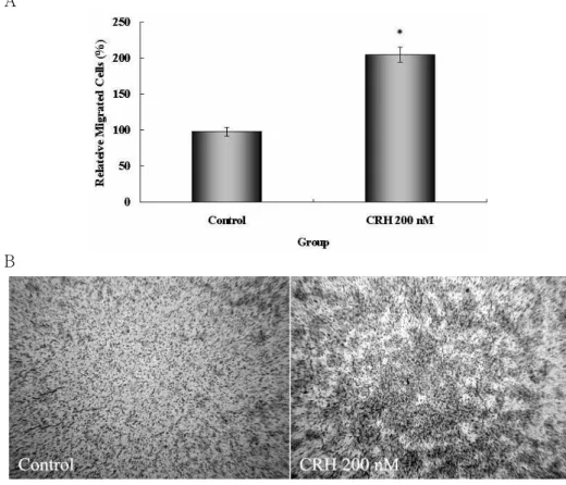
Figure 2. The enhancement of cell migration by CRH in gastric cancer cell line, MKN-28 (A) CRH-induced cell migration in MKN-28 by bar graph. MKN-28 cells were incubated with CRH 200 nM for 24 h and then used for migration assay. After cells harvest, CRH treated MKN-28 cells and no treated cells (7×10⁵) in 1.0 ml serum free media were placed in the insert. Migration cham- ber was incubated at 37oC CO2
incubator for 48h. Migrated cells were dissolved in 0.1% acetic acid and then measured at 570 nm. Data for bar graphs represent mean±
SD. *P<0.05 versus control. (B) CRH-induced cell migration in MKN-28 by photography (magni- fication, ×100). Left is control (no treated) and right is CRH treated group.
Figure 1. Expression of CRH receptor type 1 mRNA level in human gastric cancer cell line, MKN-28. HaCaT cells were used for positive control. Total RNA (5μg) was isolated from MKN-28 and HaCaT cells and examined for RT-PCR using CRH-R1 and β-actin primers. Electrophoresis was performed on 2% agarose gel.
Figure 3. CRH-induced cell migration in MKN-28 by migrated time. MKN-28 cells were treated with or without CRH 200 nM for 24 h and collected. CRH treated cells and control (no treated) cells were placed in the insert (7×10⁵/ml in serum free media) and incubated for indicated time. Migrated cells were dissolved in 0.1% acetic acid and then measured at 570 nm. Data are mean
±SD.
에서 CRH-R1의 mRNA 수준이 발현하여 CRH가 위암 세 포에 작용할 수 있는지를 확인하고자 RT-PCR을 시행하 였다. 특히, 인간 각질형성세포주인 HaCaT 세포에서도 CRH-R1이 발현한다는 것이 보고되어(24), HaCaT 세포 를 양성 대조군으로 함께 실험하였다. 결과를 통해 MKN-28에서 CRH-R1이 발현하여(Fig. 1) 스트레스 반응 에 의해 생성되는 CRH가 위암 세포에 작용할 수 있다는 것을 알았다.
CRH에 의한 세포 migration의 증가. CRH-R1을 발현하 는 MKN-28 세포의 migration에 CRH가 어떤 작용을 하 는지 확인하기 위해, CRH 200 nM을 24시간 전처리한 후, 48시간 동안 migration시켜 CRH를 처리하지 않은 세 포와 비교한 결과, CRH를 처리한 세포의 migration이 약
2배 증가하는 것을 확인했고(Fig. 2A), 현미경으로 관찰 하여 사진으로 찍은 결과 역시 같은 양상을 나타내어 (Fig. 2B) CRH가 위암세포의 migration을 증가시킨다는 것을 알았다.
CRH에 의해 증가하는 migration이 시간이 지남에 따 라 그 효과가 계속적으로 나타나는지 확인하기 위해 CRH 200 nM을 24시간동안 처리한 후, 각각 6, 12, 24,
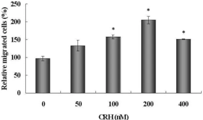
Figure 4. CRH-induced cell migration in a dose dependent man- ner. MKN-28 cells were incubated with various CRH con- centrations (0 nM~400 nM, two-fold dilution) for 24 h and then harvested. For migration assay, each cells were placed (7×10⁵/ml in serum free media) in upper chamber and then incubated at 37oC CO2 incubator for 48 h. Migrated cells were dissolved in 0.1% acetic acid and then measured at 570 nm. Data are mean±
SD. *P<0.05 versus CRH 0 nM.
48, 그리고 72시간 동안 migration을 시킨 결과 모든 시간 대에서 CRH를 처리한 세포의 migration이 처리하지 않 은 세포와 비교했을 때, 더 높음을 확인하였고 이러한 효과는 시간이 지남에 따라 점차 증가하다가 48시간 동 안 migration시켰을 때 최대가 되었다(Fig. 3).
다음으로, CRH가 위암 세포의 migration을 증가시키 는 효과를 나타내는 것에 CRH의 농도가 어떤 영향을 미 치는지 확인하기 위하여 CRH를 25 nM에서 400 nM까지 2배씩 희석하는 방법으로 24시간 동안 세포에 처리한 후, CRH의 효과가 가장 크게 나타났던 48시간 동안 migration시켜서 실험을 하였다. 그 결과, CRH에 의한 위 암 세포의 migration이 CRH의 농도가 증가함에 비례하 여 같이 증가하는 경향을 나타냈고, 200 nM에서 가장 큰 효과를 나타냈다(Fig. 4).
고 찰
스트레스는 현대인이 가지고 있는 여러 가지 질병의 원인이 되고 있는데 이번 연구에서는 한국에서 발병율 이 높다고 알려진 위암에 초점을 맞추어 스트레스 반응 의 대표적 호르몬인 CRH가 인간 위암 세포인 MKN-28 의 migration에 어떤 영향을 미치는지에 알아보았다.
CRH를 전처리 한 위암 세포는 CRH를 처리하지 않은 세 포에 비해 migration이 증가하는 결과를 나타냈다. 이러 한 경향은 migration이 되는 시간에 비례하여 증가했으 며 migration 시간이 48시간일 때 그 효과가 가장 높은 것으로 나타났다(Fig. 3). 또한 CRH 농도에도 비례하여 migration이 증가해 CRH 200 nM일 때 최대의 효과를 나 타냈다(Fig. 4). 그 동안의 많은 연구 결과들은 스트레스 와 암의 관계에 대해서 스트레스가 암의 발생을 자극하
고 암이 진행되는 단계에서 증식을 촉진시킬 뿐 아니라 세포 매개성 면역 반응을 감소시켜(9,10) 암을 제거하는 기능이 감소한다는 것에 초점을 맞추어 주장해 왔다. 또 한 스트레스와 세포의 migration 능력 사이의 관계를 봤 을 때, 스트레스에 의해 lymphocyte나 monocyte의 mi- gration이 변화한다는 보고가 발표되어(25,26) 스트레스 호르몬이 세포의 migration에 많은 영향을 미친다는 것 을 알 수 있다. 이런 내용을 바탕으로 진행된 이번 연구 결과 역시, 스트레스 호르몬이 암세포의 migration에 영 향을 미쳐 전이 과정을 촉진하여 질병의 더욱 심각하게 진행된다는 것을 보여 준다.
스트레스에 의해 작용하는 CRH는 CRH 수용체를 통 해 작용하는 것으로 알려져 있다(3-5). CRH-R2는 주로 소 화기관이나 심장 등에서 발현하고 염증반응에 관여하는 cytokine에 의해 그 발현이 감소한다는 보고가 있는데(4) 반해, CRH-R1은 스트레스에 의해 형성되는 염증성 질 환, 뇌에서 형성되는 퇴행성 신경 질환, 우울증 등에서 주로 관여하는 수용체로 알려져 있을 뿐 아니라 CRH와 의 결합력도 CRH-R1이 훨씬 높은 것으로 보고되고 있 다. 스트레스에 의한 암세포의 migration을 연구하기 위 하여 이번 실험은 사람 위암 세포인 MKN-28에서 CRH-R1 이 발현하는 것을 확인하였다(Fig. 1). 이러한 내용은 CRH와 CRH-R1 사이의 상호 작용을 억제하였을 때, CRH에 의해 증가하는 migration을 감소시킬 수 있고, 결 과적으로 암의 전이 과정을 억제하여 암의 중요한 치료 제로 사용할 수 있다는 가능성을 제시한다. CRH와 CRH-R1의 상호작용을 억제하는 방법으로는 CRH 수용 체의 길항제를 이용하거나 CRH에 대한 결합 단백질을 이용하는 것이 있는데 이런 방법은 실제 임상적으로도 이용하고 있는 방법으로써 CRH-R1에 대한 길항제를 이 용하여 CRH에 의해 유도되는 우울증을 치료한다고 보 고되고 있다(27).
CRH-R1을 억제시켜 암을 치료하려는 시도 외에 다른 치료 방법을 연구하기 위해서는 CRH가 위암 세포의 migration을 증가시키는데 어떤 과정들이 작동하는지에 대한 계속적인 연구가 필요하다. 세포의 migration에는 앞서 언급한 것처럼 세포의 주화성을 유도하는 chemokine 과 그에 선택적으로 작용하는 수용체와의 상호작용(14), 다른 세포와의 상호작용이나 세포 외 기질에 붙어 있는 데 중요하게 관여하는 부착 단백질의 발현 변화(16-18) 등 여러 가지 신호 전달과 단백질의 변화를 수반하는 복 잡한 과정이다. 이 밖에도 Rho GTPase의 활성이 세포골 격의 actin을 활성화시켜 세포의 migration을 유도하고(19), 세포의 생존과 밀접하게 연관되어 있는 신호인 MAP kinase가 세포의 migration에도 중요하게 관여한다는 연 구 결과도 발표 되었다(28). 하지만, 아직까지 CRH와 이 런 migration에 관여하는 요소들의 변화에 대한 연구가
활발하게 진행되고 있지 않아 이러한 연구를 통해 스트 레스에 의해 증가하는 세포 migration에 관여하는 요소 들을 발견하는 것이 선행되어야 하고, 그 후에 이런 요소 들의 활동을 억제하는 여러 방법을 통해 새로운 치료법 을 개발하는 데 이용할 수 있을 것이라고 생각된다. 실 제, 위암에서 migration에 관련되어 있는 여러 요소들을 살펴 보면, CXCR3, CCR7과 같은 chemokine 수용체가 위암 세포에 많이 발현되고 있음이 밝혀져 이 수용체에 선택적으로 상호 작용하는 chemokine을 발현하는 다른 조직으로 이동하여 전이를 유도한다는 내용이 보고되었 으며(29,30), integrin이나 CD44, E-cadherin과 같은 부착 단백질 역시 위암에서 많이 발현하여 세포 외 기질이나 다른 세포와의 상호 작용함으로 부착을 유도하여 mi- gration을 조절한다고 보고되어(31,32), CRH가 위암에서 위와 같은 요인들을 조절시켜 migration을 증가시킬 수 있을 것이라는 가정을 세울 수 있고 이에 대한 연구가 이루어져야 한다.
이번 연구에서는 위암이 한국인에게서 발병율이 높을 뿐 아니라, 전이도 활발하게 일어나는 암이기 때문에 위 암 세포를 가지고 실험하였다. 위암 외 다른 조직에서 발생하는 암의 경우를 살펴보면, 전이가 빠르다고 알려 진 피부암의 경우 피부 조직에서 CRH가 다량으로 발현 하고 있으며 이것이 피부암의 성장요인으로 작용하는 것으로 알려져 있지만 CRH가 피부암의 전이 과정에 어 떤 영향을 미치는지에 대한 보고는 없다. 위암이나 피부 암 같이 전이가 빈번하게 발생하는 암에서 CRH에 의한 migration의 증가가 공통적으로 동일하게 나타나는 결과 인지, 위암의 경우에만 특수하게 나타나는 현상인지에 대한 연구가 필요하고, 이런 연구를 통해 스트레스에 의 해서 암이 촉진되는 과정의 보편적인 기전을 밝혀낼 수 있다고 생각된다. 또한 암이 발생하고 전이되는 과정에 대한 우리 몸의 방어 체계인 면역 세포에 초점을 맞추어 CRH가 면역세포에 어떤 영향을 미치는지 생각해 보면, CRH가 NK 세포의 cytotoxicity 활성을 감소시켜(33) 암 에 대한 방어 능력을 감소시킨다는 등 면역 세포의 활성 을 감소시킨다는 보고는 많지만(9,10) 면역 세포의 이동 에 어떤 역할을 하는지에 대해서는 많은 연구가 진행되 지 않았다. 면역 세포가 암세포의 이동하는 것에 속도를 맞추어 활발하게 암세포가 있는 곳으로 이동한다면 전 이 과정에 중요한 방어체계로 작용할 수 있겠지만, 스트 레스에 의해서 암세포로의 면역 세포의 migration이 감 소하였고 암세포가 다른 조직으로 활발하게 migration하 는 것이라고 생각되어지므로 이 역시 연구를 통해 증명 해야 하는 과제이다.
결론적으로 이번 연구 결과는 대표적인 스트레스 호 르몬인 CRH에 의해 위암 세포의 migration을 증가시켜 위암의 전이 과정을 촉진할 수 있다는 것을 보여주었고,
CRH에 의한 전이 과정을 차단하는 방법을 통해 위암의 새로운 치료법 개발의 가능성을 제시했다.
참 고 문 헌
1. Aguilera G, Rabadan-Diehl C, Nikodemova M: Regulation of pituitary corticotropin releasing hormone receptors. Peptides 22;769-774, 2001
2. Smagin GN, Heinrichs SC, Dunn AJ: The role of CRH in behavioral response to stress. Peptides 22;713-724, 2001 3. Wang W, Ji P, Riopelle RJ, Dow KE: Functional expression
of corticotropin-releasing hormone receptor 1 in cultured rat microglia. J Neurochem 80;286-294, 2002
4. Coste SC, Heldwein KA, Stevens SL, Tobar-Dupre E, Stenzel- Poore MP: IL-1L-1 alpha and TNF alpha down-regulate CRH receptor-2 mRNA expression in the mouse heart. Endocri- nology 142;3537-3545, 2001
5. Radulovic M, Dautzenberg FM, Sydow S, Radulovic J, Spiess J: Corticotropin-releasing factor receptor 1 in mouse spleen:
expression after immune stimulation and identification of receptor-bearing cells. J Immunol 162;3013-3021, 1999 6. Pedersen WA, McCullers D, Culmsee C, Haughey NJ, Her-
man JP, Mattson MP: Corticotropin-releasing hormone pro- tects neurons against insults relevant to the pathogenesis of Alzheimer's disease. Neurobiology of Disease 8;492-503, 2001 7. Roe SY, McGowan EM, Rothwell NJ: Evidence for the
involvement of corticotropin-releasing hormone in the patho- genesis of traumatic brain injury. Eur J Neurosci 10;553-559, 1998
8. Elenkov IJ, Webster EL, Torpy DJ, Chrousos GP: Stress, corticotropin releasing hormone, glucocorticoids, and the immune/inflammatory response: Acute and chronic effects.
Annals of the Newyork Academy of Science 876;1-13,1999 9. Strausbaugh H, Irwin M: Central corticotropin-releasing hor- mones reduces cellular immunity. Brain Behav Immun 6;
11-17, 1992
10. Reiche EM, Nunes SO, Morimoto HK: Stress, depression, the immune system, and cancer. Lancet Oncol 5;617-625, 2004 11. Vrezas I, Willenberg HS, Mansmann G, Hiroi N, Fritzen R,
Bornstein SR: Ectopic adrenocorticotropin (ACTH) and cor- ticotropin-releasing hormone (CRH) production in the ad- renal gland: basic and clinical aspects. Microsc Res Tech 61;
308-314, 2003
12. Sato H, Nagashima Y, Chrousos GP, Ichihashi M, Funasak Y: The expression of corticotropin-releasing hormone in melanoma. Pigment Cell Res 15;98-103, 2002
13. Kwok CM, Wu CW, Lo SS, Shen KH, Hsieh MC, Lui WY:
Survival of gastric cancer with concomitant liver metastases.
Hepatogastroenterology 51;1527-1530, 2004
14. Balkwill F: Chemokine biology in cancer. Semin Immunol 15;49-55, 2003
15. Ueda J, Kajita M, Suenaga N, Fujii K, Seiki M: Sequence- specific silencing of MT1-MMP expression suppresses tumor cell migration and invasion: importantce of MT1-MMP as a therapeutic target for invasive tumors. Oncogene 22;8716- 8722, 2003
16. Marhaba R, Zoller M: CD44 in cancer progression: adhesion, migration and growth regulation. J Mol Histol 35;221-231, 2004
17. McGray EC, Lev DC, Bar-Eli M: Cellular adhesion pathways and metastatic potential of human melanoma. Cancer Biol Ther 1;459-465, 2002
18. Ziober BL, Silverman SS Jr, Kramer RH: Adhesive mech- anisms regulating invasion and metastasis in oral cancer. Crit Rev Oral Biol Med 12;499-510, 2001
19. Raftopoulou M, Hall A: Cell migration: Rho GTPases lead the way. Dev Biol 265;23-32, 2004
20. Maus MV, Reilly SC, Clevenger CV: Prolactin as a chemoat- tractant for human breast carcinoma. Endocrinology 140;
5447-5450, 1999
21. Mizobe K, Kishihara K, Ezz-Din El-Naggar R, Madkour GA, Kubo C, Nomoto K: Restraint stress-induced elevation of endogenous glucocorticoid suppresses migration of granulocytes and macrophages to and inflammatory locus. J Neuroimmunol 73;81-9, 1997
22. Tandon R, Khanna HD, Dorababu M, Goel RK: Oxidative stress and antioxidants status in peptic ulcer and gastric car- cinoma. Indian J Physiol Pharmacol 48;115-118, 2004 23. Hocker M, Rogenberg I, Xavier R, Henihan RJ, Wiedenmann
B, Rosewicz S, Podolsky DK, Wang TC: Oxidative stress activates the human histidine decarboxylase promoter in AGS gastric cancer cells. J Biol Chem 273;23046-23054, 1998 24. Slominski AT, Roloff B, Zbytek B, Wei ET, Fechner K, Curry
J, Wortsman J: Corticotropin releasing hormone and related peptides can act as bioregulatory factors in human keratinocytes.
In Vitro Cell Dev Biol Anim 36;211-216, 2000
25. Zhang D, Kishihara K, Wang B, Mizobe K, Kubo C, Nomoto K: Restraint stress-induced immunosuppression by inhibiting leukocyte migration and Th1 cytokine expression during the intraperioneal infection of Listeria monocytogenes. J Neuro- immunol 92;139-151, 1998
26. Genedani S, Bernardi M, Baldini MG, Bertolini A: Influence of CRF and alpha-MSH on the migration of human monocytes
in vitro 23;99-102, 1992
27. Kunzel HE, Zobel AW, Nickel T, Ackl N, Uhr M, Sonntag A, Ising M, Holsboer F: Treatment of depression with the CRH-1-receptor antagonist R122929: endocrine changes and side effects. J Psychiatr Res 37;525-533, 2003
28. Huang C, Jachbson K, Schaller MD: MAP kinases and cell migration. J Cell Sci 117;4619-4628, 2004
29. Ohshima K, Suefuji H, Karube K, Hamasaki M, Hatano B, Tutiya T, Yamaguchi T, Suzuki K, Suzumiya M: Expression of chemokine receptor CXCR3 and its ligand, mig, in gastric and thyroid marginal zone lymphomas. Possible migration and autocrine mechanism. Leuk Lymphoma 44;329-336, 2003 30. Mashino K, Sadanaga N, Yamaguchi H, Tanaka F, Ohta M,
Shibuta K, Mori M: Expression of chemokine receptor CCR7 is associated with lymph node metastasis of gastric carcinoma.
Cancer Res 62;2937-2941, 2002
31. Nejjari M, Aderson W, Pourreyron C, Jacquier MF, Scoazec JY, Remy L: The role of fibroblasts in the modulation of integrin-dependent interactions between the gastric cell line HGT-1 and fibronectin. Int J Cancer 112;560, 2004 32. Streit M, Schmidt R, Hilgefeld RU, Thiel E, Kreuser ED:
Adhesion receptors in malignant transformation and dissem- ination of gastrointestinal tumors. Recent Results Cancer Res 142;19-50, 1996
33. Irwin M, Hauger RL, Britton K: Benzodiazepines antigonize central corticotropin-releasing hormone-induced suppression of natural killer cell activity. Brain Res 63;114-118, 1993