www.krspine.org
Anterolateral and Posterior versus Posterior-Only Approaches for the Correction of Degenerative Adult
Spinal Deformity
Se-Jun Park, M.D., Ph.D., Chong-Suh Lee, M.D., Ph.D., Tae-Hoon Yum, M.D., Yunjin Nam, M.D., Jin-Sung Park, M.D.
J Korean Soc Spine Surg 2020 Mar;27(1):9-18.
Originally published online March 31, 2020;
https://doi.org/10.4184/jkss.2020.27.1.9
Korean Society of Spine Surgery
SMG-SNU Boramae Medical Center, 20, Boramae-ro 5-gil, Dongjak-gu, Seoul 07061, Korea Tel: +82-2-831-3413 Fax: +82-2-831-3414
©Copyright 2017 Korean Society of Spine Surgery pISSN 2093-4378 eISSN 2093-4386
The online version of this article, along with updated information and services, is located on the World Wide Web at:
http://www.krspine.org/DOIx.php?id=10.4184/jkss.2020.27.1.9
This is an Open Access article distributed under the terms of the Creative Commons Attribution Non-Commercial License (http://
creativecommons.org/licenses/by-nc/4.0) which permits unrestricted non-commercial use, distribution, and reproduction in any medium, provided the original work is properly cited.
Spine Surgery
© Copyright 2020 Korean Society of Spine Surgery
Journal of Korean Society of Spine Surgery. www.krspine.org. pISSN 2093-4378 eISSN 2093-4386
This is an Open Access article distributed under the terms of the Creative Commons Attribution Non-Commercial License (http://creativecommons.org/licenses/by-nc/4.0/) which permits unrestricted non-commercial use, distribution, and reproduction in any medium, provided the original work is properly cited.
9
J Korean Soc Spine Surg. 2020 Mar;27(1):9-18. https://doi.org/10.4184/jkss.2020.27.1.9
Original Article
Anterolateral and Posterior versus Posterior-Only Approaches for the Correction of Degenerative Adult Spinal Deformity
Se-Jun Park, M.D., Ph.D., Chong-Suh Lee, M.D., Ph.D., Tae-Hoon Yum, M.D., Yunjin Nam, M.D., Jin-Sung Park, M.D.
Department of Orthopedic Surgery, Samsung Medical Center, Sungkyunkwan University, School of Medicine, Seoul, South Korea
Study Design: A case-control study.
Objectives: This study was conducted to demonstrate the reliability of mini-open anterior lumbar interbody fusion (ALIF) combined with lateral lumbar interbody fusion (LLIF) followed by 2-stage posterior fixation in patients with adult spinal deformity (ASD).
Summary of Literature Review: Although the correction of ASD using LLIF has become more widespread, the amount of sagittal plane correction has been reported to be suboptimal.
Materials and Method: Thirty ASD patients who underwent ALIF with LLIF followed by 2-stage posterior fixation (AP group) were compared to 60 patients who underwent posterior-only surgery (PO group) and were matched according to age, sex, diagnosis, fusion level, pelvic incidence, and follow-up duration. Spinopelvic parameters, hospitalization data, clinical outcomes, and complications were compared between the 2 groups.
Results: Postoperative lumbar lordosis was greater in the AP group than in the PO group (p<0.001). The reduction in the sagittal vertical axis was also greater in the AP group than in the PO group (p=0.005). Postoperatively, 90.0% of the AP group had a pelvic incidence–
lumbar lordosis value within 9°, whereas only 50.0% of the PO group met that criterion (p<0.001). The operation time of the AP group was longer than that of the PO group, while estimated blood loss and red cell transfusion were lower in the AP group. Postoperative medical complications and delayed surgical complications developed more frequently in the PO group.
Conclusions: Mini-open ALIF with LLIF followed by 2-stage posterior fixation can restore sagittal balance more appropriately, with a lower rate of complications, than posterior-only surgery for the correction of ASD.
Key words: Adult spinal deformity, Anterior lumbar interbody fusion, Lateral lumbar interbody fusion, Sagittal balance, Complication
Received: August 19, 2019 Revised: September 9, 2019 Accepted: March 9, 2020 Published Online: March 31, 2020 Corresponding author: Jin-Sung Park, M.D.
ORCID ID: Se-Jun Park: https://orcid.org/0000-0002-2412-9437 Chong-Suh Lee: https://orcid.org/0000-0003-2790-9225 Jin-Sung Park: https://orcid.org/0000-0001-6517-8609 Tae-Hoon Yum: https://orcid.org/0000-0003-1968-736X Yunjin Nam: https://orcid.org/0000-0003-2294-3280 Department of Orthopedic Surgery, Samsung Medical Center, 81 Ilwon-ro Gangnam-gu, Seoul, 06351, Korea
TEL: +82-2-3410-1583, FAX: +82-2-3410-0061 E-mail: [email protected]
서론
후방 접근법은 성인 척추 변형(adult spinal deformity, ASD) 을 교정하기 위한 전통적인 수술 방법이었다. 이상적인 시상 정렬의 교정을 위해 Smith-Peterson osteotomy (SPO), pedicle subtraction osteotomy (PSO), posterior vertebral column resection (PVCR) 등의 다양한 절골술 방법이 사용되어 왔다. 하지만 이 러한 후방 접근을 통한 교정 방법은 출혈량이 많을 뿐만 아니라 신경 마비와 같은 심각한 합병증이 동반될 가능성 높으며, 특히 고령의 환자에서는 수술 후 주요 합병증 발생 위험이 높은 것으 로 알려져 있다.1-6)
최근 최소 침습적으로 성인 척추 변형을 교정하기 위한 노 력이 있으며, 특히 측방 추체간 유합술(lateral lumbar interbody fusion, LLIF)은 기존의 후방 접근법과 비교하여 합병증 발생 빈 도가 낮은 것으로 보고되고 있다.4,7,8) 또한 측방 추체간 유합술
은 간접적인 신경 감압을 통해 후궁 절제술의 필요성을 줄일 수 있는 장점도 있다.9) 하지만, 요추 전만각의 회복은 한 분절에서
10도 미만으로 시상 정렬의 교정 효과는 상대적으로 다른 장점 에 비해 크지 않다.7,10-16)
시상 정렬의 회복은 성인 척추 변형 환자에서 수술의 성공을 결정하는 중요한 요인인 만큼,17-19) 요추의 전만각을 적절하게 교정하는 것이 중요하다. 따라서 측방 추체간 유합술 단독으로 는 교정 각도가 불충분할 수 있다. 전방 추체간 유합술(anterior lumbar interbody fusion, ALIF)은 후방 추체간 유합술(posterior lumbar interbody fusion, PLIF)이나 측방 추체간 유합술보다 더 많은 전만각 교정을 얻을 수 있다.20-22) 따라서 저자들은 충분한 요추 전만각의 교정을 위해 측방 추체간 유합술과 함께 가장 마 지막 분절(제 5요추-제 1천추 와/또는 제 4요추-제 5요추)에 최 소 절개 전방 추체간 유합술(mini-open ALIF)을 시행하여 왔다.
현재까지 성인 척추 변형 환자에서 측방 추체간 유합술과 최소 절개 전방 추체간 유합술을 동시에 사용한 시상 정렬 교정에 관 한 연구는 거의 없었다.
본연구의 목적은 측방 추체간 유합술과 최소 절개 전방 추체 간 유합술을 시행한 후 단계적으로 후방 고정술을 시행한 환자 군이 후방 접근법으로만 수술을 시행한 환자군과 비교하여 시 상 정렬의 교정 정도와 합병증 발생 빈도에 있어 장점이 있는지 를 확인하고자 하였다.
대상 및 방법
본 연구는 본원 임상연구 윤리위원회 심의의 승인 후 진행한 후향적 환자 대조군 연구이다(승인번호: 2019-08-111-001). 요추 변형 후만증(lumbar degenerative kyphosis, LDK)과 요추 변 형 측후만증(lumbar degenerative kyphoscoliosis, LDKS) 환자를 대상으로 제 5요추와 제 1천추간을 포함하여 3분절 이상의 유 합술을 시행한 환자 중 최소 12개월 이상의 추시 관찰이 가능한 환자를 대상으로 하였다. 이전에 수술을 시행 받았거나 외상에 의해 후만증이 발생한 경우는 제외하였다. 2012년 1월부터 2014 년 6월까지 측방 추제간 유합술과 함께 마지막 분절에 최소 절 개 전방 추체간 유합술을 시행한 후 단계적으로 후방 고정술을 시행한 환자 30명을 AP 그룹으로 선정하였다. 2006년 1월부터 2013년 12월까지 후방 접근법으로만 교정을 시행한 124명의 환 자 중 나이, 성별, 유합 분절 수, 골반 입사각(pelvic incidence), 추 시 기간이 같은 환자 60명을 PO 그룹으로 선정하였다.
후방으로만 접근한 PO 그룹은 전통적인 정중앙 접근법으로 후방 주체간 유합술을 시행하였으며, 추체간 유합을 위해 한쪽 으로만 추간판을 제거한 후 자가골을 채운 8도 Titanium curved cage를 사용하였다. 특히 제 4요추부터 제 1천추의 경우 가능한 높이가 큰 cage를 넣으려고 하였고 척추경 나사못을 이용하여 후방 고정을 시행하였다. 추가적인 척추경을 통한 쐐기형 절골
술(pedicle subtraction osteotomy, PSO)은 11명의 환자에서 시행하 였다(Fig. 1).
AP 그룹에서는 1주 간격으로 단계적인 수술을 시행하였다.
첫번째 수술에서는 앙와위 자세로 제 5요추-제 1천추와 또는 제 4요추-제 5요추에 최소 절개 전방 추체간 유합술을 시행한 후 측면 자세 전환하여 측방 추체간 유합술을 시행하였다. 전방 추체간 유합술의 경우 12도 lordotic-angled polyetheretherketone (PEEK) cage (Syncage, Synthes Inc., West Chester, PA, USA)를 이 용하였고 측방 추체간 유합술의 경우 6도 lordotic-angled PEEK cages (Clydesdale, Medtronic Inc., Minneapolis, MN)를 사용하 였다. 모든 cage에는 allogenous chip bone 과 demineralized bone matrix (DBM, Grafton®, Medtronic Inc., Minneapolis, MN, USA)을 사용하였다. 처음 수술 후 3일째 자가공명영상 검사와 단순 방 사선 사진 촬영을 시행하여 간접 신경 감압 정도와 요추 전만각 교정 정도를 평가하였고, 추가적으로 필요한 후궁 절제술 및 전 만각 교정 정도를 결정하였다. 두번째 수술 시에 평균 1.4분절에 서 추가적인 후궁절제술을 시행하였다. 9명의 환자에서 경피적 후방 기기고정술(percutaneous pedicle fixation)을 시행하였고, 16 명 환자에서는 관절 제거(facetectomy)와 함께 관혈적 척추경 나 Fig. 1. A 54-year-old woman underwent posterior fusion surgery for degenerative lumbar kyphosis. Preoperatively, pelvic incidence was 50°, lumbar lordosis was 17°, pelvic tilt was 27°, and the sagittal vertical axis was 36 mm. Although lumbar lordosis increased to 31° and the sagittal vertical axis was 34 mm postoperatively, the correction seemed to be insufficient considering the pelvic incidence of 50°.
A B
Anterolateral and Posterior Approach for the Correction of ASD Journal of Korean Society of Spine Surgery
www.krspine.org
11
사못 고정술을 시행하였으며 5명의 환자에서 추가적인 척추경을 통한 쐐기 절골술을 시행하였다(Fig. 2).
방사선학적 평가는 수술 후 2~3주 이후와 최종 추시시 시행 한 전체 척추 전후면 방사선 사진을 통해 평가하였다. 골반 입 사각(pelvic incidence, PI), 요추 전만각(lumbar lordosis, LL), 천 추 기울기(sacral slope, SS), 골반 기울기(pelvic tilt, PT), 흉추 후 만각(thoracic kyphosis, TK), 시상면 수직축(sagittal vertical axis, SVA)은 현재 널리 쓰이는 방법을 이용하여 측정하였다.23) 관상 면 Cobb’s angle (CCA)은 전후면 방사선 사진상 최대의 각도의 Cobb’s angle로 정의하였다. 골반 입사각과 요추 전만각의 차이 (PI-LL)가 9도 이내인 경우 이상적으로 교정이 되었다고 정의 하였다.17) 추체간 유합 여부는 1년 추시시 컴퓨터 단층 촬영을 통해서 확인하였고 추체간 이식한 골조직의 완전한 연속성이 확인되었을 때로 정의하였다.24) 모든 영상 의학적 평가는 PACS (GE, Centricity®, GE healthcare IT, Barrington, IL, USA)를 이용하 여 측정하였다.
전자 의무 기록을 통해서 입원기간, 수술시간, 수술 중 출혈 량(estimated blood loss, EBL), 수술 후 출혈량, 수혈 여부를 확인 하였다. 임상적 결과는 Oswestry disability index(ODI)와 visual analogue scale (VAS)를 이용하여 평가하였다. 퇴원 전에 발생한
수술 후 내과적 합병증과 재수술을 포함한 수술과 관련된 합병 증을 추시 시간 동안 확인하였다.
통계 분석은 SPSS software (version 22.0.0; SPSS Inc., Chicago, IL, USA)를 사용하였다. 두 군간 비교를 위해 연속 변수에 대해 서는 독립표본 t-검정(independent t-test)을 이용하였고 범주형 변 수에 대해서는 카이제곱 검정(chi-square test)과 피셔정확 검정 (Fisher’s exact test)을 이용하였다. 모든 경우에서 p값이 0.05 미 만인 경우를 통계적으로 유의하다고 판단하였다.
결과
1) 연구 대상자
AP 그룹 30명과 PO 그룹 60명을 비교하였다. 두 군간 나 이, 성별, 진단명, 골반 입사각, 신체 상태(american society of Fig. 2. A 72-year-old woman underwent mini-open anterior lumbar inter-
body fusion at L5-S1 and lateral lumbar interbody fusion at L2-4, followed by posterior fixation. Preoperatively, pelvic incidence was 57°, lumbar lordosis was 24°, pelvic tilt was 31°, and the sagittal vertical axis was 106 mm. Postoperatively, lumbar lordosis increased to 54°, pelvic tilt decreased to 23°, and the sagittal vertical axis improved to 22 mm. It is noteworthy that pedicle subtraction osteotomy was not performed.
Table 1. Baseline data of matched cohorts
Characteristics AP group PO group p
Number of patients 30 60
Age (years)* 66.7±7.4 67.1±6.6 0.803
Gender (male: female)* 5:25 10:50 1.000
Diagnosis (LDK: LDKS)* 15:15 30:30 1.000
PI (°)* 55.1±8.8 52.8±12.4 0.377
No. of total fusion level* 5.4±2.2 5.7±2.2 0.593
ASA class 2.0±0.4 1.9±0.5 0.337
BMD (g/cm2) -1.1±1.2 -1.5±2.1 0.371
BMI (kg/m2) 25.8±3.0 26.3±3.3 0.284
No. of interbody fusion levels 4.0±6.4 3.3±0.9 <0.001
ALIF: LLIF 1.0:2.0 - -
No. of laminectomy levels† 1.4±1.1 3.6±0.6 <0.001
No. of patients with PSO 5 11 0.845
Iliac screw fixation 30 31 <0.001
Follow-up duration (months) 16.7±5.1
(12.0-35.6) 19.2±7.4
(12.0-42.0) 0.056
*Characteristics used for creating matched group.
†Decompression included posterior laminectomy for both central canal and foramina.
Bold p values indicate statistical significance.
ALIF indicates anterior lumbar interbody fusion,LLIF: lateral lumbar inter- body fusion,LDK: lumbar degenerative kyphosis,LDKS: lumbar degenera- tive kyphoscoliosis, ASA: American Society of Anesthesiologists, BMD:
bone mineral density, BMI: body mass index, PI: pelvic incidence, LL:lumbar lordosis, PSO: pedicle subtraction osteotomy.
A B
anesthesiologists, ASA class), 골밀도와 체질량은 두 군간에 유의 한 차이는 없었다. 총 유합 분절수는 두 군간 차이가 없었지만 추체간 유합 분절수는 AP 그룹이 4.0분절로 PO 그룹의 3.3분절 보다 더 많았다. 후궁 절제술은 PO 그룹에서는 3.6분절로 AP 그 룹의 1.4분절보다 더 많이 시행되었다. 추시 기간은 두 군간에 유의한 차이는 없었다(Table 1).
2) 방사선학적 평가
수술 전 요추 전만각, 천추 기울기, 골반 기울기, 시상면 수직 축, 흉추 후만각, 관상면 Cobb’s angle 모두 두군 간에 유의한 차 이는 없었다. 수술 후 평균 요추 전만각은 AP 그룹이 52.0도로 PO 그룹의 40.9보다 유의하게 더 컸다(p<0.001). AP 그룹은 수술 전 평균 요추 전만각은 19.9도에서 52.0도로 32.1도 교정되었으
며, PO 그룹은 26.2도에서 40.9도로 14.7도 교정되어 AP 그룹의 더 많이 교정되었다. 수술 후 천추 기울기와 골반 기울기는 AP 그룹에서 PO 그룹보다 더 많이 교정되었으며(p<0.001, p=0.001), 수술 후 시상면 수직축도 AP 그룹에서 13.8 mm로 PO 그룹 34.7 mm보다 더 많이 교정되었다(p=0.005). 수술 후 흉추 후만각과 시상면 Cobb’s angle은 두 군간에 유의한 차이는 없었다. 최종 추 시에도 요추 전만각, 천추 기울기, 시상면 수직축은 두 군간의 차이가 유의하게 유지 되었으나, 천추 기울기는 최종 추시시 두 군 간의 차이는 유의하지 않았다(Table 2).
수술 후 교정된 분절의 전만각은 유합 방법에 따라 통계적 차
Table 2. Comparison of radiographic parameters between matched groups
Parameters AP group PO group p
PI (°) - 55.1±8.8 52.8±12.4 0.377
LL (°) Preop 19.9±25.2 26.2±17.1 0.224
Postop 52.0±11.4 40.9±10.5 <0.001
Last FU 46.6±12.1 37.0±12.1 0.001
SS (°) Preop 23.5±15.2 24.9±11.4 0.627
Postop 38.4±8.0 30.7±8.9 <0.001
Last FU 33.8±8.5 29.0±9.8 0.026
PT (°) Preop 31.6±12.5 28.0±9.8 0.135
Postop 16.7±5.4 22.2±9.0 0.001
Last FU 21.3±6.8 23.8±9.7 0.157
SVA (mm) Preop 76.1±48.4 59.4±55.3 0.161
Postop 13.8±26.1 34.7±41.1 0.005
Last FU 25.6±28.9 52.9±46.9 0.001
TK (°) Preop 14.5±18.3 19.9±12.3 0.098
Postop 25.4±11.4 24.2±9.8 0.605
Last FU 27.8±13.5 27.3±10.4 0.643
CCA (°) Preop 15.5±11.3 15.1±11.2 0.836
Postop 3.2±3.5 5.3±5.7 0.153
Last FU 3.5±3.6 5.8±4.3 0.278
*Bold p values indicate statistical significance.
FU: follow-up, PI: pelvic incidence, LL: lumbar lordosis, SS: sacral slope, PT: pelvic tile, SVA: sagittal vertical axis, TK: thoracic kyphosis, CCA:
coronal Cobb’s angle.
Table 3. PI-LL mismatch between groups
AP group PO group p PI-LL (°)
Preoperative 35.2±22.9 26.7±16.4 0.045
Postoperative 3.1±7.2 12.0±11.4 <0.001
Last FU 8.5±11.0 15.9±12.6 0.006
Patients with PI-LL less than 9° (%)
Preoperative 4 (13.3%) 10 (16.7%) 0.155
Postoperative 27 (90.0%) 30 (50.0%) <0.001
Last FU 20 (66.7%) 21 (35.0%) 0.007
*Bold p values indicate statistical significance.
PI: pelvic incidence, LL: lumbar lordosis, FU: follow-up.
Fig. 3. Graph showing segmental lordosis per level according to the in- terbody fusion method. Segmental lordosis (*) and its change (†) per level were more favorable for anterior lumbar interbody fusion (ALIF) than for either lateral lumbar interbody fusion (LLIF) or posterior lumbar interbody fusion (PLIF).
Anterolateral and Posterior Approach for the Correction of ASD Journal of Korean Society of Spine Surgery
www.krspine.org
13
이를 보였다. 전방 추체간 유합술의 평균 분절 전만각은 17.4도로 8.1도인 측방 추체간 유합술과 9.1도인 후방 추체간 유합술 보다 약 2배 정도 더 컸다. 각 분절의 전만각의 교정 정도는 전 방 추체간 유합술에서 11.4도로 4.2도인 측방 추체간 유합술과 후방 추체간 유합술보다 더 많이 교정되었다(p<0.001) (Fig. 3).
수술 후 PI-LL의 불일치(mismatch)는 AP 그룹에서는 3.1도 PO 그룹은 12.0도로 두 군간의 유의한 차이를 보였다(p<0.001). 수 술 후 AP 그룹에서는 90.0%에서, PO 그룹은 50.0%에서 PI-LL이 9도 이내의 적절한 교정을 보였다 (p<0.001). 마지막 추시에서도 AP 그룹에서는 66.7%, PO 그룹은 35.0%로 AP 그룹에서도 더 좋 은 교정력을 유지하였다(p=0.007) (Table 3).
3) 임상적 결과 및 합병증
총 수술 시간은 AP그룹은 11.2 시간으로 PO 그룹의 8.6 시간 보다 더 많은 시간이 소요되었다(p<0.001). 수술 후 출혈량은 두 군간에 유의한 차이는 없었지만, 수술 중 출혈량은 AP 그룹이 PO 그룹보다 더 적었다(p<0.006). AP 그룹에서 평균 수혈량은 4.9 개로 PO 그룹의 7.0개 보다 더 적은 수혈이 필요하였다 (p<
0.043). 수술 전 ODI와 VAS점수는 두 군간에 유의한 차이는 없 었다. 최종 추시시 ODI 점수는 두 군간에 유의한 차이는 없었지
만, VAS 점수는 허리와 하지 통증 모두에서 PO 그룹보다 AP 그 룹에서 더 좋은 결과를 보여주었다(p<0.012, p<0.039) (Table 4).
수술 후 입원기간 동안 발생한 내과적 문제는 AP 그룹에서 5 명으로 PO 그룹에서 발생한 24명보다 유의하게 더 적게 발생하 였다(p=0.032). 수술 직후 발생한 수술과 관련된 합병증은 AP 그 룹에서는 9명으로 PO 그룹인 19명과 비교하였을 때 유의한 차 이는 없었다. AP 그룹에서는 경막외 혈종으로 2명, 감염으로 1 명이 재수술을 시행하였고, PO 그룹에서는 지속적인 CSF 유출 로 1명, 경막외 혈종으로 1명, 감염으로 1명, 설명은 되지 않은 지 속적인 방사통으로 1명이 재수술을 시행하였다. 추시 기간 동안 AP 그룹에서는 6명의 환자에서 근위 인접 분절 후만증(proximal junctional kyphosis, PJK)이 발생하였고 불유합이나 강봉 파열과 같은 합병증은 발생하지 않았다. PO 그룹에서는 10명의 환자에 서 근위 인접 분절 후만증이 발생하였고 불유합은 7명, 강봉 파 열은 2명에서 발생하여 3명의 환자에서 재수술을 시행하였다.
PO 그룹에서는 10명의 환자에서 근위 인접 분절 후만증은 없었 지만 시상면 수직축이 증가하는 대상실조(decompensation)가 10 명에서 발생하였으며(Fig. 4), 결과적으로 더 많은 환자에서 추 시 기간 동안의 수술 후 합병증이 발생하였다(p=0.020) (Table 5).
고찰
대부분의 고령 환자들은 동반 질환이 많고 골밀도가 좋지 않 기 때문에 척추 변형의 수술을 시행할 때 내과적으로나 외과적 으로 합병증이 발생할 위험이 높다. 이러한 합병증을 줄이기 위 해 성인 척추 변형 교정에도 측방 추체간 유합술과 같은 최소 침습적인 수술 방법이 소개되어왔다.4,7,8) 많은 연구에서 성인 퇴 행성 측만증에서 측방 추체간 유합술을 통한 관상면의 교정 정 도는 좋은 방사선학적 결과를 보고하였지만, 시상면 교정에 있 어서는 요추 전만각 회복은 한분절당 10도 미만으로 대부분의 연구에서 척추골반지표(spino-pelvic parameters)를 회복시키는 데는 제한이 있다고 보고하였다.7,8,11-13,15,16) 저자들은 이러한 결 과는 제 5요추와 제 1천추 분절을 치료하지 않았거나 후측방 유 합이나 후방추체유합으로 치료를 하였기 때문으로 생각된다. 따라서 저자들은 충분한 요추 전만각 회복을 위해 측방 추체간 유합술과 함께 마지막 분절에 최소 절개 전방 추체간 유합술을 시행해왔다.
본 연구는 측방 추체간 유합술과 마지막 분절에 전방 추체간 유합술을 시행 후 후방 고절술을 시행한 AP 그룹이 후방으로만 접근한 PO 그룹보다 시상 정렬의 회복이 더 효과적임을 보여주 였다. AP 그룹에서의 요추 전만각의 증가는 평균 32.1도로 척추 경을 통한 쐐기 절골술과 비슷한 결과를 보였다. 저자들은 이러 한 큰 교정이 가능한 이유는 전방 추체간 유합술 때문으로 생 Table 4. Comparison of hospitalization and clinical results between
groups
Parameters AP group PO group p
Hospital stay (days) 22.9±15.7 19.3±11.5 0.281 Total operation time (hours) 11.2±1.5 8.6±1.6 <0.001
EBL (L)* 1.7±1.2 2.5±1.4 0.006
Postoperative blood loss (L)* 1.2±0.6 1.3±0.4 0.365 RBC transfusion (pack)† 4.9±3.8 7.0±4.6 0.043 ODI (preoperative) 63.0±13.6 67.4±14.8 0.167 Back VAS (preoperative) 7.2±2.3 6.5±2.5 0.227 Leg VAS (preoperative) 4.6±3.4 5.2±3.0 0.362 ODI (last follow-up) 32.0±16.4 36.4±18.0 0.536 Back VAS (last follow-up) 2.8±2.1 4.1±2.3 0.012 Leg VAS (last follow-up) 2.4±2.3 3.7±3.0 0.039
*Blood loss was determined by adding both anterolateral and posterior surgeries.
†The number of transfused packs included intraoperative and postopera- tive transfusion.
Bold p-values indicate statistical significance.
EBL:estimated blood loss, RBC: red blood cell, ODI: Oswestry disability index, VAS: visual analogue scale.
각한다. 이전 연구에서 보여준 것처럼,3,20,21,25) 본연구에서도 수 술 후 유합 분절당 전만각은 전방 추체간 유합술의 경우가 측 방과 후방 추체간 유합술보다 더 좋음을 보여주었다(Fig. 3). 전 방 추체간 유합술의 전만각 교정의 장점은 특히 가장 마지막 분 절에서 시행하였을 때 그 효과가 크다. 요추 전만각은 거의 70%
가 제 4요추와 제 1천추 사이에서 이뤄지기 때문에 제 5요추와
제 1천추 또는 제 4요추와 제 1천추에서 충분한 각도를 회복하 는 것이 이상적인 시상 정렬의 회복에 중요하다. 마지막 분절에 전방 추체간 유합술을 시행하는 것은 더 큰 지렛대 효과를 가져 와 전체 시상 정렬을 회복하는데 생역학적으로 이점이 있다. 또 한 장분절 유합에서 마지막 분절에 전방 추체간 유합술을 시행 하는 것은 유합율을 더 높이기 때문에 시상 정렬의 교정 손실을 Table 5. Medical and surgical complications between groups
AO group PO group p
Medical complications
Voiding difficulty 0 6 -
Pulmonary edema 1 3 1.000
Delirium 2 4 1.000
Urinary tract infection 0 2 -
Ileus 1 1 0.613
Deep vein thrombosis 1 2 1.000
Others 0 5 -
Pulmonary thromboembolism* 0 1 -
Total 5 24 0.032
Perioperative surgical complications (reoperation)†
Dural tear 1(0) 9(1) 0.155
Transient weakness 4(0) 3(0) 0.216
Persistent weakness 1(0) 3(0) 1.000
Unexplained radiculopathy 0 1(1) -
Epidural hematoma formation 2(2) 1(1) 0.213
Deep infection 1(1) 2(1) 1.000
Total 9 19 1.000
Reoperation total 3 4 0.578
Delayed surgical complications (reoperation)‡
PJK 6(0) 10(1) 0.697
Nonunion / Rod breakage 0 7(2) -
Decompensation without PJK§ 0 10 -
Total 6 27 0.020
Reoperation total 0 3 -
*Major medical complication.
†Complications during hospitalization.
‡Complications after discharge.
§Decompensation was defined as an SVA greater than 80mm without PJK at the last follow-up.
Bold p values indicate statistical significance.
PJK: proximal junctional kyphosis.
Anterolateral and Posterior Approach for the Correction of ASD Journal of Korean Society of Spine Surgery
www.krspine.org
15
이미 여러 논문에서 전방 추체간 유합술이 후방 추제간 유합술 보다 더 큰 cage를 넣을 수 있기 때문에 유합율이 더 좋다고 보고하였다.21,28) 두번째로 시상 정렬의 불충분한 교정은 근위 인
접 분절 후만증이 발생하지 않더라고 대상실조(decompression) 가 발생할 위험을 높일 수 있기 때문이며, 특히 척추 기립근이 나 둔부 근육이 약한 환자에서는 골반 보상 능력이 부족하기 때 문에 발생 위험이 더 높다.
본연구는 몇 가지 제한점이 있다. 첫째 후향적 연구가 가지는 본질적인 약점이 있다. 둘째로, 두 술기가 시행된 시기가 다르 다. 과거에는 성인 척추 변형에 대한 모든 수술이 후방 접근으 로만 이루어진 반면, 최근에는 전방 접근법이 주로 사용되고 있 다. 이것은 추시 기간의 차이를 가져오고 결과적으로 최종 임상 결과에 영향을 줄 수 있지만 본 연구 대조군 선정에서 비슷한 추시 기간을 가진 대상자를 선정하였고 최종 추시 기간에 통계 적 차이는 없었다. 또한 본연구의 모든 환자는 동일한 방법으로 외래 추시 기간 동안 임상 결과와 방사선 평가가 이루어졌다. 셋째 AP 그룹에서는 모든 경우에 장골 나사가 추가적으로 사용 되었고 PO 그룹에서는 50%에서만 사용되었다. 골반 고정 여부 는 특히 추시 기간 동안의 수술과 관련된 합병증 및 임상 결과 에 영향을 줄 수 있다. 마지막으로 상대적으로 짧은 추시 기간 이 본 연구의 단점일 수 있다. 근위 인접 분절 후만증, 가관절증 (psudoarthrosis), 대상실조와 같은 변형 교정 후 발생할 수 있는 합병증에 대해서는 추가적인 장기 추시 연구가 필요하다. 줄일 수 있다. 이처럼 성인 척추 변형을 교정하는데 마지막 분
절에 전방 추체간 유합술을 시행하는 것이 여러 장점이 있지만 단점도 있다. 전방 추체간 유합술과 측방 추체간 유합술을 위해 서로 다른 자세에서 다른 절개가 필요하기 때문에 본 연구에서 보여진 것처럼 수술 시간이 더 오래 걸릴 수 있다. 하지만 수술 중 출혈량은 더 적었고 필요한 수혈도 더 적었다. 또한 본 연구 는 단계적으로 전방 접근과 후방 접근 수술을 시행하는 것이 후 방으로만 접근하는 것보다 더 좋은 방사선학적 결과와 더 적은 합병증을 일으킨다는 보고를 뒷받침하여 준다.26) AP 그룹에서 출혈량이 적은 것은 모든 추체간 유합이 후방 접근과 다르게 전 방으로 이루어지기 때문에 경막외 혈관 손상을 피할 수 있으며, 단계적 수술을 통해서 간접 감압 효과를 확인하고 추가적인 후 궁 절제술의 필요성을 줄일 수 있기 때문이다. 본 연구에서 AP 그룹에서 후궁 절제술은 1.4분절에서 시행되어 PO 그룹의 3.6 분절보다 더 적었지만 하지 통증에 대한 VAS 점수는 PO 그룹의 VAS 점수보다 더 좋은 결과를 보였다. 허리 통증에 대한 VAS 점수도 PO 그룹이 더 높았다. 이는 PO 그룹에서 근위 인접 분절 후만증, 불유합 또는 대상실조가 더 많이 발생한 것이 원인으로 생각되며, 또한 AP 그룹의 경우 일부 환자에서 경피적 후방 고 정술만 시행한 경우가 허리 통증을 줄이는데 영향을 주었을 수 있다. 수술 후 ODI 점수는 두군간에 유의한 차이는 없었다. 이는 두군간 최종 추시시 골반 기울기에는 유의한 차이가 없었고, 두 그룹 모두에서 대부분의 환자가 시상 수직축이 50 mm 미만이었 기 때문으로 생각된다.
본연구는 측방 추체간 유합술과 최소 절개 전방 추체간 유합 술 후 단계적으로 후방 고정술을 시행하는 것이 후방접근으로 만 수술하는 경우보다 덜 침습적인 술식 인지를 비교하기 위해 수술 후 합병증 발생 빈도를 비교하였다. 입원 기간 동안 AP 그 룹에서는 5개의 경미한 내과적 합병증만 발생한 것에 반해서 PO 그룹에서는 23개의 경미한 내과적 합병증과 1개의 중요한 합병증에 발생하였다. 하지만 수술 직후 합병증은 두 군간에 유 의한 차이는 없었으며 이로 인한 재수술 빈도도 통계적 차이는 없었다. 하지만 추시 기간 동안의 수술과 관련된 합병증은 근위 인접 분절 후만증를 제외한 불유합과 강봉 부러짐은 PO 그룹에 서 AP 그룹보다 더 많이 발생하였다. 특히 제 1천추 위쪽 끝에 서부터 80 mm 이상 시상면 수직축이 증가한 경우를 대상실조 (decompensation)라고 정의 하였을 때,27) 근위 인접 분절 후만증 없이 대상실조의 발생이 AP 그룹에서는 한명도 없었지만 PO 그 룹에서는 10명의 환자에서 발생하였다. 이러한 방사선학적 결 과는 골반 기울기에서 큰 회복을 보이지 못한 PO 그룹에서 많 이 발생하였다(Fig. 4). 원인으로는 첫째 PO 그룹에서는 제 5요 추와 제 1천추 분절에서 전방 추체간 유합술보다 불유합이 되 기 쉽고 이로 인해 전만각의 소실이 발생할 수 있기 때문이다.
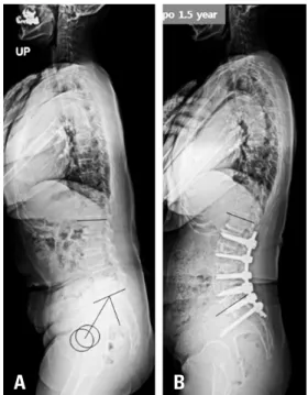
Fig. 4. A 75-year-old woman underwent posterior correction for lumbar degenerative kyphosis. Preoperatively, pelvic incidence was 75° and lumbar lordosis was 41°. At 1 month postoperatively, lumbar lordosis had increased to 54°. With time, the patient’s sagittal balance became de- compensated even without proximal junctional kyphosis. It is noteworthy that despite the decompensation, pelvic tilt did not increase and sacral slope did not decrease.
A B C D
결론
성인 척추 변형 교정에 있어서 측방 추체간 유합술과 함께 마 지막 분절에 최소 절개 전방 추체간 유합술 후 후방 고정 방법 은 후방 접근만으로 수술을 시행하는 것과 비교하였을 때 적은 합병증으로 효과적인 시상 정렬 교정을 가능하게 해주었다.
REFERENCES
1. Lenke LG, Sides BA, Koester LA, et al. Vertebral column resection for the treatment of severe spinal deformity.
Clin Orthop Relat Res. 2010 Mar;468(3):687-99. DOI:
10.1007/s11999-009-1037-x. Epub 2009 Sep 1.
2. Daubs MD, Lenke LG, Cheh G, et al. Adult spinal defor- mity surgery: complications and outcomes in patients over age 60. Spine (Phila Pa 1976). 2007 Sep 15;32(20):2238- 44. 10.1097/BRS.0b013e31814cf24a.
3. Cho KJ, Suk SI, Park SR, et al. Complications in posterior fusion and instrumentation for degenerative lumbar sco- liosis. Spine (Phila Pa 1976). 2007 Sep 15;32(20):2232-7.
10.1097/BRS.0b013e31814b2d3c.
4. Uribe JS, Deukmedjian AR, Mummaneni PV, et al. Com- plications in adult spinal deformity surgery: an analysis of minimally invasive, hybrid, and open surgical tech- niques. Neurosurg Focus. 2014 May;36(5):E15. Doi:
10.3171/2014.3.FOCUS13534.
5. Lee CS, Chung SS, Choi SW, et al. Critical length of fu- sion requiring additional fixation to prevent nonunion of the lumbosacral junction. Spine (Phila Pa 1976). 2010 Mar 15;35(6):E206-11. DOI: 10.1097/BRS.0b013e3181bfa518.
6. Kim KT, Lee SH, Suk KS, et al. Outcome of pedicle sub- traction osteotomies for fixed sagittal imbalance of multiple etiologies: a retrospective review of 140 patients. Spine (Phila Pa 1976). 2012 Sep 1;37(19):1667-75. DOI: 10.1097/
BRS.0b013e3182552fd0.
7. Phillips FM, Isaacs RE, Rodgers WB, et al. Adult degenera- tive scoliosis treated with XLIF: clinical and radiographical results of a prospective multicenter study with 24-month follow-up. Spine (Phila Pa 1976). 2013 Oct 1;38(21):1853- 61. DOI: 10.1097/BRS.0b013e3182a43f0b.
8. Isaacs RE, Hyde J, Goodrich JA, et al. A prospective, non- randomized, multicenter evaluation of extreme lateral inter- body fusion for the treatment of adult degenerative scoliosis:
perioperative outcomes and complications. Spine (Phila Pa 1976). 2010 Dec 15;35(26 Suppl):S322-30. DOI: 10.1097/
BRS.0b013e3182022e04.
9. Oliveira L, Marchi L, Coutinho E, et al. A radiographic as- sessment of the ability of the extreme lateral interbody fu- sion procedure to indirectly decompress the neural elements.
Spine (Phila Pa 1976). 2010 Dec 15;35(26 Suppl):S331-7.
DOI: 10.1097/BRS.0b013e3182022db0.
10. Manwaring JC, Bach K, Ahmadian AA, et al. Manage- ment of sagittal balance in adult spinal deformity with minimally invasive anterolateral lumbar interbody fusion:
a preliminary radiographic study. J Neurosurg Spine. 2014 May;20(5):515-22. DOI: 10.3171/2014.2.SPINE1347.
Epub 2014 Mar 1.
11. Baghdadi YM, Larson AN, Dekutoski MB, et al. Sagit- tal balance and spinopelvic parameters after lateral lumbar interbody fusion for degenerative scoliosis: a case-control study. Spine (Phila Pa 1976). 2014 Feb 1;39(3):E166-73.
DOI: 10.1097/BRS.0000000000000073.
12. Castro C, Oliveira L, Amaral R, et al. Is the lateral trans- psoas approach feasible for the treatment of adult de- generative scoliosis? Clin Orthop Relat Res. 2014 Feb 1;39(3):E166-73. DOI: 10.1097/BRS.0000000000000073.
13. Acosta FL, Liu J, Slimack N, et al. Changes in coronal and sagittal plane alignment following minimally inva- sive direct lateral interbody fusion for the treatment of degenerative lumbar disease in adults: a radiographic study. J Neurosurg Spine. 2011 Jul;15(1):92-6. DOI:
10.3171/2011.3.SPINE10425. Epub 2011 Apr 8.
14. Tempel ZJ, Gandhoke GS, Bonfield CM, et al. Radio- graphic and clinical outcomes following combined lateral lumbar interbody fusion and posterior segmental stabiliza- tion in patients with adult degenerative scoliosis. Neurosurg Focus. 2014 May;36(5):E11. DOI: 10.3171/2014.3.FO- CUS13368.
15. Johnson RD, Valore A, Villaminar A, et al. Pelvic param- eters of sagittal balance in extreme lateral interbody fusion for degenerative lumbar disc disease. J Clin Neurosci. 2013 Apr;20(4):576-81. DOI: 10.1016/j.jocn.2012.05.032.
Epub 2013 Jan 30.
16. Costanzo G, Zoccali C, Maykowski P, et al. The role of minimally invasive lateral lumbar interbody fusion in sagittal balance correction and spinal deformity. Eur Spine J. 2014
Anterolateral and Posterior Approach for the Correction of ASD Journal of Korean Society of Spine Surgery
www.krspine.org
17
Oct;23 Suppl 6:699-704. DOI: 10.1007/s00586-014-3561-y. Epub 2014 Sep 13.
17. Schwab F, Patel A, Ungar B, et al. Adult spinal deformity- postoperative standing imbalance: how much can you tolerate? An overview of key parameters in assessing alignment and planning corrective surgery. Spine (Phila Pa 1976). 2010 Dec 1;35(25):2224-31. DOI: 10.1097/
BRS.0b013e3181ee6bd4.
18. Glassman SD, Berven S, Bridwell K, et al. Correlation of radiographic parameters and clinical symptoms in adult scoliosis. Spine (Phila Pa 1976). 2005 Mar 15;30(6):682-8.
10.1097/01.brs.0000155425.04536.f7.
19. Bridwell KH, Glassman S, Horton W, et al. Does treatment (nonoperative and operative) improve the two-year quality of life in patients with adult symptomatic lumbar scoliosis:
a prospective multicenter evidence-based medicine study.
Spine (Phila Pa 1976). 2009 Sep 15;34(20):2171-8. DOI:
10.1097/BRS.0b013e3181a8fdc8.
20. Watkins RGt, Hanna R, Chang D, et al. Sagittal alignment after lumbar interbody fusion: comparing anterior, lateral, and transforaminal approaches. J Spinal Disord Tech. 2014 Jul;27(5):253-6. DOI: 10.1097/BSD.0b013e31828a8447.
21. Hsieh PC, Koski TR, O’Shaughnessy BA, et al. Anterior lumbar interbody fusion in comparison with transforaminal lumbar interbody fusion: implications for the restoration of foraminal height, local disc angle, lumbar lordosis, and sagittal balance. J Neurosurg Spine. 2007 Oct;7(4):379-86.
10.3171/SPI-07/10/379.
22. Dorward IG, Lenke LG, Bridwell KH, et al. Transforami- nal versus anterior lumbar interbody fusion in long defor- mity constructs: a matched cohort analysis. Spine (Phila Pa 1976). 2013 May 20;38(12):E755-62. DOI: 10.1097/
BRS.0b013e31828d6ca3.
23. Vaz G, Roussouly P, Berthonnaud E, et al. Sagittal mor- phology and equilibrium of pelvis and spine. Eur Spine J.
2002 Feb;11(1):80-7. 10.1007/s005860000224.
24. Christensen FB, Laursen M, Gelineck J, et al. Interob- server and intraobserver agreement of radiograph inter- pretation with and without pedicle screw implants - The need for a detailed classification system in posterolateral spinal fusion. Spine. 2001 Mar 1;26(5):538-43. DOI:
10.1097/00007632-200103010-00018.
25. Hasegawa K, Homma T. One-stage three-dimensional correction and fusion: a multilevel posterior lumbar in- terbody fusion procedure for degenerative lumbar ky- phoscoliosis. Technical note. J Neurosurg. 2003 Jul;99(1 Suppl):125-31.
26. Kim KT, Lee SH, Lee JH, et al. Three Different Meth- ods in Deformity Correction of Degenerative Flat Back: A Single Surgeon’s Experience with 64 Consecutive Cases.
Asian Spine J. 2015 Jun;9(3):361-9. DOI: 10.4184/
asj.2015.9.3.361. Epub 2015 Jun 8.
27. Kim YJ, Bridwell KH, Lenke LG, et al. Sagittal thoracic decompensation following long adult lumbar spinal in- strumentation and fusion to L5 or S1: causes, prevalence, and risk factor analysis. Spine (Phila Pa 1976). 2006 Sep 15;31(20):2359-66. 10.1097/01.brs.0000238969.59928.73.
28. Voor MJ, Mehta S, Wang M, et al. Biomechanical evalua- tion of posterior and anterior lumbar interbody fusion tech- niques. J Spinal Disord. 1998 Aug;11(4):328-34.
퇴행성 성인 척추 변형 교정에 대한 전측 -후방 접근법과 후방 접근법의 비교
박세준 • 이종서 • 염태훈 • 남윤진 • 박진성 성균관대학교 의과대학 삼성서울병원 정형외과교실
연구 계획: 환자 대조군 연구
목적: 성인 척추 변형 환자에서 측방 추체간 유합술과 최소 절개 전방 추체간 유합술을 시행한 후 단계적으로 후방 고정술을 시행하는 것이 신뢰할만한 술식 인지를 확인하고자 하였다.
선행 연구문헌의 요약: 성인 척추 변형 환자에서 측방 추체간 유합술을 이용한 교정 수술이 증가하고 있지만 시상 정렬의 교정 정도는 충분하지 못하다.
대상 및 방법: 측방 추제간 유합술과 함께 전방 추체간 유합술을 시행한 후 단계적으로 후방 고정술을 시행한 AP 그룹 30명의 환자와 나이, 성별, 진단명, 유합 분절 수, 골반 입사각, 추시 기간이 같은 PO 그룹 60명의 환자를 비교하였다. 척추골반지표, 입원기간, 임상 결과와 합병증 발생을 비교 분석하였다.
결과: 수술 후 요추 전만각은 AP 그룹이 PO 그룹보다 유의하게 더 컸다(p<0.001). 시상면 수직축 교정 정도는 AP 그룹에서 PO 그룹보다 더 많이 교정되 었다(p=0.005). 수술 후 AP 그룹에서는 90.0%에서, PO 그룹은 50.0%에서 PI-LL이 9도 이내의 적절한 교정을 보였다(p<0.001). 총 수술 시간은 AP그룹 이 PO 그룹보다 더 많은 시간이 소요되었지만 수술 중 출혈량과 수혈량은 AP 그룹에서 더 적었다. 수술 후 발생한 내과적 합병증과 수술과 관련된 합병 증은 PO 그룹에서 더 많이 발생하였다.
결론: 성인 척추 변형 교정에 있어서 측방 추체간 유합술과 함께 최소 절개 전방 추체간 유합술 후 후방 고정 방법은 후방 접근만으로 수술을 시행하는 것 과 비교하였을 때 적은 합병증으로 효과적인 시상 정렬 교정을 가능하게 해주었다.
색인 단어: 성인 척추 변형, 전방 추체간 유합술, 측방 추체간 유합술, 시상 정렬, 합병증 약칭 제목: 성인 척추 변형 교정에 대한 전측-후방 접근
접수일: 2019년 8월 19일 수정일: 2019년 9월 9일 게재확정일: 2020년 3월 9일 교신저자: 박진성
서울시 강남구 일원로 81 성균관대학교 의과대학 삼성서울병원 정형외과교실
TEL: 02-3410-1583 FAX: 02-3410-0061 E-mail: [email protected]