CORROSION SCIENCE AND TECHNOLOGY, Vol.8, No.6(2009), pp.223~226
223
Self-healing Coatings for an Anti-corrosion Barrier in Damaged Parts
Soo Hyoun Cho
699 Gumho-dong Gwangyang-si, Jeonnam, 545-090, KOREA
Surface Technology Research Group, POSCO Technical Research Laboratories (Received October 14, 2009; Revised December 29, 2009; Accepted December 29, 2009)
Polymer coatings are commonly applied to metal substrates to prevent corrosion in aggressive environments such as high humidity and under salt water. Once the polymer coating has been breached, for example due to cracking or scratches, it loses its effectiveness, and corrosion can rapidly propagate across the substrate.
The self-healing system we will describe prevents corrosion by healing the damage through a healing reaction triggered by the actual damage event. This self-healing coating solution can be easily applied to most substrate materials, and our dual-capsule healing system provides a general approach to be compatible with most common polymer matrices. Specifically, we expect an excellent anti-corrosion property of the self-healing coatings in damaged parts coated on galvanized metal substrates.
Keywords : self-healing, coating, anti-corrosion, metal substrate, healing agent, PDMS polymerization, tin catalyst
†Corresponding author: [email protected]
1. Introduction
Polymers with the ability to autonomically respond to damage and heal (i.e. self-heal) have been recently de- monstrated.1)-8) Self-healing is accomplished by incorporat- ing functional constituents (healing agent and catalyst) within a polymer matrix. These phases are incorporated either in microencapsulated form or as phase-separated regions. When the material is damaged, the microcapsules rupture and release their contents into the damaged region through capillary action. As the healing agent contacts the catalyst, polymerization is initiated and the damage is repaired.
A chemically stable self-healing materials system has recently been demonstrated6) based on the tin catalyzed polycondensation of phase separated droplets containing hydroxyl end-functionized polydimethylsiloxane (HOPDMS) and polydiethoxysiloxane (PDES).9) Because side re- actions are limited, organotin catalysts are highly desirable for curing PDMS based systems, even in open air.9),10) The organotin catalsyst is contained within polyurethane mi- crocapsules embedded in a matrix and is released when the capsules are broken by mechanical damage. This sys- tem possesses a number of important advantages over the previous self-healing methodology including that a) the healing chemistry remains stable in humid or wet environ-
ments, b) the chemistry is stable to elevated temperature (>100 ℃) enabling healing in higher temperature thermo- set systems, c) the components are widely available and comparatively low in cost, and d) the concept of phase separation of the healing agent greatly simplifies process- ing, as the healing agent can now be simply mixed into the polymer matrix.
Although the mechanical properties of the resultant cross-linked siloxanes are not very excellent, the mechan- ical strength of the healed matrix is of a secondary im- portance in a coating system, compared to chemical stabil- ity and passivating ability. Furthermore, the stability to water and air is of critical importance for practical realiza- tion of self-healing coatings, and was a prime motivation for this study.
2. Experimental
Microcapsules containing tin catalysts in chlorobenzene, were microencapsulated within polyurethane capsules.
PDMS healing-agent-filled ureaformaldehyde microcap- sules were formed following the published procedure6) with the following modifications. 250 mL of reaction mix- ture was heated to 55 ℃ and stirred at 700 rpm. 60 mL of a mixture of the PDMS healing agent was added. After 4 h, the reaction mixture was cooled to room temperature, and the microcapsules were separated.
All coatings were applied to 75 mm×150 mm steel
SOO HYOUN CHO
224 CORROSION SCIENCE AND TECHNOLOGY Vol.8, No.6, 2009
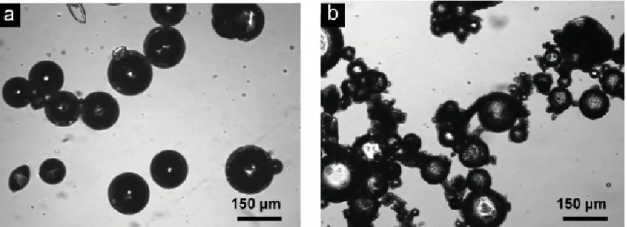
Fig. 1. Optical microscoy images of a) tin catalyst containing PU microcapsules and b) PDMS healing agent-filled UF microcapsules.
a b
Fig. 2. Experimental results of (a) Size histogram and (b) thermogravimetric analysis for both microcapsules.
sheets, using a micrometer-controlled doctor blade. The epoxy-amine self-healing coating is composed of epoxy mixed with 12 wt% diethylenetriamine, 3 wt% adhesion promoter, (3-trimethoxysilylpropyl) dimethylene triamine, 3 wt% tin catalyst-containing microcapsules, and 14 wt%
of PDMS-containing microcapsules. The coating solution was mixed by mechanical stirring, followed by degassing under vacuum. Samples were coated on the substrate with a thickness of 100 ㎛, and cured at room temperature for 24 h and 30 ℃ for 24 h.
3. Results and discussion
Self-healing coatings are composed of microencap- sulated catalysts and phase separated or encapsulated heal- ing-agent droplets in a matrix on a metallic substrate. No reactions take place between the HOPDMS and PDES pri- or to exposure to the catalyst in a matrix. When the self- healing coating layer is damaged by cracking or scratches,
the catalyst released from microcapsules and the healing agent wets the damaged plane. Diffusive mixing event of healing agent and catalyst follows in the damaged region.
Finally, the damage of coating layer is healed by cross- linked PDMS, which protects the substrate from the environment.
The size of tin catalyst filled capsules average 90 ㎛ in diameter (Fig. 1a and Fig. 2a), and the average diameter of this mirocapsules was a strong function of stirring rate during the interfacial polymerization process. The PDMS healing agent can be introduced by either phase separated liquid droplets in a matrix or PDMS filled microcapsules (Fig. 1b). In case of phase separated droplets, the diameter was not a strong function of stirring rate, and did not change significantly when samples were stirred between 100 and 2000 rpm.
Thermogravimetric analysis (TGA) exhibit a primary weight loss starting near the boiling point of chlorobenzene (131 ℃), and a secondary weight loss starting at 225 ℃,
SELF-HEALING COATINGS FOR AN ANTI-CORROSION BARRIER IN DAMAGED PARTS
CORROSION SCIENCE AND TECHNOLOGY Vol.8, No.6, 2009 225
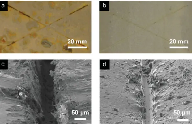
Fig. 3. Corrosion and morphological evaluation of self-healing coatings. a,b) Optical images after 120h immersion in salt water of a) control sample, and b) self-healing coating. c,d) SEM images of the scribed region of the control coating c) and the self-healing coating after healing d).
a b
Fig. 4. Electrochemical evaluation of self-healing coatings. a) Schematic diagram of electrochemical test. b) Current versus time for scribed control and self-healed sample.
corresponding to thermal decomposition of the polyur- ethane shell (Fig. 2b). Thus, we estimate the synthesized microcapsules do not rapidly leak below the boiling point of the solvent and should have good thermal stability under common working environments.
The self-healing function of coating system is evaluated through corrosion testing of damaged and healed coated steel samples compared to control samples (Fig. 3). The
PDMS based healing chemistry holds great promise for non-structural applications like coatings where simply fill- ing or sealing the crack and protecting the substrate from harsh environments is as prime importance. To this end we have begun evaluation of self-healing coatings for steel substrates. A control sample containing the adhesion pro- moter and neat vinyl ester resin shows obvious evidence of substrate corrosion, most prevalently within the groove
SOO HYOUN CHO
226 CORROSION SCIENCE AND TECHNOLOGY Vol.8, No.6, 2009
of the scribed regions, but also extending across the sub- strate surface (Fig. 3a). In dramatic contrast, the self-heal- ing samples show no visual evidence of corrosion after 120 h exposure in salt water (Fig. 3b). Separate control tests reveal that the presence of both the healing agent and catalyst are necessary for self-healing functionality.
Removal of either phase results in a coating which cor- rodes rapidly, providing a clear indication that simple re- flow of one of the phases into the crack is not sufficient to prevent corrosion.
Scanning electron microscopy (SEM) imaging of the scribe region in control and self-healing coatings reveals the morphology of the repaired coating (Fig. 3c and 3d).
Flow of healing agent and catalyst into the scribe and re- coating (passivation) of the substrate is readily apparent.
The damage is significantly (~40%) filled by polymerized healing agent in the self-healing coating, while the scribe extends about 15 ㎛ into the metal substrate in the control sample. Profilometry measurements and energy-dispersive spectroscopy of nickel coated cross-sectional samples (post-healing) confirmed these findings.
Electrochemical testing provides further evidence of passivation of the substrate by self-healing coatings. In these experiments, the coated metal substrate serves as one electrode in an electrochemical cell (Fig. 4a). The steady- state conduction between the coated metal substrate and a platinum electrode held at 3 V through an aqueous elec- trolyte (1 M NaCl) is measured (Fig. 4b). The current pass- ing through the control and self-healing polymer coatings before scribing are nearly identical, ~0.34 μA cm-2. After scribing, samples are allowed to heal and are tested in the electrochemical cell. The current passing through the scribed control samples is quite large (26.6-58.6 mA cm-2, three samples), compared to the undamaged state, and we note rapid gas evolution from the scribed region during the test. The self-healing samples show a dramatically re- duced current (12.9 μA cm-2-1.4 mA cm-2, four samples), and no gas evolution is observed from the self-healing sample.
4. Summary
Self-healing has the potential to extend the lifetime and increase the reliability of thermosetting polymers used in a wide variety of applications. The materials system pre- sented in this paper greatly extends the capability of self-
healing polymers by introducing a new, environmentally stable healing chemistry and demonstrating the concept of phase separating healing agents in a polymer matrix.
Self-healing coatings utilizing this materials system have shown very promising results for enhanced corrosion pro- tection of steel substrates exposed to salt water. For con- firming general compatibility, it was also investigated the anti-corrosion property of commercialized coating solution with the dual microcapsule based self-healing coatings. By incorporating active tin catalyst, we show that autonomic corrosion protection can be obtained by self-healing under ambient environmental conditions. Multilayered coatings can also be formulated to provide self-healing functionality while maintaining extreme tolerances on surface finish, specific requirements for engineered primers, or unique surface chemistries (e.g., self-cleaning). We believe the microcapsule motif also provides a delivery mechanism for multifunctional chemical agents, which provide healing as well as corrosion inhibitors,11) antimicrobial agents,12) or other functional chemicals.
References
1. S.R. White, N.R. Sottos, J. Moore, P. Geubelle, M.
Kessler, E. Brown, S, Suresh, and S. Viswanathan, Nature, 409, 794 (2001).
2. E.N. Brown, N.R. Sottos, S.R. White, Experimental Mechanics, 42, 372 (2002).
3. M.R. Kessler, N.R. Sottos, and S.R. White, Composites Part A: Applied Science and Manufacturing, 34, 743 (2003).
4. J.D. Rule, E.N. Brown, N.R. Sottos, S.R. White and J.S. Moore, Advanced Materials, 17, 205 (2005).
5. E.N. Brown, S.R. White, and N.R. Sottos, Composites Science and Technology, Special Anniversary Issue, 65, 2474 (2005).
6. S.H. Cho, H.M. Andersson, S.R. White, N.R. Sottos, P.V. Braun, Advanced Materials, 18, 997 (2006).
7. S.H. Cho, S.R. White, P.V. Braun, Advanced Materials, 21, 645 (2009).
8. A.S. Jones, J, Rule, J.S. Moore, S. R. White, N.R. Sottos, Chemistry of Materials, 18, 1312 (2006).
9. G.B. Shah, J. Appl. Poly. Sci., 70, 2235 (1998).
10. F.W. Van der Weij, Macroml. Chem., 181, 2541 (1980).
11. V. S. Sastri, Corrosion Inhibitors: Principles and Applications, Wiley, New York, 1998.
12. A. Bryskier, Antimicrobial Agents: Antibacterials and Antifungals, ASM Press, Washington, 2005.