Does Obesity Affect the Severity of Exercise-Induced Muscle Injury?
Jooyoung Kim1, Jin Hwan Yoon2,*
1Office of Academic Affairs, Konkuk University, Chungju; 2Department of Sport Science, College of Life Science and Nano Technology, Hannam University, Daejeon, Korea
This literature review investigates the effects of obesity on exercise-induced muscle injury and reexamines the potential mechanisms of exercise-induced muscle injury related to obesity. Several studies reported that high body mass index and percent body fat can significantly affect the markers of muscle injury after exercise, includ- ing maximal strength, delayed onset muscle soreness, creatinine kinase level, and myoglobin level. The potential mechanisms resulting in these outcomes include structural changes in the cell membrane induced by high fat levels, increased inflammatory responses due to adipose tissues, reduced muscle satellite cell activation and myogenesis due to lipid overload, differences in muscle fiber distributions, and sedentary behaviors. These mechanisms, however, must be verified through more research. As obesity is a potential risk factor increasing the severity of exercise-induced muscle injuries, the exercise intensity and duration for obese patients must be carefully selected, and a preconditioning intervention (e.g., low-intensity eccentric training) may be considered before or during the early stages of the exercise program.
Key words: Adipose tissue, Body mass index, Creatine kinase, Exercise, Inflammation, Obesity
Received September 21, 2020 Reviewed November 21, 2020 Accepted January 2, 2021
* Corresponding author Jin Hwan Yoon
https://orcid.org/0000-0001-9026-6416 Department of Sport Science, College of Life Science and Nano Technology, Hannam University, 70 Hannam-ro, Daedeok-gu, Daejeon 34430, Korea Tel: +82-42-629-7990
Fax: +82-42-629-8402 E-mail: yoonjh@hnu.kr
INTRODUCTION
Obesity is a disease that has become an important issue world- wide and in health-related policies.
1Regular participation in exer- cise is an important component of an obesity treatment program
2and significantly improves the body composition by reducing weight and body fat while increasing lean body mass; therefore, it has been known to effectively prevent hypertension, type 2 diabetes, meta- bolic syndrome, and cardiovascular diseases.
3-5However, exercising at an intensity beyond one’s physical abili- ties or performing unaccustomed exercises that involve eccentric muscle contractions has been reported to induce muscle injuries.
6,7Muscle injuries reduce muscle function and significantly increase not only the leakage of proteins such as creatinine kinase (CK) and myoglobin (Mb) into the bloodstream, but also the activation lev-
els of cells associated with inflammatory responses.
8,9Furthermore, they induce delayed onset muscle soreness (DOMS), which hin- ders recovery, interferes with regular exercise, and negatively affects one’s psychological health.
10-12Several studies have reported that the higher the levels of the body composition markers such as body mass index (BMI) and percent body fat (%BF), the more severe the muscle injury after ec- centric exercise and the more delayed the recovery.
13-16Kim and So
14reported that individuals with a high BMI had greater loss of maximal strength and increase in the severity of DOMS and CK levels after eccentric exercise. Margaritelis et al.
16also reported that a group with high %BF had greater reduction in muscle function and increase in DOMS and CK levels after eccentric exercise than did a group with lower %BF.
While studies on obesity and exercise have mostly focused on
Copyright © 2021 Korean Society for the Study of Obesity
This is an Open Access article distributed under the terms of the Creative Commons Attribution Non-Commercial License (https://creativecommons.org/licenses/by-nc/4.0/) which permits unrestricted non-commercial use, distribution, and reproduction in any medium, provided the original work is properly cited.
2017-03-16 https://crossmark-cdn.crossref.org/widget/v2.0/logos/CROSSMARK_Color_square.svg
Does Obesity Affect the Severity of Exercise-Induced Muscle Injury?
Jooyoung Kim1, Jin Hwan Yoon2,*
1Office of Academic Affairs, Konkuk University, Chungju; 2Department of Sport Science, College of Life Science and Nano Technology, Hannam University, Daejeon, Korea
This literature review investigates the effects of obesity on exercise-induced muscle injury and reexamines the potential mechanisms of exercise-induced muscle injury related to obesity. Several studies reported that high body mass index and percent body fat can significantly affect the markers of muscle injury after exercise, includ- ing maximal strength, delayed onset muscle soreness, creatinine kinase level, and myoglobin level. The potential mechanisms resulting in these outcomes include structural changes in the cell membrane induced by high fat levels, increased inflammatory responses due to adipose tissues, reduced muscle satellite cell activation and myogenesis due to lipid overload, differences in muscle fiber distributions, and sedentary behaviors. These mechanisms, however, must be verified through more research. As obesity is a potential risk factor increasing the severity of exercise-induced muscle injuries, the exercise intensity and duration for obese patients must be carefully selected, and a preconditioning intervention (e.g., low-intensity eccentric training) may be considered before or during the early stages of the exercise program.
Key words: Adipose tissue, Body mass index, Creatine kinase, Exercise, Inflammation, Obesity
Received September 21, 2020 Reviewed November 21, 2020 Accepted January 2, 2021
* Corresponding author Jin Hwan Yoon
https://orcid.org/0000-0001-9026-6416 Department of Sport Science, College of Life Science and Nano Technology, Hannam University, 70 Hannam-ro, Daedeok-gu, Daejeon 34430, Korea Tel: +82-42-629-7990
Fax: +82-42-629-8402 E-mail: yoonjh@hnu.kr
changes in body composition and physical fitness, limited studies have been conducted on muscle injuries that occur after exercise in patients with obesity. Therefore, this literature review investigates the effects of obesity on exercise-induced muscle injuries by exam- ining previous research and re-examining the potential mechanisms of exercise-induced muscle injuries related to obesity.
EXERCISE-INDUCED MUSCLE INJURY
As previously explained, suddenly engaging in intense or unac- customed exercise after a long period of physical inactivity and per- forming exercises that involve eccentric muscle contractions can lead to muscle injuries.
6Eccentric muscle contraction is one of many types of muscle contractions that form body movements. It refers to contractions in which the activated muscle lengthens and is a major cause of exercise-induced muscle injuries.
17Linnamo et al.
18and Morgan et al.
19reported that eccentric muscle contractions do not occur completely during the recruitment of muscle fibers for maximal strength during exercise and are thus more likely to cause muscle injury than isotonic or isometric muscle contractions.
In general, the mechanisms of exercise-induced muscle injuries are divided into primary and secondary muscle damages. Primary muscle damage refers to the morphological changes caused by mus- cle length changes and muscle tension. Proske and Morgan
20re- ported that sarcomere lengths exceed the normal range of contrac- tion during an eccentric muscle contraction, and the resulting ten- sion causes muscle injury. Repeated eccentric muscle contractions lead to sarcomere injuries, including those of the Z-disk, I band, and A band that result in “Z-disk streaming.”
20Furthermore, Lo- monosova et al.
21and Raastad et al.
22reported that additional inju- ries can occur not only in the sarcolemma and sarcoplasmic reticu- lum but also in cytoskeletal elements such as desmin, dystrophin, vimentin, nestin, and lamin.
The causes of secondary muscle damage, which occurs after pri- mary muscle damage, include loss of homeostasis of calcium ions and increased inflammatory responses and oxidative stress. Mur- phy and Lamb
23and Vissing et al.
24reported that without the reup- take of calcium ions released to the cytosol back into the sarcoplas- mic reticulum by Ca-ATPase, excitation-contraction coupling, which induces muscle contraction, becomes impaired, thus leading to the
activation of calpain, a Ca
2+-dependent protease, and elevated in- flammatory responses in the injured muscle. Peake et al.
25reported that the activities of neutrophils and macrophages, which are leu- kocytes, increase in the event of a muscle injury. Neutrophils con- tribute to increasing the risk of muscle injury by secreting chemoat- tractants that promote phagocytosis and proinflammatory cyto- kines.
26Proinflammatory cytokines additionally activate the pro- duction of substrates that are toxic to neutrophils and generate free radicals that induce oxidative stress.
27Macrophages are divided into M1 and M2, playing complex roles in inflammatory responses. M1 macrophages are abundant in ne- crotic muscle fibers one day after neutrophil infiltration and are ac- tivated by proinflammatory cytokines such as tumor necrosis factor-α (TNF-α) and interleukin (IL)-1β.
28Activated M1 macrophages produce and release more than 100 different substrates, including prostaglandin and proinflammatory cytokines, to strengthen the inflammatory response.
29On the other hand, M2 macrophages are observed after inflammatory responses and mediate tissue repair and growth by increasing the levels of growth-promoting factors, including fibroblast growth factor, insulin-like growth factor 1, trans- forming growth factor-β1, and cytokines.
30EFFECT OF OBESITY ON EXERCISE-INDUCED MUSCLE INJURY
Several studies have reported that obesity can affect the markers associated with exercise-induced muscle injury.
14,15,31-33Salvadori et al.
32performed an exercise test in 10 healthy subjects with a mean BMI of 22 kg/m
2and 11 obese patients with a mean BMI of 41 kg/m
2using a cycle ergometer and reported that the obese patients showed higher increases in CK levels than did the healthy subjects.
Paschalis et al.
33divided 22 healthy women into a lean group (BMI,
18.5–24.9 kg/m
2; mean BMI, 21.2 kg/m
2) and an overweight
group (BMI, 25.0–33.0 kg/m
2; mean BMI, 29.5 kg/m
2) and in-
structed them to perform eccentric exercise of the knee extensor
using an isokinetic dynamometer. In comparison of the muscle in-
jury markers between the two groups, the authors
33found a greater
reduction in muscle torque and greater DOMS during palpation
and walking in the overweight group than in the lean group. The
overweight group also showed significantly higher CK levels from
24 to 72 hours after exercise than the lean group.
Paschalis et al.
34reported similar results in their study in 2013.
They divided 32 healthy women into two groups according to BMI to perform eccentric exercise. They reported that the overweight group with a high BMI (mean BMI, 29.4 kg/m
2) showed negative changes in the parameters associated with muscle function and had increased DOMS and CK levels compared with the lean group with a normal BMI (mean BMI, 22.0 kg/m
2). Kim and So
14divid- ed 40 healthy male university students into a normal BMI (18.5–
22.9 kg/m
2) group and a high BMI (≥25 kg/m
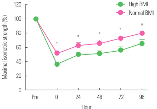
2) group to perform eccentric exercise of the elbow flexor using a modified preacher curl machine. They observed significant changes in the maximal isomet- ric strength, DOMS, and CK levels after eccentric exercise in both groups. However, the high BMI group showed a higher reduction in maximal isometric strength immediately after exercise than did the normal BMI group and showed a delayed recovery throughout the whole period after the exercise (Fig. 1). The high BMI group also showed higher DOMS severity (Fig. 2) and CK levels.
On the other hand, Comstock et al.
31reported that BMI does not affect post-exercise muscle injury. They divided 19 healthy males into groups according to BMI and instructed them to per- form acute resistance exercise. They reported no significant chang- es in the levels of CK or Mb, or DOMS severity according to BMI.
However, the examined muscle injury markers and their measure- ment timings in Comstock’s study
31differed from those in previous studies.
14,33For instance, the mean CK level was ≥ 1,000 U/L after
exercise in both the high BMI and lean groups in the studies by Kim and So
14and Paschalis et al.,
33whereas the mean CK level was 188 U/L in the high BMI (> 35 kg/m
2) group and 195.8 U/L in the lean (BMI, < 25 kg/m
2) group at 24 hours after exercise, with- out any significant difference as compared with the baseline value in the study by Comstock et al.
31These CK results may indirectly indicate that the resistance exercise assigned to the subjects did not sufficiently induce muscle injury. Furthermore, because Comstock et al.
31only measured muscle injury markers until 24 hours after exercise, the changes in these markers that occurred after the 24 hours could not be determined. Studies on exercise-induced mus- cle injury generally measure changes in muscle injury markers until 96 hours after exercise.
35,36In their recent study that used an eccentric exercise protocol for the elbow flexor similar to the one used by Kim and So,
14Yoon and Kim
15reported that while BMI can be used to make a simple diag- nosis of obesity, it does not accurately represent the amount of adi- pose tissues; thus, they examined the association between %BF and muscle injury. They divided 30 healthy male university students into a high %BF (≥ 20%) group and a low %BF (≤ 15%) group ac- cording to their %BF and instructed them to perform high-intensi- ty eccentric exercise. They reported that the high %BF group showed high CK and Mb levels during the post-exercise recovery period as compared with the low %BF group (Fig. 3). These results were similar to those of Margaritelis et al.,
16who induced muscle injury in 39 healthy women by subjecting them to knee extensor exercise
Figure 1. Change of maximal isometric strength after eccentric exercise according to body mass index (BMI). Normal BMI, 18.5–22.9 kg/m2; high BMI, ≥ 25 kg/m2.
*Significant difference between groups (P< 0.05); †Significant between group (P< 0.01). Data from Kim and So.14
120 100 80 60 40 20 0
Maximal isometric strength (%)
Pre 0 24 48 72 96 Hour
High BMI Normal BMI
†
†
* *
*
Figure 2. Change of delayed onset muscle soreness (DOMS) after eccentric exer- cise according to body mass index (BMI). Normal BMI, 18.5–22.9 kg/m2; high BMI,
≥ 25 kg/m2. *Significant difference between groups (P< 0.05). Data from Kim and So.14
60
40
20
0
DOMS (mm)
0 24 48 72 96
Hour
High BMI Normal BMI
*
using an isokinetic dynamometer and reported that the overweight group with a %BF ≥ 30.1% had higher CK levels than did the lean group.
The increased CK and Mb levels observed in the studies by Yoon and Kim
15and Margaritelis et al.
16are an indirect indication of mus- cle membrane disruption after exercise.
37In relation to this, Knob- lauch et al.
38reported that obese mice showed a greater myofiber membrane disruption after downhill running as an eccentric exer- cise than did lean mice, suggesting that obese mice are more sus- ceptible to mechanical injury. Although these results were obtained from an animal experiment, they allow visualization of the mor- phological changes of injured myofibers after eccentric exercise on light microscopy, which are the direct evidence of the changes in CK and Mb levels after eccentric exercise. In contrast, Totsuka et al.
39divided 15 young males into a high responder group (CK level,
> 500 IU) and a low responder group (CK level, < 300 IU) ac- cording to the CK level after endurance exercise and reported that
%BF had no effect on the CK level in both groups. However, the two groups had %BF of 15.6% and 17.4%, respectively, and did not comprise obese participants.
POTENTIAL MECHANISMS RELATED TO EXERCISE-INDUCED MUSCLE INJURY AND
OBESITY
Obesity refers to the excessive lipid accumulation in adipose or non-adipose tissues (e.g., skeletal muscle)
40and can cause structural
changes in the cell membrane, making it vulnerable to mechanical tension.
38A few studies reported that high levels of fat cause the saturation of fatty acyl chains in the sarcolemma to increase the phospholipid packing density and membrane rigidity.
41-43Such structural changes may aggravate cell membrane disruption in re- sponse to a mechanical stimulus from exercise, consequently in- creasing the CK and Mb levels.
As obesity increases, the levels of circulating inflammatory mole- cules and macrophage cell infiltration via the effect of proinflam- matory cytokines released from adipose tissues, it can cause chron- ic low-grade inflammation.
44The M1 macrophages of macrophage cells exhibit proinflammatory characteristics that are more activat- ed by high fat levels.
45Therefore, obesity-induced increases in in- flammatory responses and M1 macrophages in injured muscles can promote cell membrane disruption (Fig. 4).
A few studies reported the association between inflammatory re- sponses and cell membrane disruption.
36,46Kanda et al.
36examined changes in various inflammation markers after eccentric exercise and reported that increased levels of neutrophil migratory activity after exercise are positively correlated with changes in Mb levels.
Kawamura et al.
46reported that increased IL-6 concentration after eccentric exercise significantly correlated with the peripheral neu- trophil count and serum CK activity. Dutra et al.
47and Tajra et al.
48provided evidence suggesting that obesity affects cell membrane disruption after exercise. Dutra et al.
47reported that excess body fat is associated with increased levels of inflammatory markers such as IL-7 and C-reactive protein levels in postmenopausal women. Tajra
Figure 3. Change of creatine kinase (A) and myoglobin (B) after eccentric exercise according to body fat percentage (%fat). High %fat, ≥ 20%; low %fat, ≤ 15%. Data from Yoon and Kim.1530,000
20,000
10,000
0
Creatine kinase (U/L)
0 24 48 72 96 Hour
High %fat Low %fat A
4,000
3,000
2,000
1,000
0
Myoglobin (ng/mL)
0 24 48 72 96 Hour
High %fat Low %fat B
et al.
48reported high CK levels in the recovery period in elderly obese women with high IL-6 levels after exercise.
Obesity-induced inflammatory responses are highly likely to con- tribute to the increase in post-exercise DOMS severity. Although the causes of DOMS are unclear, a major hypothesis suggests in- flammatory responses as one cause.
9,49Increased levels of bradyki- nin and prostaglandin during inflammatory response can stimulate types III and IV afferent nerve fibers near the muscle, thus induc- ing DOMS.
9A few studies
50,51reported that body fat is associated with musculoskeletal pain and suggested that the upregulation of cytokines (TNF-α) secreted by adipose tissues and the resulting systemic inflammation may be involved in this association.
However, further research on the relationship between obesity and post-exercise DOMS is needed. While a few studies reported higher DOMS severity levels in obese individuals after performing eccentric exercise,
14,33they did not directly measure inflammation markers. Miles et al.
52,53reported that obesity-induced increases in the levels of inflammation markers are not associated with DOMS.
Miles et al.
52divided subjects into a normal-weight (BMI, ≤25 kg/m
2; mean BMI, 22.4 kg/m
2) group and an overweight (BMI, 25–30 kg/m
2; mean BMI, 27.1 kg/m
2) group and instructed them to per- form eccentric exercise of the elbow flexor using an isokinetic dy- namometer. They reported higher levels of sTNFR1 and IL-6 at 12–24 hours and 8 hours after exercise, respectively, in the over-
weight group than in the normal-weight group, but no significant difference in DOMS between the two groups. In their study in 2016, Miles et al.
53divided subjects into a low waist circumference (Lo-WC; ≤ 80 cm) group and a high waist circumference (Hi-WC;
> 80 cm) group according to waist circumference, which is a mark- er of abdominal obesity, and examined changes in the markers of muscle injury after downhill or uphill running exercise. They re- ported higher IL-6 levels during the recovery period in the Hi-WC group than in the Lo-WC group regardless of the type of exercise but no significant difference in DOMS between the two groups.
The reason for the conflicting results of the studies is unclear, and further research is necessary to draw a clear conclusion.
A study reported that lipid overload disrupts the activation of muscle satellite cells, thereby reducing myogenesis.
54Regeneration after muscle injury requires the activation of muscle stem and sat- ellite cells.
55Satellite cells are called the stem cells of muscles and exist between the sarcolemma and basement membrane of termi- nally differentiated muscle fibers.
56They usually stay dormant but induce myogenesis through activation, proliferation, differentia- tion, and fusion in the event of a muscle injury.
57However, in- creases in insulin or leptin resistance and levels of proinflamma- tory cytokines in individuals with obesity disrupt satellite cell pro- liferation and, subsequently, myogenesis.
58Based on these facts, an obesity-induced reduction in satellite cell activation and myo-
Figure 4. Potential mechanisms related to exercise-induced muscle injury and obesity.Maximal isometric strength ↓ Delayed onset muscle soreness ↑
Creatine kinase, myoglobin ↑ Excessive lipid accumulation
Phospholipid packing density and membrane rigidity ↑
Inflammatory responses and M1 macrophages infiltration ↑
Activation of muscle satellite cells ↓
Loss of calcium homeostasis ↑