大 韓 不 妊 學 會 誌 : 第 32 卷 第 1 號 2005 Kor. J. Fertil. Steril., Vol. 32, No. 1, 2005, 3
근이영양증에 대한 착상전 유전진단에서 Duplex-nested PCR과 Fluorescent PCR 방법의 효용성
성균관대학교 의과대학 삼성제일병원 생식생물학 및 불임연구실1, 유전학연구실2, 산부인과학교실3
이형송1·최혜원1·임천규1·박소연2·김진영3·궁미경3·전진현1*·강인수3
Efficacy of Duplex-nested PCR and Fluorescent PCR in the Preimplantation Genetic Diagnosis for Duchenne Muscular Dystrophy
Hyoung-Song Lee
1, Hye Won Choi
1, Chun Kyu Lim
1, So Yeon Park
2, Jin Young Kim
3, Mi Kyoung Koong
3, Jin Hyun Jun
1*, Inn Soo Kang
31
Laboratory of Reproductive Biology and Infertility,
2Laboratory of Medical Genetics,
3
Department of Obstetrics and Gynecology, Samsung Cheil Hospital and Women's Healthcare Center, Sungkyunkwan University School of Medicine
Objective: Preimplantation genetic diagnosis (PGD) is reserved for couples with a risk of transmitting a serious and incurable disease, and hence avoids the undesirable therapeutic abortion. In this study, we evaluated the efficacy of PGD for Duchenne muscular dystrophy (DMD) cases by the fluorescent PCR with polymorphic linked markers and the conventional duplex-nested PCR methods.
Methods: Biopsy of one or two blastomeres was done from the embryos fertilized by ICSI on the third day after fertilization. We performed two cases of PGD-DMD by the duplex-nested PCR for the causative mutation loci and the SRY gene on Y chromosome. The triplex fluorescent PCR for the mutation loci, the SRY gene and the polymorphic microsatellite marker on X chromosome was applied for two cases of PGD-DMD.
Results: By the duplex-nested PCR, successful diagnosis rate was 95.5% (21/22), but we could not discriminate the female embryos whether normal or carrier in this X-linked recessive disease. However, the triplex fluorescent PCR method showed 100% (27/27) of successful diagnosis rate, and all female embryos (n=17) were distinguished normal (n=10) from carrier (n=7) embryos. Unaffected and normal embryos were transferred into mother's uterus after diagnosis. A healthy normal male was achieved after PGD with the duplex-nested PCR method and a twin, a male and a female, were delivered with triplex fluorescent PCR method. The normality of dystrophin gene was confirmed by amniocentesis and postnatal genetic analysis in all offsprings.
Conclusion: The fluorescent PCR with polymorphic marker might be useful in improving the
연 락 저 자: 이형송, 우) 100-380, 서울시 중구 묵정동 1-19, 삼성제일병원 생식생물학 및 불임연구실 Tel: (02) 2000-7635, Fax: (02) 2265-5621, e-mail: [email protected]
주관책임자: 전진현, 우) 100-380, 서울시 중구 묵정동 1-19, 삼성제일병원 생식생물학 및 불임연구실 Tel: (02) 2000-7592, Fax: (02) 2265-5621, e-mail: [email protected]
*본 논문은 2004년 대한불임학회 제47차 추계학술대회 구연부문 우수발표상을 수상하였음.
specificity and reliability of PGD for single gene disorders.
Key Words: Preimplantation genetic diagnosis (PGD), Duchenne muscular dystrophy (DMD), Duplex- nested PCR, Fluorescent PCR, Polymorphic marker
최근 인간의 체외수정 및 배아이식술 분야에서, 유전적으로 이상이 있는 환아를 출산할 확률이 높은 부부들에게 정상아의 출산 기회를 제공하기 위해서 착상전 유전진단 (preimplantation genetic diagnosis, PGD) 방법이 도입, 적용되고 있다. 이러한 착상전 유전진단은 기존의 산전진단에 비하여 유전적으로 비정상인 배아의 이식을 배제하고 정상적인 배아만 을 선별적으로 이식하여, 유전질환을 갖는 태아의 임신과 유산으로 인한 정신적, 육체적 부담을 감소 시킬 수 있다.
현재의 착상전 유전진단의 개념은 1968년 Ed- wards와 Gardner1에 의해 처음으로 제시되었으며, 그 후 착상전 유전진단의 성공적인 임상적용은 1990년 Handyside 등2에 의해 최초로 보고되었다.
이들은 X-연관 유전질환에서 중합효소연쇄반응 (polymerase chain reaction, PCR) 방법을 이용하여 Y 염색체의 특정 부위를 증폭시켜 성별을 구분하는 방 법으로 여성배아만을 이식함으로써 유전질환에 이 환되지 않은 여아의 출산에 성공하였다. 이 후 착상 전 유전진단은 습관성 유산 등 임신 예후가 좋지 않은 불임부부를 대상으로 한 염색체 수적 이상 (aneuploidy) 검사와 cystic fibrosis3와 β-globin gene defect4,5 같은 여러 단일 유전자 이상 질환의 위험 이 있는 부부를 대상으로 전 세계적으로 널리 시행 되고 있다. 이러한 착상전 유전진단은 임신 후에 시 행되는 유전질환에 대한 산전 진단 (prenatal diag- nosis)을 대체할 수 있는 가장 이상적인 방법으로 생각되고 있다.
본 연구실에서도 수년 전 부터 착상전 유전진단 을 시행하고 있으며, 대부분 염색체의 수적 이상이 나 구조적 이상을 대상으로 형광직접보합법 (fluo- rescent in situ hybridization, FISH)을 이용하여 시행 한 사례들을 보고하였다.6~9 또한, 최근 PCR 방법 을 이용하여 근이영양증, ornithine transcarbamylase (OTC) deficiency, lactic acidosis 그리고 epidermolysis bullosa (EB) 등 단일 유전자 이상 질환에서 착상전 유전진단 시행 결과를 보고한 바 있다.10,11
근이영양증 (Duchenne muscular dystrophy, DMD) 은 가장 유병율이 높은 신경근 질환으로써 신생 남 아 3,500명당 1명의 빈도로 발생하며, 환아는 점진 적인 근육의 약화로 결국 20세를 전후하여 사망하 는 것으로 보고되고 있다.12 근이영양증은 X 염색체 단완 (Xp21)에 존재하는 dystrophin 유전자의 이상 으로 인해 발생하는 X-연관 열성 유전질환이며,13 dystrophin 유전자는 79개의 exons이 약 2.5 Mb의 genomic DNA에 걸쳐 있는 현재까지 밝혀진 유전자 중 가장 큰 것으로 알려져 있다.14 근이영양증에 대 한 착상전 유전진단 시 기존에 사용하였던 성염색 체에 대한 형광직접보합법이나 duplex-nested PCR 방법으로는 여성배아의 경우 해당 배아의 유전형 이 정상인지 보인자인지 명확하게 구분할 수가 없 었다. 이에 저자들은 이러한 문제점을 해결하기 위 해 dystrophin 유전자와 연관되어 있는 polymorphic marker를 동정하고, 이를 착상전 유전진단에 적용 하여 배아의 유전형을 정확하게 진단할 수 있는 방 법을 시도하였다. 본 연구에서는 duplex-nested PCR 방법과 최근에 새로 확립한 polymorphic marker를 이용한 triplex fluorescent PCR 방법을 적용한 근이 영양증에 대한 착상전 유전진단의 효용성과 임상 결과를 살펴보고자 한다.
연구 대상 및 방법
1. 연구 대상
연구 대상은 총 4 가계였으며, 이들은 근이영양 증을 앓고 있는 아들이나 친척을 두고 있었다. 그 중 case 4를 제외한 나머지 3 cases는 기존의 Cham- berain's primer set나 Begg's primer set으로 결실되어 있는 부위를 확인할 수 있었다 (data not shown). 그 러나 case 4의 경우 기존의 primer sets으로 결실 부 위를 확인할 수 없었기 때문에 환자의 가족을 대상 으로 여러 가지 marker를 이용한 linkage analysis를 수행하여 보인자인 어머니와 환아인 아들의 돌연변 이 allele를 확인할 수 있었다 (Figure 1).
2. Genomic DNA의 분리와 single lymphocyte preparation
각 부부와 질환이 있는 아들 또는 친척들의 혈액 에서 AquaPure Genomic DNA kit (Bio-Rad Laborato- ries, Hercules, CA, USA)을 이용하여 genomic DNA 를 추출한 후 사용 전까지 -20℃에 보관하였으며, 이를 이용하여 각 가계에서 dystrophin 유전자에 대 한 돌연변이 여부를 재확인하였다. 착상전 유전진단 을 시행하기 전 각 primer의 PCR 조건, amplification rate 그리고 allele drop-out (ADO) rate를 조사하기 위하여 각 환자의 혈액으로부터 Ficoll-Paque density gradient separation (Ficoll-PaqueTM PLUS, Amersham Biosciences AB, Uppsala, Sweden) 방법으로 lympho- cytes를 분리하였다. 배양접시에 Ca2+ /Mg2+-free pho-
sphate buffered saline (PBS)로 소적을 만들고 그 소 적에 lymphocytes 일부를 넣어 희석한 후 현미경 하에서 micropipette을 이용하여 각각의 lymphocyte 를 5 µl의 lysis buffer (200 mM KOH, 50 mM DTT)가 들어 있는 각각의 PCR tube에 넣었다. 이렇게 준비 된 single lymphocyte tube는 사용 전까지 -70℃에 보 관하였다.
3. 난자의 채취 및 배아의 배양
Gonadotrophin-releasing hormone (GnRH) agonist, human menopausal gonadotrophins (hMG)와 human follicular stimulating hormone (hFSH)을 이용하여 과 배란을 유도하였다. 초음파로 난자의 크기를 관찰 하여 최소한 2개 이상의 난포가 18 mm 이상, 17-β estradiol의 농도가 500 pg/ml 되었을 때 human cho- Male Partner
(Normal Male)
Female Partner (Carrier Female)
Son (Affected Male)
Normal Mutant Normal Mutant Allele Allele Allele Allele
3'CA STR44 Polymorphic Marker
A
B
C
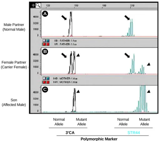
Figure 1. Electropherogram of the dinucleotide repeat analysis for genetic diagnosis of Duchenne muscular dystrophy.
The length of the repeats (base pairs) is represented on the top of electropherogram. Electropherogram A (top): the unaffected male partner; B (middle): the carrier female partner; C (bottom): the affected son. Arrows and arrow heads indicate normal and mutant alleles, respectively.
rionic gonadotrophins (hCG)를 10,000 IU 주사하였 다. hCG 주사 후 34시간에 질식초음파를 이용하여 난자를 채취하였다. 채취된 난자는 G-Fert 배양액 (Vitrolife Sweden AB, Kungsbacka, Sweden)에 넣어, 37℃, 5% CO2, 95% 공기 중 배양기에서 3~4시간 배양하였다. 배양 후 성숙된 난자를 대상으로 세포 질내 정자주입술을 시행하였으며, 16~18시간 후에 수정을 확인하였다. 수정이 확인된 수정란은 48시간 동안 배양한 후 할구 생검을 실시하였다.
4. 할구 생검 (blastomere biopsy)
배양 3일째 6~10 세포기의 배아를 대상으로 할 구 생검을 시행하였다. 생검 과정을 용이하게 하기 위하여 먼저 배아를 Ca2+ /Mg2+이 들어 있지 않은 배양액 (EB-10, Vitrolife Sweden AB, Kungsbacka, Sweden)에 5분간 처리하였다. Biopsy pipette 내에 적 당량의 acid Tyrode 용액 (pH 2.4)을 채우고 holding pipette으로 배아를 고정한 후, acid Tyrode 용액으로 투명대의 일부를 제거하였다. 투명대 제거 후 뚜렷 한 1개의 핵을 갖는 할구 1~2개를 생검하였다. 할 구 생검 시에 상태가 양호한 6-세포기 이상의 배아 에서는 2개의 할구를 생검하였다. 할구를 생검한 후 배아는 3~4회 수세한 후 배양액으로 옮겨, 배아이 식 전까지 배양하였다.
5. Single cell lysis 및 duplex -nested PCR analysis
준비된 single lymphocyte 또는 생검 할구는 alka- line lysis buffer를 이용하여 용해시키고, neutralization buffer (900 mM Tris-HCl, pH 8.3, 300 mM KCl, 200 mM HCl)를 넣어 중화시켰다. Duplex-nested PCR 방법에 있어서 첫번째 PCR의 경우, 10 mM Tris-HCl (pH 8.3), 50 mM KCl, 1.5 mM MgCl2, 각 0.2 mM dNTPs, 10 pmol primer pairs, 1 unit Taq DNA poly- merase를 혼합한 후 전체 반응액이 30 µl가 되게 하 였다. PCR은 DNA thermal cycler (ABI 2700, Applied Biosystems, Foster City, CA, USA)에서 수행하였으 며, 96℃에서 10분, 94℃에서 40초 (처음 10 cycles 에서는 96℃에서 40초), 각 primer의 annealing tem- perature에서 1분, 72℃에서 1분의 cycle을 25회 수 행한 후 최종적으로 72℃에서 10분간 반응시켰다.
일차 PCR 반응이 끝난 후 그 산물의 1 µl를 사용 하여 nested PCR을 진행하였다. Nested PCR의 반응 조건은 일차 PCR과 동일하게 수행하였으며, 그 산 물은 2% agarose gel 전기영동법으로 확인하였다. 또 한, 착상전 유전진단의 오진 원인 중 하나인 오염 여부를 확인하기 위하여 PCR 시 2개 이상의 nega- tive control tube를 포함시켰으며 biopsy 당시에도 배 아를 배양하였던 배양액만을 수획하여 동일하게 실 험함으로써 오염 여부를 확인하였다.
6. Fluorescent PCR and fragment analysis PCR 조건이 서로 다른 primer를 사용하기 때문에 일차 PCR의 경우는 기존의 duplex-nested PCR 방법 과 동일한 방법으로 수행하였다. 일차 PCR의 증폭 산물 1 µl를 사용하여 triplex fluorescent nested PCR 을 진행하였으며 10 mM Tris-HCl (pH 8.3), 50 mM KCl, 1.5 mM MgCl2, 각 0.2 mM dNTP, 1 pmol FAM (HEX or NED) - labelled primers (Applied Biosystems, Foster City, CA, USA), 1 unit Taq DNA polymerase를 혼합한 후 20 µl가 되게 하였다. 반응조건은 94℃에 서 10분, 94℃에서 40초, 각 primer의 annealing tem- perature에서 1분, 72℃에서 1분의 cycle을 40회 수 행한 후 최종적으로 72℃에서 10분간 반응시켰다.
증폭산물은 ABI 3100 Avant automatic genetic analyzer (Applied Biosystems, Foster City, CA, USA)를 이용하 여 capillary electrophoresis한 후, GeneScan Analysis software version 3.7 (Applied Biosystems, Foster City, CA, USA)과 GeneScan-ROX1000 Size Standard (App- lied Biosystems, Foster City, CA, USA)를 이용하여 PCR fragment analysis를 수행하였다.
결 과
1. Preclinical single lymphocyte test
착상전 유전진단을 실제로 임상에 적용하기 이전 에 환자와 그 가족들의 genomic DNA를 이용하여 유전검사 결과를 재확인하였으며, 그 결과를 토대 로 각각의 nested primer와 fluorescent primer를 합성 하였다. Genomic DNA가 아닌 single cell level에서 의 amplification rate와 ADO rate 그리고 해당 pri- mer의 효율성을 점검하기 위하여 single lymphocyte
test를 수행하였다. 그 결과 평균 90% 이상의 ampli- fication rate를 나타내었으며 10% 이내의 ADO rate 가 확인되어 임상에 적용 가능한 것으로 판단하였 다 (data not shown).
2. Duplex-nested PCR 방법을 이용한 착상전 유전진단
1) 사례 1
Dystrophin exon 45가 결실되어 있는 근이영양증 아들이 있는 부부로서 착상전 유전진단을 위해 본 원에 내원하였다. 회수된 난자 (n=8) 중에서 6개의 성숙난자를 대상으로 세포질내 정자주입술을 수행 하였다. 총 6개의 배아로부터 1개씩의 할구를 생검 하였으며 그 중 2개의 배아에서는 할구의 핵이 관 찰되지 않았지만 이후의 분석 과정을 동일하게 진 행하였다. 각각의 할구를 dystrophin 유전자의 결실 부위인 exon 45와 Y 염색체 특이적인 Sry를 증폭시 키는 일반적인 duplex-nested PCR 방법을 사용하여 진단한 결과 예상과 같이 생검 당시 핵이 관찰되지 않았던 2개의 할구에서는 PCR 산물을 관찰할 수 없었으며, 나머지 4개의 할구로부터는 2개의 정상 남성배아와 2개의 여성배아를 확인할 수 있었다. 그 러나 여성배아의 경우 보인자인지 완전한 정상 유 전형을 가지고 있는 지는 구분할 수 없었다 (Figure 2). 이 중 2개의 배아를 이식하였으며, 그 후 β-hCG
검사 결과 양성으로 확인되었으며, 임신 17주 후 양 수검사에서 태아가 염색체 및 dystrophin 유전자에 대해 정상임을 확인하였다. 최근 임신 36주 만에 제 왕절개를 시행하여 2.8 kg의 건강한 남아를 분만하 였으며 신생아로부터 혈액을 채취하여 유전검사를 시행한 결과 정상으로 진단되었다 (Table 1).
2) 사례 2
근이영양증 아들이 있는 부부로서 아들의 경우 dystrophin 유전자의 exons 6~17이 결실되어 있었으 며 부인은 보인자로 확인되었다. 이 부부의 경우 과 거에 본원에서 형광직접보합법을 이용한 성별검사 방법으로 착상전 유전진단으로 실시하여 건강한 여 아를 출산한 바 있으며, 다시 아이를 원하여 내원을 하였다. 첫번째 PCR-PGD cycle에서는 과거 착상전 유전진단 시 동결하였던 20개의 동결란 중 12개를 융해하였으며, 12개 모두 생존하여 8개의 배아로부 터는 각 1개씩, 4개의 6 세포기 이상의 배아로부터 는 2개씩의 할구를 생검하여 총 16개의 할구를 대 상으로 dystrophin 유전자의 결실 부위인 exon 17과 Y 염색체 특이적인 Sry를 증폭하는 duplex-nested PCR 방법을 사용하여 착상전 유전진단을 수행하였 다. 그 결과 1개의 배아에서는 PCR 산물을 관찰할 수 없었으며, 나머지 11개의 배아로부터는 2개의 정상 남성배아와 3개의 돌연변이 남성배아, 그리고 6개의 여성배아를 확인할 수 있었다. 이 중 정상적 Figure 2. The results of the preimplantation genetic diagnosis for Duchenne muscular dystrophy using duplex-nested PCR in case 1 (A) and case 2 (B). A: The PCR products of dystrophin exon 45 and Sry were detected in embryo 1, 2, 4, 5 and embryo 4, 5, respectively. There were no PCR products in all negative controls. B: The PCR products of dystro- phin exon 17 and Sry were detected in embryo 1, 2, 3, 4, 6, 7, 9, 10 and embryo 4, 5, 8, 10, 11, respectively. The PCR products for Sry in the blastomere 4-1 and the PCR products for Sry and dystrophin exon 17 in the blastomere 12 were not detected.
인 배아 3개를 이식하였으나 임신에는 성공하지 못 하였다 (Figure 2). 그 후 두 번째 PCR-PGD cycle에 서도 남은 동결란 8개를 융해하여 6개가 생존하여 생검을 수행하였으며 이들 중 4개로부터는 2개씩의 할구를 생검하여 총 10개를 대상으로 착상전 유전 진단을 수행한 결과 2개의 정상 남성배아, 1개의 돌 연변이 남성배아 그리고 3개의 여성배아로 확인되 어 4개 배아를 이식하였지만 임신에는 성공하지 못 하였다 (Table 1).
3. Fluorescent PCR을 이용한 착상전 유전진단 1) 사례 3
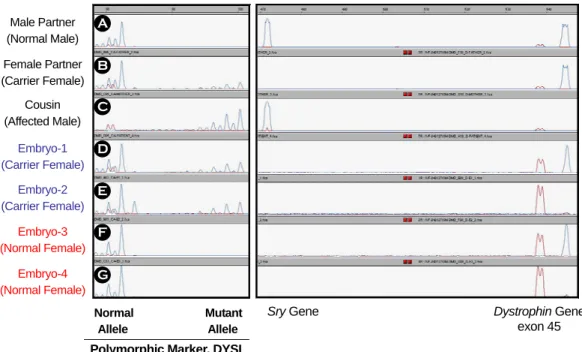
친언니의 아들이 dystrophin 유전자의 exon 45~50 이 결실되어 있는 근이영양증 가계이기 때문에 내 원한 사례로서, 유전검사 결과 부인의 경우 조카와 친언니의 돌연변이 allele을 가지고 있는 것으로 확 인되었다. 이 부부의 경우 34개의 난자를 회수하여 29개를 대상으로 세포질내 정자주입술을 수행하였 으며 이 중 22개의 수정란을 얻어 7개는 동결하였 다. 총 15개의 배아를 대상으로 각각 1개씩의 할구
를 생검하였다. 기존의 duplex-nested PCR 방법과 fluorescent PCR 방법을 병행하기 위해 dystrophin exon 45 부위, Y 염색체 특이적인 Sry, 그리고 poly- morphic marker인 DYSI 부위를 동시에 증폭시키는 triplex PCR을 수행하였다. Sry와 dystrophin exon 45 를 증폭시키는 duplex-nested PCR 방법을 이용하여 2개의 정상 남성배아와 1개의 돌연변이 남성배아 그리고 12개의 정상과 보인자의 구분이 명확하지 않은 여성배아를 진단할 수 있었다. 일차 PCR 산물 을 대상으로 polymorphic marker인 DYSI를 포함한 triplex fluorescent PCR 결과 4개의 정상 남성배아와 1개의 돌연변이 남성배아 그리고 총 10개의 여성배 아 중 6개의 정상 여성배아와 4개의 보인자 여성배 아가 진단되었다 (Figure 3). 총 10개의 정상 배아 중 4개를 이식하여 쌍태임신이 확인되었으며, 산전 양 수검사 결과 정상 염색체와 dystrophin 유전자를 가 지고 있는 것으로 확인되었으며, 제왕절개를 통해 건강한 남아 및 여아를 분만하였다. 출산 후 신생아 들의 유전검사 결과 정상으로 진단되었다 (Table 1).
Male Partner (Normal Male) Female Partner (Carrier Female)
Cousin (Affected Male)
Embryo-1 (Carrier Female)
Embryo-2 (Carrier Female)
Embryo-3 (Normal Female)
Embryo-4 (Normal Female)
Normal Mutant Allele Allele
Sry Gene Dystrophin Gene
exon 45 Polymorphic Marker, DYSI
A B C D E F G
Figure 3. Electropherogram of the dinucleotide repeat marker (DYSI) analysis, Sry, and dystrophin exon 45 for pre- implantation genetic diagnosis of Duchenne muscular dystrophy in case 3. The approximate length of the repeats (base pairs) is represented on the top of electropherogram. Electropherogram A (top): the unaffected male partner; B: the carrier female partner; C the affected son; D and E: the carrier female embryos; F and G: the normal female embryos.
2) 사례 4
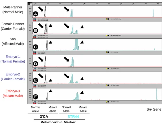
본 사례의 경우 근이영양증 환자인 아들을 대상 으로 Chamberain's primer set와 Begg's primer set 등 총 18개의 primer로 근이영양증을 검사하였으나 결 실 부위가 확인되지 않은 사례로서, 연관분석을 통 해 아들의 돌연변이 allele과 부인의 allele이 같음을 확인하였으며, dystrophin 유전자와 연관되어 있는 polymorphic marker를 이용해서 착상전 유전진단을 실시하였다 (Figure 1). Polymorphic markers로는 3'- CA와 STR44 marker를 사용하였으며 Y 염색체 특 이적인 Sry primer를 같이 증폭하는 triplex fluore- scent PCR 방법을 수행하였다. 총 11개의 배아를 대 상으로 할구 생검을 시행하였으며, 2개의 배아에서 는 1개씩의 할구, 나머지 9개의 배아로부터는 2개 씩의 할구를 생검하였다. 착상전 유전진단 결과 정 상 남성배아 2개, 돌연변이 남성배아 2개, 정상 여
성배아 4개, 그리고 보인자 여성배아 3개로 각각 진단되었으며 정상배아 4개를 이식하였으나 bioche- mical pregnancy로 확인되었다 (Figure 4, Table 1).
고 찰
착상전 유전진단의 도입 초기에는 주로 형광직접 보합법을 이용한 염색체 구조적 이상이라든가 수적 이상에 대한 정보만을 이용하여 정상 혹은 보인자 의 가능성이 있는 배아의 이식을 시도하였었다. 형 광직접보합법 외에 PCR을 이용한 착상전 유전진단 은 여러 가지 문제점으로 인하여 초기에는 그 결과 가 비효율적이거나 명확하지 않은 경우가 많았다.
그러나 이러한 문제점들을 해결하기 위하여 nested PCR이나 multiplex PCR 등 여러 가지 방법들이 고 안되어 시행되었으며, 그 외에도 Taq polymerase의 Male Partner
(Normal Male)
Female Partner (Carrier Female)
Son (Affected Male)
Embryo-1 (Normal Female)
Embryo-2 (Carrier Female)
Embryo-3 (Mutant Male)
Normal Mutant Normal Mutant Allele Allele Allele Allele
3'CA STR44 Polymorphic Marker
Sry Gene A
B
C
D
E
F
Figure 4. Electropherogram of the dinucleotide repeat markers (3'CA and STR44) analysis and Sry for preimplan- tation genetic diagnosis of Duchenne muscular dystrophy in case 4. The approximate length of the repeats (base pairs) is represented on top of the electropherogram. Electropherogram A (top): the unaffected male partner; B: the carrier female partner; C: the affected son; D: the normal female embryo; E: the carrier female embryo; F: the affected embryo.
Arrows and arrow heads indicate normal and mutant alleles, respectively.
다양화, cell lysis 방법의 발전, 여러 가지 PCR 조 건의 개선 등 다방면에서 발전이 이루어짐에 따라 현재에는 높은 PCR 특이성과 낮은 ADO rate를 나 타내는 조건이 확립되어 PCR을 이용한 착상전 유전 진단의 신뢰성을 높이는데 크게 기여하고 있다.15,16
근이영양증과 같은 X 연관 유전질환의 경우, 초 기에는 임상적으로 비정상적인 표현형을 보일 수 있는 남아를 배제하기 위해 성별검사에 해당하는 착상전 유전진단이 시행되었으며, 이로 인하여 환자 가 될 가능성이 있는 남아의 출산을 피할 수 있었 다. 그러나 이러한 진단방법은 남성배아의 50%는 정상일 수 있음에도 불구하고 모든 남성배아가 이 식되지 못하고 폐기될 수 있는 단점이 있었다. 또 한, 본 연구의 case 4와 같이 dystrophin 유전자의 결실 부위가 확인되지 않은 경우에 linkage analysis 를 통한 착상전 유전진단이 보고되기도 하였다.17 한편 Hussey 등18 (1999)이 보고한 방법에 의하면
dystrophin 유전자의 결실 exon과 성별검사를 위한 Sry 유전자를 함께 증폭시키는 duplex PCR 방법을 사용하여 남성배아에 대한 유전검사를 시행하여 이 식 가능 배아수를 증가시킬 수 있었다. 그러나 이러 한 방법의 도입에도 불구하고 보인자 또는 정상 여 성배아의 정확한 유전자형을 구분할 수 없었다.
위와 같은 문제점을 해결하고 PCR을 이용한 착 상전 유전진단의 가장 큰 문제점인 ADO rate를 줄 이기 위해 linked polymorphic marker를 결실 부위 와 함께 증폭하는 방법을 시도하였다. 또한, 일반적 인 PCR 방법보다 민감도가 높은 방법으로 알려진 fluorescent PCR 방법의 도입 역시 진단의 특이성을 높여줄 뿐 아니라 ADO를 줄일 수 있는 장점이 있 다. 따라서 본 연구팀에서는 과거의 형광직접보합법 이나 PCR 방법을 이용한 성별검사법 그리고 성별 검사와 동시에 특이적인 원인 유전자를 증폭시키는 duplex-nested PCR 방법의 문제점을 해결하기 위해 Table 1. Clinical outcome of the PGD for Duchenne muscular dystrophy cases
Duplex-nested PCR Fluorescent PCR
Case-1 Case-2 Case-3 Case-4
Mutation loci Exon 45 Exon 6~17 Exon 45~50 Not found
Informative markers ND ND DYS I STR44/3'CA
Female partner's age (years) 34 28 34 42
No of biopsied embryos (blastomeres*) 4 (4) 18 (26) 16 (16) 11(20) Diagnosis rate per blastomere 100% 94.4% 100% 100%
Normal 2 4 4 2
No. of male embryos
Affected 0 4 2 2
Normal 6 4
No. of female embryos
Carrier 2 9
4 3 No. of transferable embryos 2 (50%) 4 (22%) 10 (63%) 6 (55%)
No. of transferred embryos 2 4 4 4
Results of pregnancies Singleton - Twin Biochemical
Results of amniocentesis 46, XX (normal for dystrophin)
- 46, XY & 46, XX (normal for dystrophin) Delivery outcome Female
(2.8 kg) - Male/Female
(2.8 kg/2.6 kg) - ND: Not determined, *blastomeres with nucleus
linked polymorphic marker를 이용한 fluorescent PCR 방법을 착상전 유전진단에 적용하였다.
본 연구에서의 효용성를 각 방법 별로 분석해 보 면, duplex-nested PCR 방법을 사용한 사례 1과 2의 경우 총 22개 배아 중 21개의 배아를 진단하여 95.5%의 진단 성공률을 나타내었으며, linked poly- morphic marker를 triplex fluorescent PCR 방법을 사 용한 사례 3과 4의 경우 총 27개 배아 모두 진단에 성공하여 100%의 진단 성공률을 나타내어 진단 성 공률에는 큰 차이를 보이지 않았다. 그러나 duplex- nested PCR 방법을 사용한 사례들에서 여성배아의 경우 정상과 보인자 여부가 확실하게 진단되지 않 아 보인자를 원하지 않는 부부에게는 이식할 수 없 었다. 따라서 실제 이식 가능 배아수는 전체 진단배 아의 28.6% (6/21)로 나타났다. 반면에 linked poly- morphic marker를 사용하여 돌연변이 allele의 유전 여부를 정확하게 확인할 수 있었던 사례 3과 4의 경우 기존의 duplex-nested PCR 방법을 사용하여 17 개의 여성배아 중 10개 (58.8%)의 배아를 정상 여 성배아로 정확히 진단함으로써 전체 진단배아 중 59.3% (16/27)의 이식 가능 배아수를 확보할 수 있 었으며 성 (sex)에 관계없이 정상으로 진단된 배아 를 이식할 수 있었다. 이와 같은 효율적인 결과는 일반적인 PCR과 agarose gel 전기영동보다 더 민감 한 fluorescent PCR과 모세관 전기영동을 이용한 방 법의 적용으로 가능하였으며 또한, 각 가계에 유용 한 linked polymorphic markers를 이용하였기에 가능 하였다고 생각된다. 결과적으로 근이영양증에 대한 착상전 유전진단을 통해 duplex-nested PCR 방법을 시행한 2 사례 중 1 사례가 임신에 성공하였으며, fluorescent PCR 방법을 사용한 2사례 중 1 사례는 쌍둥이 임신에 성공하였으며 1 사례는 biochemical pregnancy를 나타내었다. 이들 임신된 사례의 경우 각각 임신 17주에 양수검사와 분만 후 신생아의 혈 액을 채취, 분석하여 dystrophin 유전자의 정상 여부 를 최종적으로 확인하였다.
본 논문의 연구 결과는 근이영양증 질환을 대상 으로 국내에서 최초로 시도된 duplex-nested PCR 방 법과 triplex fluorescent PCR 방법으로 정상 임신 및 출산에 성공한 착상전 유전진단 사례이다. 특히, polymorphic marker를 이용한 fluorescent PCR 방법
의 도입으로 여성배아의 정확한 유전형을 확인하여 여성배아의 일부 (이론상 50%)를 이식 가능한 정상 배아로 정확히 진단함으로써 이식 가능한 배아의 숫자를 증가시킬 수 있었다. 이러한 유용한 poly- morphic marker를 이용한 fluorescent PCR은 단일 유 전자 이상에 대한 착상전 유전진단의 특이성과 신 뢰도를 높일 수 있는 효과적인 방법으로 생각된다.
참 고 문 헌
1. Edwards RG, Gardner RL. Choosing sex before birth.
New Scientists 1968; 38: 218-20.
2. Handyside AH, Kontogianni EH, Hardy K, Winston RML. Pregnancies from biopsied human preimplan- tation embryos sexed by Y-specific DNA amplifica- tion. Nature 1990; 344: 768-70.
3. Coutelle C, Williams C, Handyside H, Hardy K, Winston R, Williamson R. Genetic analysis of DNA from single human oocytes: a model for preimplan- tation diagnosis of cystic fibrosis. Br Med J 1989;
299: 2-24.
4. Holding C, Monk M. Diagnosis of beta-thalassaemia by DNA amplification in single blastomeres from mouse preimplantation embryos. Lancet 1989; 2:
532-5.
5. Pickering S, McConell J, Johnson M, Braude P. Re- liability of detection by polymerase chain reaction of the sickle-cell containing region of the β-globin gene in single human blastomeres. Hum Reprod 1992; 7: 630-6.
6. 임천규, 한미현, 전진현, 송견지, 김정욱, 박소 연 등. 균형 전좌 또는 Robertsonian 전좌 보 인자의 체외수정 및 배아이식술에서 형광직접 보합법을 이용한 착상전 유전자진단의 임상적 적용. 대한산부인과학회지 2000; 43: 1147-53.
7. 김진영, 임천규, 송인옥, 유근재, 양광문, 한국 선 등. 유전질환 및 염색체 이상의 예방을 위 한 착상전 유전진단의 결과. 대한불임학회지 2002; 29: 269-78.
8. 임천규, 민동미, 이형송, 변혜경, 박소연, 류현 미 등. 형광직접보합법을 이용한 착상전 유전
진단 기법의 최적화와 경험 축적에 의한 임신 율의 향상. 대한불임학회지 2004; 31: 29-38.
9. Lim CK, Jun JH, Min DM, Lee HS, Kim JY, Koong MK, et al. Efficacy and clinical outcome of preim- plantation genetic diagnosis using FISH for couples of reciprocal and Robertsonian translocations: the Korean experience. Prenat Diagn 2004; 24: 556-61.
10. 최수경, 이은호, 이호준, 전진현, 강인수, 백은 찬 등. 근이양증 가계에서의 PEP-PCR을 이용 한 착상전 유전자진단. 대한불임학회지 1996;
23: 109-14.
11. 이형송, 최혜원, 임천규, 민동미, 변혜경, 김진 영 등. OTC 효소결핍증, 수포성 표피박리증 및 lactic acidosis 가계에서 duplex nested PCR 방법을 이용한 착상전 유전진단: OTC 효소결 핍증 가계에서의 정상아 임신 및 출산. 대한산 부인과학회지 2004; 47: 708-18.
12. Roberts RG. Dystrophin, its gene, and the dystro- phinopathies. Adv Genet 1995; 33: 177-231.
13. Koenig M, Hoffman EP, Bertelson CJ, Monaco AP, Feener C, Kunkel LM. Complete cloning of the Du- chenne muscular dystrophy (DMD) cDNA and pre- liminary genomic organization of the DMD gene in normal and affected individuals. Cell 1987; 50: 509
-17.
14. Roberts RG, coffey AJ, Bobrow M, Bentley DR.
Exon structure of the human dystrophin gene. Geno- mics 1993; 16: 536-8.
15. Piyamongkol W, Bermudez MG, Harper JC, Wells D. Detailed investigation of factors influencing amp- lification efficiency and allele drop-out in single cell PCR: implications for preimplantation genetic diag- nosis. Mol Hum Reprod 2003; 9: 411-20.
16. Sermon K. Current concepts in preimplantation ge- netic diagnosis (PGD): a molecular biologist's view.
Hum Reprod Update 2003; 8: 11-20.
17. Lee SH, Kwak IP, Cha KE, Park SE, Kim NK, Cha KY. Preimplantation diagnosis of non-deletion Du- chenne muscular dystrophy (DMD) by linkage poly- merase chain reaction analysis. Mol Hum Reprod 1998; 4: 345-9.
18. Hussey ND, Donggui H, Froiland DA, Hussey DJ, Haan EA, Matthews CD et al. Analysis of five Du- chenne muscular dystrophy exons and gender de- termination using conventional duplex polymerase chain reaction on single cells. Mol Hum Reprod 1999; 5: 1089-94.