ISSN 2234-3806 • eISSN 2234-3814
158 www.annlabmed.org http://dx.doi.org/10.3343/alm.2012.32.2.158 Ann Lab Med 2012;32:158-161
http://dx.doi.org/10.3343/alm.2012.32.2.158
Case Report
Diagnostic Hematology
A Case of Rosai-Dorfman Disease with Highly Elevated Serum Ferritin
Sung-Yoon Rew, M.D.1, Hee-Chang Jang, M.D.1, Kyung-Hwa Park, M.D.1, Jae-Sook Ahn, M.D.1, Ga-Eon Kim, M.D.2, Yoo-Duk Choi, M.D.2, and Sook-In Jung, M.D.1
Departments of Internal Medicine1 and Pathology2, Chonnam National University Medical School, Gwangju, Korea
Sinus histiocytosis with massive lymphadenopathy, also known as Rosai-Dorfman disease is a rare disorder characterized by proliferation of distinctive histiocytes within lymph node sinuses and lymphatics, sometimes involving extranodal sites. However, clinical suspicion is difficult and there is also a lack of useful diagnostic markers for this disorder prior to histological confirmation. High elevation of serum ferritin is known to be a useful diagnos- tic marker for various hematologic diseases, including hemophagocytic lymphohistiocyto- sis and lymphoma. Here, we report a case of fever of unknown origin that presented along with highly elevated serum ferritin (5,780 ng/mL), and was finally diagnosed as Rosai-Dor- fman disease by lymph node biopsy.
Key Words: Ferritin, Fever of unknown origin, Sinus histiocytosis with massive lymphade- nopathy, Rosai-Dorfman disease
Received: April 22, 2011
Revision received: September 28, 2011 Accepted: November 21, 2011 Corresponding author: Hee-Chang Jang Department of Internal Medicine, Chonnam National University Hospital, 671 Jebong-no, Dong-gu, Gwangju 501-757, Korea
Tel: 82-62-220-6296 Fax: 82-62-225-8578 E-mail: [email protected]
© The Korean Society for Laboratory Medicine.
This is an Open Access article distributed under the terms of the Creative Commons Attribution Non-Commercial License (http://creativecom- mons.org/licenses/by-nc/3.0) which permits un- restricted non-commercial use, distribution, and reproduction in any medium, provided the origi- nal work is properly cited.
INTRODUCTION
Sinus histiocytosis with massive lymphadenopathy (SHML), also known as Rosai-Dorfman disease [1], is a rare pathological condi- tion characterized by fever and lymph node enlargement. Some- times the disease involves extra-nodal sites such as the skin, upper respiratory tract, bone, and retro-orbital tissue [2-6]. Al- though most patients have prominent bilateral cervical lymph node involvement, some cases do not have such findings and present as fever of unknown origin (FUO). Presently, neutro- philia, high erythrocyte sedimentation rate (ESR), elevation of serum lactate dehydrogenase (LDH), rouleaux formation of red blood cells, polyclonal hypergammaglobulinemia, and hyperme- tabolism in lymph nodes and spleen on positron emission to- mography-computed tomography (PET-CT) [7] are suggestive laboratory findings for the diagnosis. However, a useful, specific marker (prior to pathological diagnosis) suggestive of Rosai-Dor- fman disease is still lacking for patients with unexplained fever,
lymphadenopathy, or skin rash.
Serum ferritin is a useful marker for the diagnosis of hemato- logic disorders. Highly elevated serum ferritin (>500 ng/mL) is included in the diagnostic criteria of hemophagocytic lympho- histiocytosis, and is also observed in lymphoma, leukemia, and connective tissue disorders such as systemic lupus erythemato- sus and adult-onset Still’s disease [8]. Recently, the diagnostic utility of ferritin in SHML has been suggested, based on a highly elevated serum ferritin level in a case [9].
Here, we describe a patient with FUO, skin rash, and highly elevated serum ferritin, who was finally diagnosed as having Ro- sai-Dorfman disease by lymph node biopsy.
CASE REPORT
A 40-yr-old Korean woman presented with fever and generalized erythematous rash, which had persisted for 17 and 14 days, re- spectively, prior to her visiting Chonnam National University Hos-
ISSN 2234-3806 • eISSN 2234-3814
Rew S-Y, et al.
Rosai-Dorfman disease with high ferritin
159
http://dx.doi.org/10.3343/alm.2012.32.2.158 www.annlabmed.org
pital (Gwangju, Republic of Korea), a 1,000-bed teaching hospi- tal and referral center. She had visited local clinics and taken le- vofloxacin and metronidazole for 3 days, and doxycycline for 4 days for suspected uteritis or Tsutsugamushi disease. However, the fever and rash did not improve. She had no history of comor- bidity. On physical examination, generalized small macular erup- tions on her upper and lower extremities and trunk, palpable bi- lateral and cervical and inguinal lymph nodes, and fever (38.1°C) were observed. The initial laboratory examination showed a white blood cell count of 10,000/µL (neutrophils 78.1%, eosinophils 1.5%, lymphocytes 13.6%, and monocytes 6.4%); hemoglobin level of 10.1 g/dL, and platelet count of 319×103/µL. Aspartate aminotransferase and alanine aminotransferase were mildly ele- vated to 90 U/L and 41 U/L, respectively. LDH was elevated to 1,808 U/L; C-reactive protein (CRP), to 9.2 mg/dL; and ESR, to 76 mm/h. Serologic tests for Orientia tsutsugamushi, Leptospira, and Hantaan virus showed negative results. The serum iron level was 11 µg/dL, total iron-binding capacity was 234 µg/dL, and transferrin saturation was <15%, which are compatible with
anemia of inflammation. However, the serum ferritin level was elevated to 5,780 ng/mL. Polyclonal hypergammaglobulinemia was also detected (by serum protein electrophoresis). Two sets of blood cultures were performed, but no organisms were iso- lated. Test results for autoimmune markers, including antinu- clear antibody and rheumatoid factor, were negative. A tubercu- losis-specific interferon-γ release assay (Quantiferon Tb; Cellestis Limited, Chadstone, Australia) gave a negative result.
Abdominal CT showed mild splenomegaly (maximum diame- ter, 13.5 cm). Thoracic CT showed scanty bilateral pleural effu- sion without enhancement or thickening. There was no mass, consolidation, or lymphadenopathy involving the thorax. How- ever, PET-CT showed hypermetabolism in the spleen and lymph nodes in the cervical, abdominal, and inguinal areas (Fig. 1A).
Moderate normocytic normochromic anemia and red cell rouleaux formation were observed on peripheral blood smear examination. Superficial perivascular dermatosis was reported from a skin biopsy. Bone marrow biopsy, including iron staining, showed normocellular marrow and no abnormal findings. Tissue
A B
Fig. 1. (A) PET/CT scan. Diffuse hypermetabolism in the spleen (maximum standardized uptake value, 4.6) and hypermetabolism in the neck, inguinal, and intra-abdominal lymph nodes (maximum standardized uptake value, 8.4-10.6) were observed. (B) Histological results of a right inguinal lymph node biopsy. Left upper image: Prominent wide sinuses are observed (shown in the circled area) (H&E stain,×100).
Right upper image: These sinuses are composed of histiocytes, lymphocytes, and plasma cells. Histiocytes engulfing lymphocytes are not- ed (emperiopolesis, indicated by arrow) (H&E stain,×400). Left lower image: On immunohistochemistry, the inter-digitating dendritic cells are positive for S-100 (×100). Right lower image: Biopsied tissue stains negative for CD1a immunohistochemical stain (×100). Lymph node tissue also had cells positive for CD68 and CD163 (antigens positive for monocytes/macrophages).
Abbreviations: PET/CT, positron emission tomography-computed tomography; CD, cluster of differentiation.
Rew S-Y, et al.
Rosai-Dorfman disease with high ferritin
160 www.annlabmed.org http://dx.doi.org/10.3343/alm.2012.32.2.158 diagnosis for lymphadenopathy was performed using material
from the right inguinal lymph node, which had the highest maxi- mum standardized uptake value on PET-CT. Upon histological examination of an excisional lymph node biopsy, SHML was di- agnosed (Fig. 1B). Molecular clonality assays for the diagnosis of lymphoma, including immunoglobulin heavy chain gene rear- rangement and T-cell receptor rearrangement, showed negative results.
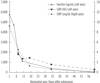
The fever began to subside without medication after 2 weeks of admission. She was discharged on day 18 of hospitalization, and her fever and skin rash were completely resolved. The lev- els of LDH, ferritin, and CRP also normalized with time (Fig. 2).
DISCUSSION
SHML, also known as Rosai-Dorfman disease, is a rare disorder of unknown etiology and is usually associated with lymph node enlargement at various superficial or deep sites. It can also in- volve extranodal sites, commonly the skin, upper respiratory tract, and bone. The involvement of the kidney, lower respiratory tract, or liver has been shown to indicate a poor prognosis. Clini- cal features of SHML mimic lymphoma, but the prognosis is dif- ferent. Treatment is not necessary in most cases [10]. However, in the presence of vital organ compression and/or extranodal lo- calization with important clinical signs, surgical debulking, ra- diotherapy, or chemotherapy are necessary [2].
SHML can be diagnosed by pathologic examination. It should
be distinguished from other lymphohistiocytic infiltrative dis- eases, such as lymphoma, Langerhans cell histiocytosis, hemo- phagocytic lymphohistiocytosis, and other reactive histiocytosis.
SHML shows emperiopolesis on lymph node biopsy and fea- tures of immunohistochemical staining that differ from other diseases: SHML is positive for S-100 and negative for CD1a. Al- though Langerhans cell histiocytosis is also positive for S-100, it is additionally characterized by Birbeck granules and positive staining for CD1a, which are not seen in SHML. And reactive histiocytosis shows no emperiopolesis [9].
Some abnormal laboratory findings, previously reported to be associated with SHML, such as an elevated ESR and polyclonal hypergammaglobulinemia, rouleaux formation of red blood cells and elevation of serum LDH [9], were also observed in the cur- rent case. A highly elevated serum ferritin level in a febrile pa- tient limits the diagnostic possibilities to rheumatic disorders and hemato-oncologic disorders [8]. Among the neoplastic eti- ologies of FUO, highly elevated ferritin levels suggest a myelo- proliferative disorder, pre-leukemia, lymphoma, or other lym- phoreticular malignancies. Among the rheumatic etiologies of FUO, highly elevated serum ferritin levels are associated with flare of systemic lupus erythematosus, adult-onset Still’s dis- ease, or temporal arteritis [8]. The diagnostic utility of ferritin in febrile SHML has been suggested by Cunha et al. [9] based on a highly elevated serum ferritin level in one case. There have been other cases of Rosai-Dorfman disease without elevated or minimally elevated serum ferritin levels [11, 12]. However, in these cases, the patients had no fever when the ferritin was measured. The current case also shows that highly elevated se- rum ferritin level can be useful for narrowing the differential di- agnostic categories and suspicion of SHML in FUO patients.
However, the diagnostic usefulness of serum ferritin level in fe- brile Rosai-Dorfman disease needs to be determined based on data from more cases.
In summary, the current case suggests that Rosai-Dorfman disease should also be suspected in patients with unexplained fever, lymphadenopathy, or skin rash and highly elevated serum ferritin level.
Authors’ Disclosures of Potential Conflicts of Interest
No potential conflict of interest relevant to this article was re- ported.
Fig. 2. Serum LDH, ferritin, and CRP were highly elevated initially, rapidly decreased during the initial 2 weeks of hospitalization, and then normalized gradually over time.
Abbreviations: LDH, lactate dehydrogenase; CRP, C-reactive protein.
7,000 6,000 5,000 4,000 3,000 2,000 1,000 0
14 12 10 8 6 4 2 0 Horizontal axis: Days after admission
1 6 11 16 21 26 31 36 41 46 51 56 Ferritin (ng/mL Left axis) LDH (U/L Left axis) CRP (mg/dL Right axis)
Rew S-Y, et al.
Rosai-Dorfman disease with high ferritin
161
http://dx.doi.org/10.3343/alm.2012.32.2.158 www.annlabmed.org
REFERENCES
1. Rosai J and Dorfman RF. Sinus histiocytosis with massive lymphadenop- athy, a pseudolymphomatous benign disorder. Analysis of 34 cases. Can- cer 1972;30:1174-88.
2. Pulsoni A, Anghel G, Falcucci P, Matera R, Pescarmona E, Ribersani M, et al. Treatment of sinus histiocytosis with massive lymphadenopathy (Rosai-Dorfman disease): report of a case and literature review. Am J Hematol 2002;69:67-71.
3. Chen J, Tang H, Li B, Xiu Q. Rosai-Dorfman disease of multiple organs, including the epicardium: an unusual case with poor prognosis. Heart Lung 2011;40:168-71.
4. Payne JF, Srivastava SK, Wells JR, Grossniklaus HE. Rosai-Dorfman disease simulating nodular scleritis and panuveitis. Arch Ophthalmol 2011;129:518-20.
5. Molina-Garrido MJ and Guillén-Ponce C. Extranodal rosai-dorfman dis- ease with cutaneous and periodontal involvement: a rare presentation.
Case Rep Oncol 2011;4:96-100.
6. Ju J, Kwon YS, Jo KJ, Chae DR, Lim JH, Ban HJ, et al. Sinus histiocyto-
sis with massive lymphadenopathy: a case report with pleural effusion and cervical lymphadenopathy. J Korean Med Sci 2009;24:760-2. 7. Hock AT, Long MT, Sittampalam K, Eng DN. Rosai-Dorfman disease:
FDG PET/CT findings in a patient presenting with pyrexia and cervical adenopathy. Clin Nucl Med 2010;35:576-8.
8. Cunha BA. Fever of unknown origin (FUO): diagnostic importance of serum ferritin levels. Scand J Infect Dis 2007;39:651-2.
9. Cunha BA, Durie N, Selbs E, Pherez F. Fever of unknown origin (FUO) due to Rosai- Dorfman disease with mediastinal adenopathy mimicking lymphoma: diagnostic importance of elevated serum ferritin levels and polyclonal gammopathy. Heart Lung 2009;38:83-8.
10. Komp DM. The treatment of sinus histiocytosis with massive lymphade- nopathy (Rosai-Dorfman disease). Semin Diagn Pathol 1990;7:83-6. 11. Jabali Y, Smrcka V, Pradna J. Rosai-Dorfman disease: successful long-
term results by combination chemotherapy with prednisone, 6-mercap- topurine, methotrexate, and vinblastine: a case report. Int J Surg Pathol 2005;13:285-9.
12. Sacchi S, Artusi T, Selleri L, Temperani P, Zucchini P, Vecchi A, et al. Si- nus histiocytosis with massive lymphadenopathy: immunological, cyto- genetic and molecular studies. Blut 1990 ;60:339-44.