INTRODUCTION
Natural killer (NK) cell neoplasms are a group of rare but highly malignant tumors with a broad spectrum of morpho- logic, immunophenotypes and clinical features. The WHO classification of NK cell neoplasms encompasses 3 distinct
entities: 1) aggressive NK cell leukemia, 2) extranodal NK/T- cell lymphoma, nasal type [1], and 3) blastic NK cell lym- phoma [2]. NK cell tumors are prevalent in Asia and Cen- tral and South America. NK cells were originally described as large granular lymphocytes with natural cytotoxicity against tumor cells. NK cells were later recognized as a se- parate lymphocyte lineage [3]. NK cells represent a lineage of non-T lymphocytes and non-B lymphocytes that medi- ate a major histocompatibility complex-nonrestricted cyto- toxicity against tumor cells and bacterial or viral infected cells [4]. They constitute 10 to 15% of human peripheral blood lymphocytes and express CD2, cytoplasmic CD3e and
194 DOI 10.3343/kjlm.2009.29.3.194
194
결핵성 림프절병증으로 오진된 자연세포독성세포 백혈병 1예
A Case of Natural Killer Cell Leukemia Misdiagnosed as Tuberculous Lymphadenopathy
A-jin Lee, M.D.
1, Sang-Gyung Kim, M.D.
1, Chang Ho Jeon, M.D.
1, Hun Suk Suh, M.D.
1, Ghil Suk Yoon, M.D.
2, and An-Na Seo, M.D.
2Departments of Laboratory Medicine1, Catholic University Hospital of Daegu, Daegu; Departments of Anatomical Pathology2, Kyoungbuk National University Hospital, Daegu, Korea
이아진
1∙김상경
1∙전창호
1∙서헌석
1∙윤길숙
2∙서안나
2대구가톨릭대학교 의과대학 진단검사의학교실1, 경북대학교 의과대학 해부병리학교실2
194 194
Natural killer (NK) cell neoplasms are a group of rare but highly malignant tumors. We report here one case of NK cell leukemia. A 54-yr-old woman presented with a 2-month history of progressive left neck mass. Based on the positive result of tissue PCR for Mycobacterium tuberculosis, she was at first diagnosed with tuberculous lymphadenopathy. After two weeks, she developed generalized lymphadenopathy, hepatosplenomegaly, fever and anemia. Subsequent evaluation was performed including bone marrow aspiration and biopsy. Peripheral blood smear showed leukoerythroblastic features with 31% blasts. Bone marrow was packed with agranular blastoid cells, which were periodic acid-Schiff (PAS) positive and myeloperoxidase (MPO) negative. Immunophenotyping showed that these cells were positive for CD45 and HLA-DR, whereas negative for CD3, CD5, CD7, CD10, CD13, CD14, CD19, CD20, CD22, CD33, CD34, and CD61. Because of the absence of the markers of T-cell, B-cell, and myeloid lineage-specific antigens, we added CD16/56 for the immunophenotyping and the blasts were positive (94%). The tumor cells of biopsied lymph node were only positive for CD56, consistent with NK cell lymphoma. Epstein-Barr virus (EBV) was not detected by RNA in situ hybridiza- tion. Culture for M. tuberculosis was negative. Thus this patient was diagnosed with blastic NK cell lymphoma/leukemia involving bone marrow and lymph node. (Korean J Lab Med 2009;29:194-8)
Key Words : Natural killer cell, Natural killer cell leukemia, Tuberculosis, Polymerase chain reaction
Received :August 13, 2008 Manuscript No :KJLM2160 Revision received :February 2, 2009
Accepted :April 13, 2009
Corresponding author :Sang-Gyung Kim, M.D.
Departments of Laboratory Medicine, Catholic University Hospital of Daegu, 3056-6 Daemyung-4-dong, Nam-gu, Daegu 705-718, Korea
Tel : +82-53-650-4145, Fax : +82-53-653-8672 E-mail : sgkim@cu.ac.kr
CD56 without T cell receptor (TCR) gene rearrangement [5, 6]. The CD56 antigen is regarded as a marker of NK cell leukemia because it is consistently expressed although not specific for NK cells [7]. The etiology of NK cell neoplasm is not well understood but is considered to be strongly asso- ciated with Epstein-Barr virus (EBV). We report here the clinical, laboratory, and immunophenotypic findings of NK cell leukemia of a 54-yr-old woman.
CASE REPORT
A 54-yr-old woman presented with a 2-month history of progressive left neck mass. One month later, right neck and scalp masses were observed. Fine needle aspiration of supraclavicular nodes performed at that time showed find- ings consistent with tuberculosis and tissue PCR for M. tu- berculosiswas positive. Impression was then tuberculous lymphadenopathy. Two weeks later, she developed febrile and chilling sensation accompanied by cough and sputum.
Because of the persistence and progression of the symptoms, she was referred to a pulmonologist for further evaluation and management. On physical examination, there were generalized palpable lymphadenopathy and hepatospleno- megaly. There were palpable masses on both mandibular angles, which were hard and movable. Hemoglobin was 9.1 g/dL, white blood cell count was 20.2×10⁹/L with 89.5%
lymphocytes and 5.2% neutrophils, and platelet count was 76×109/L. Peripheral blood smear showed leukoerythrob- lastic features with 31% atypical blastoid cells (Fig. 1). Aspar- tate aminotransferase was elevated to 63 IU/L and lactate dehydrogenase was elevated to 2,939 IU/L. Gram stain and acid fast bacilli (AFB) stain were negative. Culture of spu- tum for M. tuberculosiswas negative after examinations for 8 weeks. Chest x-ray showed bronchiectasis of the left lower lobe. Computed tomography scan of the chest, abdo- men and pelvis showed multiple enlarged lymph nodes in the lower neck, both supraclavicular, right paratracheal, paraaortic, subcarinal, both hilar, both axillary, portocav- al, both common iliac and both inguinal areas, probably consistent with malignant lymphoma.
Bone marrow aspiration showed 80% to 90% cellularity,
hypercellular for her age, with adequate megakaryocytes.
M:E ratio was 1.3:1. Approximately 90% of hematopoietic cells were blastoid cells (mononuclear cells). These cells were irregular in size and shape with ovoid to irregular nuclei, irregular nuclear membrane, reticular pattern chromatin, inconspicuous nucleoli, and scanty agranular cytoplasm, compatible with L2 morphology by Frech-American-British classification (Fig. 2). The cytochemistry was positive only for periodic acid-Schiff (PAS) (Fig. 3). Immunophenotyping of the blastoid cells of bone marrow aspirates was positive for CD45 and HLA-DR but negative for other T cells, B cells, and myeloid markers: CD3, CD5, CD7, CD10, CD13, CD14, CD19, CD20, CD22, CD33, CD34, and CD61. We added CD16/
56 for the immunophenotyping because blasts of this case were not positive for any lymphoid or myeloid antigen mark- er in our routine immunophenotyping panel for leukemia.
Blasts were positive (94%) with mean fluorescence inten- sity of 11.0. There were no EBV-encoded RNAs by RNA in situ hybridization. The PCR for TCR gene rearrangement was not done. The histologic appearance of the biopsied lymph node showed a diffuse infiltrate of polymorphous tumor cells. Immunohistochemical staining of this speci- men revealed that the tumor cells were only positive for CD56, and negative for CD3, CD20, and terminal deoxynu- cleotidyl transferase (TdT) (Fig. 4). The cytogenetic finding was 46, XX normal karyotype. With these findings, she was diagnosed with blastic NK cell lymphoma/leukemia involv- ing bone marrow, and lymph node. For further manage- ment, she was transferred to a community cancer center.
DISCUSSION
We reported one case of blastic NK cell lymphoma/leukemia with clinical, morphological and immunophenotypic fea- tures. At first, it was diagnosed as tuberculous lymphade- nopathy according to the positive result of tissue PCR for M. tuberculosis. PCR is a powerful and reliable technique for rapid diagnosis of M. tuberculosiswith a reported sen- sitivity of 55-95% in culture positives, and 100% in both smear- and culture-positive clinical specimens [8, 9]. In a previous study, tissue PCR was found to be highly sensi-
tive (94.4%), although less specific (38.2%) [10, 11]. There can be problems in the PCR technique. PCR may be influ- enced by the processing technique. PCR may be false pos- itive due to contamination. In addition, positive PCR results of M. tuberculosishave been reported from sarcoidosis le- sions, which may not have clinical relevance [12]. In our case, Gram stain and AFB stain were negative. Culture was examined weekly for 8 weeks and there were no visible colonies. Therefore, this tissue-PCR result may be false positive.
NK cell tumors are a heterogeneous group of disorders.
These are classified into three groups: 1) aggressive NK cell leukemia, 2) extranodal NK/T-cell lymphoma, nasal type
[1], and 3) blastic NK cell lymphoma [2]. Among these, extra- nodal NK/T-cell lymphoma, nasal type is the most common type and is characterized by extranodal presentation, pleo- morphic cells with angioinvasion and angiodestruction, azu- rophillic granules, and a strong association with EBV. Extra- nodal NK/T-cell lymphomas are considered to originate from mature NK cells and express CD56, CD2, and cyto- plasmic CD3ε, but not surface CD3, CD5, CD16 or CD57 [2,7].
Imamura et al. characterized aggressive NK cell leukemia first as a catastrophic, systemic disease and more preva- lent in Asians than in Caucasians [6]. It is highly malig- nant and rapidly progressive. Clinical features are fever, systemic symptoms, liver dysfunction, hepatosplenomegaly, and systemic lymphadenopathy. Cutaneous lesions are un- Fig. 1. Peripheral blood smear shows atypical blastoid cells (Wright
stain, ×400).
Fig. 2. Tumor cells in bone marrow aspirate smear show lympho- blastoid features with high nuclear cytoplasmic ratio and incon- spicuous nucleoli. Intracytoplasmic azurophilic granules are absent (Wright stain, ×400).
Fig. 3. Blasts are periodic acid-Schiff (PAS) positive (×1,000).
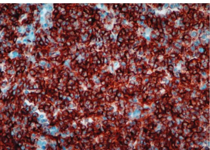
Fig. 4. Immunohistochemical staining of lymph node shows neo- plastic cells with expression of the natural killer (NK) cell antigen, CD56 (×400).
common. The neoplastic cells are slightly larger than nor- mal large granular lymphocytes (LGLs). The cytoplasm is pale or slightly basophilic with fine or coarse azurophilic granules. The nuclei have inconspicuous or distinct nucle- oli with slightly immature chromatin.
Chronic NK cell lymphocytosis is a chronic expansion of mature looking NK cells (≥600/μL) in the peripheral blood for ≥6 months. This is characterized by a chronic, indolent course. In rare cases, the disease is transformed into aggres- sive NK cell leukemia [13].
Precursor lymphoblastic lymphoma/leukemia (LBL) ex- pressing NK-cell associated antigens is a rare entity [14].
In a study by Sheibani et al., six tumors that expressed CD16 and CD57 in addition to terminal deoxynucleotidyl trans- ferase (TdT), CD2, and CD4 were identified among 38 pati- ents who were screened for LBL. These tumors, were group- ed as “NK-LBL” [14]. Subsequently, CD56 has been recog- nized as a sensitive marker for NK cells. The patients fre- quently presented with leukemia and lymphadenopathy without skin involvement. These tumors were negative for EBV [12].
Our case was diagnosed with blastic NK cell lymphoma/
leukemia. In our case, the leukemic cells expressed CD56 in the absence of the markers of T-cell, B-cell, and myeloid lineage-specific antigens. These cells involved bone mar- row, peripheral blood and lymph node. There was no involve- ment of skin or nasal cavity. Morphologically, these cells were blasts rather than large granular lymphocytes. Intra- cytoplasmic azaurophilic granules were absent. EBV was negative. These findings are unusual in aggressive NK cell leukemia. Aggressive NK cell leukemia is associated with EBV, whereas blastic NK cell lymphoma is not related to EBV. Azurophilic granules in the cytoplasm are present in aggressive NK cell leukemia, whereas they are absent in blastic NK cell lymphoma. Consequently, this case was clas- sified as blastic NK cell lymphoma/leukemia.
Although NK cell neoplasm is a very rare entity, clinicians should always recognize this disease. NK cell neoplasms show overlapping clinical and pathologic features with other lymphomas. When morphology of tumor cells is blastic, and immunophenotyping of these cells has no myeloid or lym-
phoid lineage specific markers, it is necessary to add NK cell specific marker for immunophenotyping. CD56 is not lineage-specific and can be expressed by other neoplasms.
However, specific markers for NK cells, such as CD161, CD- 117, and CD94 [15] are not routinely tested in clinical hema- tology laboratory. These markers will be useful for clari- fying blastic NK cell leukemia.
REFERENCES
1. Jaffe ES and Ralfkiaer E. Mature T-cell and NK-cell neoplasms: intro- duction. In: Jaffe ES, Harris NL, et al. eds. World Health Organiza- tion Classification of Tumours: Pathology and genetics of tumours of hematopoietic and lymphoid tissue. Lyon, France: IARC Press, 2001:191-4.
2. Chan JK, Jaffe ES, et al. Extranodal NK/T-cell lymphoma, nasal type.
In: Jaffe ES, Harris NL, et al. eds. World Health Organization Clas- sification of Tumours: Pathology and genetics of tumours of hemato- poietic and lymphoid tissue. Lyon, France: IARC Press, 2001:204-7.
3. Trinchieri G. Biology of natural killer cells. Adv Immunol 1989;47:
187-376.
4. Spits H, Lanier LL, Phillips JH. Development of human T and nat- ural killer cells. Blood 1995;85:2654-70.
5. Robertson MJ and Ritz J. Biology and clinical relevance of human natural killer cells. Blood 1990;76:2421-38.
6. Chan JK, Wong KF, et al. Aggressive NK-cell leukemia. In: Jaffe ES, Harris NL, et al. eds. World Health Organization Classification of Tumours: Pathology and genetics of tumours of hematopoietic and lymphoid tissue. Lyon, France: IARC Press, 2001:198-200.
7. Kwong YL. Natural killer-cell malignancies: diagnosis and treatment.
Leukemia 2005;19:2186-94.
8. Lau SK, Wei WI, Hsu C, Engzell UC. Efficacy of fine needle aspira- tion cytology in the diagnosis of tuberculous cervical lymphadenopa- thy. J Laryngol Otol 1990;104:24-7.
9. Yang HY, Lee HJ, Park SY, Lee KK, Suh JT. Comparison of in-house polymerase chain reaction assay with conventional techniques forthe detection of Mycobacterium tuberculosis. Korean J Lab Med 2006;26:
174-8. (양희영, 이희주, 박수연, 이광길, 서진태. 결핵 진단에 있어서 In- house 중합효소연쇄반응법과 전통적인 진단 방법간의 비교검토. 대한 진단검사의학회지 2006;26:174-8.)
10. Singh KK, Muralidhar M, Kumar A, Chattopadhyaya TK, Kapila
K, Singh MK, et al. Comparison of in house polymerase chain reac- tion with conventional techniques for the detection of Mycobacteri- um tuberculosis DNA in granulomatous lymphadenopathy. J Clin Pathol 2000;53:355-61.
11. Goel MM, Ranjan V, Dhole TN, Srivastava AN, Mehrotra A, Kush- waha MR, et al. Polymerase chain reaction vs. conventional diag- nosis in fine needle aspirates of tuberculous lymph nodes. Acta Cytol 2001;45:333-40.
12. Vago L, Barberis M, Gori A, Scarpellini P, Sala E, Nebuloni M, et al.
Nested polymerase chain reaction for Mycobacterium tuberculosis IS6110 sequence on formalin-fixed paraffin-embedded tissues with
granulomatous diseases for rapid diagnosis of tuberculosis. Am J Clin Pathol 1998;109:411-5.
13. Oshimi K. Leukemia and lymphoma of natural killer lineage cells.
Int J Hematol 2003;78:18-23.
14. Sheibani K, Winberg CD, Burke JS, Nathwani BN, Blayney DW, Van de Velde S, et al. Lymphoblastic lymphoma expressing natural killer cell-associated antigens: a clinicopathologic study of 6 cases. Leuk Res 1987;11:371-7.
15. Freud AG and Caligiuri MA. Human natural killer cell development.
Immunol Rev 2006;214:56-72.