ISSN 2234-3806 • eISSN 2234-3814
Ann Lab Med 2012;32:307-311
http://dx.doi.org/10.3343/alm.2012.32.4.307
Ring Chromosome 5 in Acute Myeloid Leukemia Defined by Whole-genome Single Nucleotide
Polymorphism Array
Jungwon Huh, M.D.1, Yeung Chul Mun, M.D.2, Wha Soon Chung, M.D.1, and Chu Myong Seong, M.D.2
Departments of Laboratory Medicine1, and Internal Medicine2, Ewha Womans University School of Medicine, Seoul, Korea
Chromosomes forming a corresponding ring cannot be clearly defined by conventional cy- togenetics or FISH. Karyotypic analyses using whole-genome single nucleotide polymor- phism arrays (SNP-A) may result in the identification of previously cryptic lesions and al- low for more precise definition of breakpoints. We describe a case of AML with metaphase cells bearing -5, del(11)(q22), and +r. With SNP-A, a 5p-terminal deletion (11 megabases [Mb]), a 5q-terminal deletion (27 Mb), an 11q-interstitial deletion (29 Mb), and a 21q gain (3 Mb) were identified. Therefore, the G-banded karyotype was revised as 46, XY, r(5)(p15. 2q33.2), del(11)(q14.1q23.2), dup(21)(q22.13q22.2)[18]/46,XY[2]. SNP-A could be a pow- erful tool for characterizing ring chromosomes in which the involved chromosomes or bands cannot be precisely identified by conventional cytogenetics or FISH.
Key Words: Ring, Chromosome 5, Single nucleotide polymorphism, Array, AML
Received: October 28, 2011 Revision received: March 2, 2012 Accepted: May 17, 2012
Corresponding author: Jungwon Huh Department of Laboratory Medicine, Ewha Womans University School of Medicine, Seoul 158-710, Korea
Tel: +82-2-2650-5287 Fax: +82-2-2650-5091 Email: JungWonH@ewha.ac.kr
Co-Corresponding author: Chu Myong Seong Department of Internal Medicine, Ewha Womans University School of Medicine, Seoul 158-710, Korea
Tel: +82-2-2650-5015 Fax: +82-2-2650-5062 Email: cmseong@ewha.ac.kr
© The Korean Society for Laboratory Medicine.
This is an Open Access article distributed under the terms of the Creative Commons Attribution Non-Commercial License (http://creativecom- mons.org/licenses/by-nc/3.0) which permits unrestricted non-commercial use, distribution, and reproduction in any medium, provided the original work is properly cited.
INTRODUCTION
Chromosome 5 abnormalities, which are involved in various re- arrangements (including deletions, translocations, insertions, and rings), are a common aberration that is associated with AML or MDS [1-3]. Acquired ring chromosomes have been observed as part of a complex karyotype or, less frequently, as the sole change in human neoplasias [4, 5].
Sometimes, chromosomes that form a corresponding ring can- not be clearly defined by conventional cytogenetics (CC) owing to the complexity of the rearrangements, the suboptimal band-
ing quality, and/or the shortage of material. FISH studies with region-specific probes provide only partial information that is con- fined to the target regions that are examined. Single nucleotide polymorphism arrays (SNP-A) were initially developed for geno- typing DNA sequence polymorphisms and determining genome- wide allelic information. Because the strength of the hybridiza- tion signal is proportional to the copy numbers of the genomic region corresponding to a given probe, copy number changes can also be identified by SNP-A [6, 7]. Karyotypic analyses us- ing high-density, whole-genome SNP-A may result in the better resolution of chromosomal defects, the identification of previ-
ISSN 2234-3806 • eISSN 2234-3814
ously cryptic lesions, and more precise definitions of the break- points [6, 7].
Here, we describe a case of AML with a ring chromosome 5, resulting in terminal deletions of both 5p and 5q. To the best of our knowledge, this is the first report of a ring chromosome 5, in which the breakpoint and deleted segments were defined by genome-wide SNP-A-based karyotyping.
CASE REPORT
1. Case historyA 55-yr-old male presented with complaints of dizziness and dyspnea. The initial laboratory findings revealed a leukocyte count of 3.4×10⁹/L, a hemoglobin level of 8.1 g/dL, and a plate- let count of 37×109/L. On a peripheral blood smear, 63% imma- ture cells were found with 2% segmented neutrophils and 35% lymphocytes. The bone marrow aspirate was hypercellular and replaced by 90% blasts. The remaining cells were lymphocytes.
Megakaryocytes were decreased in number. Accordingly, our case did not have sufficient non-blast cell elements in the bone marrow to adequately assess for multilineage dysplasia. The bi- opsy could not be evaluated due to an inadequate specimen.
Flow cytometry showed that the blasts were positive for cytoplas- mic myeloperoxidase, CD13, CD33, CD34, HLA-DR, and CD7 and negative for CD3, CD5, CD19, CD20, CD22, CD10, CD56, and CD14. Cytochemistry demonstrated that nonspecific ester- ase was positive. Finally, the patient was diagnosed with AML with myelodysplasia-related changes based on the 2008 WHO classification, because he had a MDS-related cytogenetic ab- normality, the -5/del(5q). He was treated with induction chemo- therapy involving idarubicin and arabinofuranosyl cytidine (Ara- C). Complete hematologic remission with a normal karyotype was achieved. After the second consolidation chemotherapy (daunorubicin+Ara-C), the patient received an autologous pe- ripheral stem cell transplantation and remained in remission status for 3 months. However, 9 months after the diagnosis, re- lapse occurred (Table 1). Even after 3 reinduction therapies, re- mission could not be achieved and clonal evolution developed.
The proportion of blasts increased up to more than 80% by 12 months after the diagnosis. The patient subsequently refused to receive continuous treatment and was lost to follow up.
2. CC, SNP-A, and FISH
For the cytogenetic analysis, unstimulated short-term cultures were set up using bone marrow aspirates, and at least 20 meta- phases were analyzed. G-banded karyotype results were descri-
bed according to the International System for Human Cytoge- netic Nomenclature (ISCN) 2009 [8].
We applied a genome-wide SNP 6.0 array (Genome-Wide Human SNP array 6.0; Affymetrix Inc., Santa Clara, CA, USA) using genomic DNA according to the manufacturer’s instruc- tions, and the data were analyzed using Genotyping Console 3.1 software (Affymetrix Inc.). Aberrations that were identified by SNP-A were described according to ISCN 2009 [8]. In order to detect the somatic origin of the copy number alterations that were distinguished from constitutional polymorphic copy num- ber variants (CNVs), we adopted our stringent and conservative algorithm. We selected only the lesions that were more than 100 kb and with more than 10 SNP/CNV probe markers of the array involved within those regions. The lesions that were then identi- fied by SNP-A were compared with the Database of Genomic Variants (http://projects.tcag.ca/variation/) and with an internal control series from healthy normal controls with Korean ethnici- ties (N =200) in order to exclude known CNVs. For the copy neutral loss of heterozygosity (CN-LOH), we excluded homozy- gous stretches of the DNA region that were less than 25 mega- bases (Mb) in the interstitial chromosomal regions, except for those encompassing the telomeric regions of the chromosome, according to the algorithm adopted in the previous studies [6].
Interphase FISH studies were done on the bone marrow aspi- rates using commercially available probes (BCR/ABL1, RUNX1/ RUNX1T1, PML/RARA, CBFB/MYH11, MLL, EGR1/D5S23, D5S- 721, and CEP7/D7S486; Abbott Laboratories, Abbott Park, IL, USA), according to the manufacturer’s instructions.
The cytogenetic analysis showed that 90% of the metaphase Table 1. Follow-up data of G-banded karyotype and FISH
Date G-banded karyotype 5p15.2 deletion by FISH (%) Feb. 2010 46 ,XY,r(5)(p15.2q33.2),del(11)(q14.1q23.2)[18]/
46,XY[2]
65
Mar. 2010 46,XY[20] 1.8
Apr. 2010 46,XY[20] 1.8
Nov. 2010 46 ,XY,add(4)(q32),r(5)(p15.2q33.2),del(11)
(q14.1q23.2)[4]/ 3
46,idem,?del(5)(q21q31)[2]/46,XY[14]
Dec. 2010 46,XY,r(5)(p15.2q33.2),del(11)(q14.1q23.2)[1]/ 16 46,idem,add(4)(q32)[1]/46,XY[18]
Mar. 2011 46 ,XY,add(4)(q32),r(5)(p15.2q33.2),del(11) (q14.1q23.2)[18]/46,Y,der(X)t(X;4) (q13;p16),der(4)t(X;4)(q13;p16)add(4)(q32), r(5)(p15.2q33.2), del(11)(q14.1q23.2)[2]
73
Fig. 1. G-banded karyogram showing 46,XY,r(5)(p14q35),del(11) (q13q22). Arrows stand for abnormal chromosome 5 and abnormal chromosome 11, respectively.
1
6
13
19
2
7 8
14
20
3
9
15 16
21
4
10
17
22
5
11 12
18
X Y
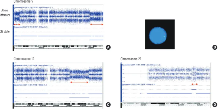
Chromosome 5 Allele
difference
CN state
A B
Chromosome 11 Chromosome 21
C D
Fig. 2. Single nucleotide polymorphisms array (SNP-A) and FISH results.
By SNP-A, the allele difference at the top of each panel represents genotyping calls or the frequency of the A and B alleles. The CN state re- presents the copy number status (CN=1, copy number 1; CN=2, copy number 2; and CN=3, copy number 3). (A) A decrease in the copy number of 5p15.33-p15.2 (423,554-11,003,428) and 5q33.2-q35.3 (153,225,007-180,652,396). (B) Interphase FISH using EGR1/D5S23, D5S721. One green (D5S23, D5S721) and 2 red (EGR1) signal patterns suggested deleted 5p15.2 and retained 5q31 regions. (C) A de- crease in the copy number of 11q14.1-q23.2 (84,514,776-113,765,153). (D) An increased copy number of 21q22.13-q22.2 (37,018,986- 39,492,610).
cells were bearing -5, del(11)(q22), +r in 20 metaphase cells an- alyzed (Fig. 1). Genomic aberrations identified by SNP-A were arr 5p15.33p15.2 (423,554-11,003,428)×1, 5q33.2q35.3 (153,225,007- 180,652,396)×1, 11q14.1q23.2 (84,514,776-113,765,153)×1, and
21q22.13q22.2 (37,018,986-39,492,610) ×3. These results indi- cated a 5p-terminal deletion (11 Mb), a 5q-terminal deletion (27 Mb), an 11q-interstitial deletion (29 Mb), and a 21q gain (3 Mb) (Fig. 2A, C, D). Therefore, the G-banded karyotype was finally revised as 46,XY,r(5)(p15.2q33.2),del(11)(q14.1q23.2),dup(21) (q22.13q22.2)[18]/46,XY[2].
As for the chromosome 5 abnormality, FISH revealed nuc ish (D5S721×1, D5S23×1, EGR1×2)[129/200]. This result corre- sponded to the signal patterns of the 5p15.2 deletion in 65% of the nuclei examined, whereas 2 EGR1 (early growth response 1) signals were indicative of no deletion of 5q31 on both chromo- somes 5 (Fig. 2B). This finding was compatible with the result that the deleted region of 5q that was identified by SNP-A was more telomeric to 5q31, which was spanned by the commercially available EGR1/D5S23, D5S721 FISH probe.
Interphase FISH using the commercial MLL (mixed lineage leukemia) probe demonstrated normal hybridization patterns of the 11q23 regions, indicating that the deleted region of 11q by SNP-A was located more proximal to the MLL gene. FISH using BCR/ABL1, RUNX1/RUNX1T1, PML/RARA, CBFB/MYH11, and
CEP7/D7S486 probes demonstrated normal hybridization signal patterns.
During the follow up, additional chromosomal abnormalities, including add(4)(q32) were found, suggesting clonal evolution (Table 1).
DISCUSSION
In this case, cytogenetic analysis showed metaphase cells bear- ing monosomy 5 and a ring chromosome. By SNP-A, 5p- and 5q-terminal deletions could be identified. However, FISH showed a normal hybridization pattern for the 5q31 region, although the common deleted region of the long arm of chromosome 5 is known to be located on 5q31 [1, 2]. Finally, in the present case, r(5) resulting from the partial deletion of the distal part of the short and long arms of the chromosome 5 and 5q31 chromo- somal regions were included in part of the ring chromosome.
Similar to our case, another study reported that monosomy 5 that was detected by CC was not true monosomy 5. Instead, some regions of chromosome 5 were retained in part of the ring or derivative chromosomes, as determined by molecular cytoge- netics [3].
Ring chromosome formation may occur through breaks in the chromosome arms and fusion of the proximal broken ends, leading to the loss of distal material [1, 5]. In addition, rings may also be formed by telomere dysfunction that triggers fusion of the reactive chromosome ends without a major loss of genetic material [1, 5]. In our case, chromosome 5 appeared to be bro- ken and fused at the 5p15.2 and 5q33.2 subtelomeric bands, leading to the formation of r(5).
A review of the Mitelman database revealed 12 cases carrying r(5) with variable breakpoints and the loss of various parts of chromosome 5 [1]. Most of the ring chromosomes were embed- ded in highly complex karyotypes. This suggests that the ring chromosomes were secondary changes in the course of disease progression. Rings were reported to be associated with a poor prognosis in most, but not all, cases [4, 5]. It cannot be deter- mined whether the complex karyotype or the presence of a ring chromosome itself influences the prognosis. In our case, relapse occurred 9 months after diagnosis and progressed to refractory AML with clonal evolution, suggesting that r(5) may be associ- ated with poor prognosis. More data needs to be collected to definitively establish the role and impact of a ring chromosome.
One advantage of SNP-A over other cytogenetic techniques is its higher level of resolution (less than 1 Mb), which allows the detection of cryptic lesions that can harbor clinically relevant
genes [6, 7]. Because of the limited resolution of CC, lesions less than 2 Mb are mostly undetectable with CC. In some complex karyotypes, especially those containing marker or ring chromo- somes like in our case, it is often impossible to determine the exact nature of the chromosomal rearrangements with certainty based on the banding pattern alone. In the present study using SNP-A, we could determine the exact nature of the r(5) rearrange- ments and define the precise breakpoint of r(5) and del(11q).
Furthermore, the cryptic lesion of the 21q gain (3 Mb) could be detected by SNP-A, and it was not identified by CC.
In particular, another advantage of SNP-A over other genomic analysis techniques, such as CC, is its ability to identify the CN- LOH through the simultaneous measurement of DNA copy num- ber and the detection of genotype calls [6, 7, 9]. CN-LOH can- not be recognized by CC, FISH, or array-based comparative ge- nomic hybridization (aCGH) because CN-LOH occurs without concurrent changes in the gene copy number. Somatic CN-LOH results from mitotic homologous recombination events, or it may represent an attempt to correct for the loss of chromosomal ma- terial, followed by reduplication of the remaining chromosome.
The existence of CN-LOH has aroused considerable interest in the field of cancer genetics because CN-LOH appears to be an important mechanism by which cells are rendered homozygous for preexisting mutations [6, 7, 9]. For example, CN-LOH of 9p is associated with JAK2 homozygous mutations [6, 7, 9]. In our case, CN-LOH lesions were not found.
In conclusion, in the present case, r(5) resulted from partial deletion of the distal part of the short and long arms of chromo- some 5. The breakpoint and deleted segments of ring chromo- some 5 could be precisely defined by whole-genome SNP-A.
Therefore, SNP-A could be a powerful tool for characterizing the cytogenetic material of various rearranged chromosomes, espe- cially when the involved chromosomes or bands cannot be iden- tified by CC or commercially available FISH.
Authors’ Disclosures of Potential Conflicts of Interest
No potential conflicts of interest relevant to this article were re- ported.
Acknowledgement
This study was supported by a grant (2009) from Ewha Womans University School of Medicine, Mokdong Hospital.
REFERENCES
1. Dessen P and Huret JL (Eds.). Chromosome. Atlas genet cytogenet on- col haematol. http://AtlasGeneticsOncology.org/Indexbychrom/idxa_5. html (Updated on Dec 2011).
2. Mitelman F, Johansson B, Mertens F (Eds.). Mitelman database of chro- mosome aberrations and gene fusions in cancer. http://cgap.nci.nih.gov/
Chromosomes/Mitelman (Updated on Nov 2011).
3. Herry A, Douet-Guilbert N, Morel F, Le Bris MJ, De Braekeleer M. Re- defining monosomy 5 by molecular cytogenetics in 23 patients with MDS/
AML. Eur J Haematol 2007;78:457-67.
4. Gebhart E. Ring chromosomes in human neoplasias. Cytogenet Genome Res 2008;121:149-73.
5. Gisselsson D, Höglund M, Mertens F, Johnsson B, Dal Cin P, Van den Berghe H, et al. The structure and dynamics of ring chromosomes in human neoplastic and non-neoplastic cells. Hum Genet 1999;104:315- 25.
6. Maciejewski JP, Tiu RV, O’Keefe C. Application of array-based whole ge- nome scanning technologies as a cytogenetic tool in haematological malignancies. Br J Haematol 2009;146:479-88.
7. Dutt A and Beroukhim R. Single nucleotide polymorphism array analy- sis of cancer. Curr Opin Oncol 2007;19:43-9.
8. Shaffer LG, Slovak ML, et al., eds. ISCN 2009: an international system for human cytogenetic nomenclature. Basel: Karger, 2009.
9. Makishima H and Maciejewski JP. Pathogenesis and consequences of uniparental disomy in cancer. Clin Cancer Res 2011;17:3913-23.