https://doi.org/10.4174/astr.2018.94.4.183 Annals of Surgical Treatment and Research
Prognosis of preoperative positron emission tomography uptake in hepatectomy patients
Jong Man Kim1, Choon Hyuck David Kwon1, Jae-Won Joh1, Dong Hyun Sinn2, Gyu-Seong Choi1, Seung Woon Paik2
1Department of Surgery, Samsung Medical Center, Sungkyunkwan University School of Medicine, Seoul, Korea
2Division of Gastroenterology, Department of Medicine, Samsung Medical Center, Sungkyunkwan University School of Medicine, Seoul, Korea
INTRODUCTION
Liver resection is a curative treatment for management of solitary liver cancer; however, tumor recurrence after curative surgery has limited treatment options. Tumor size, intrahepatic metastasis, microvascular invasion, serum AFP, and serum proteins induced by vitamin K antagonistII (PIVKAII), CRP, and serum alkaline phosphatase have been reported as risk factors associated with early recurrence [14].
The most commonly used method for evaluating oncologic
patients is F18fluoro2deoxyglucose positron emission tomography (18FFDG PET), which is based on glucose metabolism processes that are enhanced in rapidly growing cells and thus cause increased 18FFDG uptake [5]. PETCT is increasingly being used in the field of oncology not only for detecting and staging malignant tumors but also for monitoring therapy response and differentiating malignant from benign lesions [57]. PETCT has been used as a predictor of outcomes in various cancers. In addition, PETCT has been applied for the detection and evaluation of hepatocellular carcinoma (HCC).
Reviewed January February March April May June July August September October November December
Received June 15, 2017, Revised August 6, 2017, Accepted August 22, 2017 Corresponding Author: Jae-Won Joh
Department of Surgery, Samsung Medical Center, Sungkyunkwan University School of Medicine, 81 Irwon-ro, Gangnam-gu, Seoul 06351, Korea Tel: +82-2-3410-3466, Fax: +82-2-3410-0040
E-mail: [email protected]
ORCID code: https://orcid.org/0000-0003-4823-6218
Copyright ⓒ 2018, the Korean Surgical Society
cc Annals of Surgical Treatment and Research is an Open Access Journal. All articles are distributed under the terms of the Creative Commons Attribution Non- Commercial License (http://creativecommons.org/licenses/by-nc/4.0/) which permits unrestricted non-commercial use, distribution, and reproduction in any medium, provided the original work is properly cited.
Purpose: Preoperative F-18-fluoro-2-deoxy-glucose positron emission tomography (18F-FDG PET) imaging results appear to predict tumor recurrence and patient survival. The present study compared outcomes between PET-positive and PET- negative groups with HBV-related hepatocellular carcinoma (HCC) who underwent curative hepatectomy and assessed the prognostic value of positive PET-CT for HCC recurrence and death.
Methods: This study included patients who underwent liver resection of solitary HCC between 2007 and 2014 based on preoperative radiological images. There were 133 patients in the PET-positive group and 93 in the PET-negative group.
Results: There were no statistically significant differences in baseline, perioperative, or pathologic characteristics between the 2 groups except HBsAg titer, tumor size, and presence of bile duct tumor thrombi. Multivariate analysis showed that tumor size >3.5 cm and HBsAg titer >1,000 cutoff index were predisposing factors of positive PET-CT. Disease-free survival and overall survival rate at 1, 3, and 5 years were 76.3%, 64.4%, 60.3% and 96.8%, 91.1%, 85.1% in the PET-nega- tive group, respectively, compared with 70.7%, 62.2%, 58.9% and 98.5%, 97.0%, 97.0% in the PET-positive group (P = 0.547 and P = 0.046). Multivariate analysis showed that positive PET-CT was closely associated with increased patient survival, but was not related to HCC recurrence.
Conclusion: These results suggest that positive PET findings are not a predisposing factor for recurrence of HBV-related HCC patients, but appear to be associated with improved patient survival. Further prospective studies are needed to confirm the prognostic value of 18F-FDG PET in these patients.
[Ann Surg Treat Res 2018;94(4):183-189]
Key Words: Hepatocellular carcinoma, Hepatitis B virus, Outcome
PETCT is useful for evaluating patients with unexplained increased AFP after locoregional therapy for HCC and in differentiating malignant and benign portal vein thrombosis [8,9]. However, PETCT is not currently included as an HCC diagnostic method because of its suboptimal sensitivity (<50%) for detection of new HCC. In a series of patients who underwent liver resection for HCC, preoperative positive PETCT imaging results were associated with poor tumor differentiation and appeared to predict poor outcomes such as tumor recurrence and death [10]. However, there are few studies on the prognostic value of 18FFDG PETCT in patients with HBVrelated HCC.
The purpose of the present study was to compare the out
comes between PETpositive and PETnegative groups with HBVrelated HCC who underwent curative hepatic resection and to assess the prognostic value of positive PETCT for HCC recurrence and death.
METHODS
Patients
This study included patients who underwent surgical resection of solitary HCC based on preoperative radiological images between July 2007 and September 2014. This study was approved by the Institutional Review Board of Samsung Medical Center (SMC201608161001). A total of 226 patients with HBV
related HCC underwent curative liver resection in our hospital.
HCC was proved based on pathology after hepatectomy.
Exclusion criteria were as follows: mixed HCC and cholangio
carcinoma on pathology; age <18 years; history of liver resection, transarterial chemoembolization, radiofrequency ablation (RFA), or percutaneous ethanol injection; history of radiation;
concurrent intraoperative RFA; fibrolamellar HCC; lack of PET
CT evaluation in the preoperative period; or loss to followup after hepatectomy. Demographic, preoperative laboratory, and pathologic data collected from electronic medical records were retrospectively reviewed.
None of the patients received postoperative adjuvant therapy before recurrence. All patients received antiviral therapy after liver resection. The procedures used for surveillance after liver resection have been described previously [1].
Surgery and pathology
Surgical and pathological procedures used after liver resec
tion have been described previously [4,11]. Major hepatectomy was defined as resection of 3 or more segments, and minor hepatectomy was defined as resection of fewer than 3 seg
ments. Postoperative histological assessment included maxi
mum tumor size, encapsulation, intrahepatic metastasis, multi
centric occurrence, microvascular invasion, serosal involvement, and cirrhosis. Intrahepatic metastasis and multicentric occu
rrence were defined based on guidelines from the Liver Cancer
Study Group of Japan [12]. The histologic grade of HCC was assigned according to the EdmonsonSteiner system as well dif ferentiated (grade I), moderately differentiated (grade II), or poorly differentiated (grades III, IV) [13].
FDG PET-CT procedures
The PDF PETCT procedure in our hospital was previously described [14]. All FDG PETCT imaging was performed with dedicated PETCT scanners (Discovery STE, GE Healthcare, Milwaukee, WI, USA) at Samsung Medical Center. All patients fasted for at least 6 hours prior to intravenous administration of FDG. A blood glucose level ≤140 mg/dL was required before administering FDG. Approximately 5.5 MBq/kg of FDG was administered intra venously for the Discovery STE. Foci of increased metabolic acti vity were compared between normal surrounding tissues and tumor tissue and visually interpreted using a 2point grading score of (1) positive and (2) negative.
Statistical analysis
All statistical analyses were performed using IBM SPSS Statistics ver. 23.0 (IBM Co., Armonk, NY, USA). Continuous variables are described as median with range. Categorical variables are expressed as number and percentage of patients.
Fisher exact test was conducted to evaluate differences in the frequencies of categorical variables between the groups. Mann
Whitney U analysis was conducted to evaluate differences in continuous variables between the two groups. Binary logistic regression analysis using significant factors (P < 0.1) was used to predict positive PETCT in the preoperative period. The KaplanMeier survival method was performed to evaluate dif
ferences in patient survival between the 2 groups. Cox regre
ssion analysis was performed to identify prognostic factors of patient survival. All tests were 2sided, and statistical signifi
cance was defined as P < 0.05.
RESULTS
Baseline characteristics
The baseline characteristics of the PETnegative and PET
positive groups are summarized in Table 1. The median HBsAg titer was 4,435 cutoff index (COI) (range, 1–9,758 COI) in the PETpositive group and 3,319 COI (range, 1–20,093 COI) in the PETnegative group (P = 0.009). There were no statistically significant differences in sex, age, white blood cells, lympho
cyte to neutrophil ratio, monocyte to neutrophil ratio, hemo
globin level, platelet count, liver function tests, HBV DNA level, presence of HBeAg, or indocyanine green value between the 2 groups. The median AFP and PIVKAII levels were 14.1 ng/dL (range, 1.3–50,488.1 ng/dL) and 48 mAU/mL (range, 8–41,631 mAU/mL), respectively, in the PETnegative group compared with 18.3 ng/dL (range, 1.3–200,000 ng/mL) and 56 mAU/mL
(range, 12–67,612 mAU/mL) in the PETpositive group. AFP and PIVKAII levels were not significantly different between the groups.
Perioperative and pathologic characteristics
The incidence of laparoscopic resection and major liver resection was not significantly different between PETpositive and PETnegative groups. Median tumor size was 3.3 cm (range, 1.0–16.5) in the PETpositive group and 2.8 cm (range, 0.3–16.0) in the PETnegative group (P = 0.036). There were no statistically significant differences in tumor grade, necrosis, encapsulation,
microvascular invasion, portal vein tumor throm bosis, serosal involvement, intrahepatic metastasis, multi centric occurrence, or cirrhosis between the 2 groups. The incidence of bile duct tumor thrombi was 6.5% in the PETnegative group and 0.8% in the PETpositive group (P = 0.020). Tumor size > 3.5 cm (odds ratio [OR], 2.291; 95% confidence interval [CI], 1.130–4.645; P = 0.0022) and HBsAg titer > 1,000 COI (OR, 4.354; 95% CI, 1.932–
9.813; P < 0.001) were predisposing factors of positive PETCT in multivariate analysis (Table 2).
Table 1. Baseline, perioperative and pathologic characteristics
Characteristic Negative PET CT (n = 93) Positive PET CT (n = 133) Pvalue Baseline characteristics
Male sex 69 (74.2) 104 (78.2) 0.525
Age (yr) 53 (28–76) 55 (32–76) 0.053
White blood cells (/µL) 5,370 (2,070–13,610) 5,260 (1,370–11,970) 0.855
LNR 0.631 (0.051–1.458) 0.649 (0.136–1.737) 0.320
MNR 0.133 (0.026–0.283) 0.135 (0.014–0.325) 0.900
Hemoglobin (g/dL) 14.2 (10.0–17.2) 14.7 (8.2–17.9) 0.161
Platelet (/µL) 151,000 (44,000–380,000) 162,000 (60,000–324,000) 0.707
Total bilirubin (mg/dL) 0.6 (0.2–2.3) 0.6 (0.2–2.2) 0.291
AST (U/L) 31 (15–177) 29 (13–244) 0.408
ALT (U/L) 31 (5–158) 30 (8–302) 0.248
ALP (U/L) 76 (35–287) 71 (41–245) 0.175
INR 1.05 (0.91–1.34) 1.04 (0.90–1.60) 0.059
Albumin (g/dL) 4.3 (3.2–5.2) 4.4 (3.3–5.1) 0.167
Creatinine (mg/dL) 0.86 (0.40–5.64) 0.85 (0.50–3.50) 0.958
ICGR15 (%) 10.1 (1.2–24.8) 10.0 (1.4–21.4) 0.479
HBV DNA (IU/mL) 153 (12–43,075,048) 232 (12–200,000,000) 0.527
HBeAg (COI) 26 (28.0) 47 (35.3) 0.252
AFP >200 ng/mL 18 (20.7) 42 (31.6) 0.089
PIVKAII >40 mAU/mL 49 (52.7) 71 (53.4) 0.974
HBsAg titer >1,000 COI 58 (62.4) 101 (75.9) <0.001
Perioperative and pathologic characteristics
Laparoscopic approach 15 (16.1) 22 (16.5) 0.856
Extent of operation, major 37 (39.8) 51 (38.3) 0.890
Tumor size >3.5 cm 34 (36.6) 64 (48.1) 0.172
Grades 3 and 4 12 (12.9) 19 (14.3) 0.846
Necrosis 35 (37.6) 64 (48.1) 0.164
Hemorrhage 44 (47.8) 67 (50.4) 0.786
Encapsulation 81 (87.1) 122 (91.7) 0.272
Microvascular invasion 45 (48.4) 74 (55.6) 0.343
PVTT 6 (6.5) 11 (8.3) 0.799
BDTT 6 (6.5) 1 (0.8) 0.020
Serosal involvement 1 (1.1) 5 (3.8) 0.405
Intrahepatic metastasis 11 (11.8) 12 (9.0) 0.510
Multicentric occurrence 6 (6.5) 3 (2.3) 0.166
Cirrhosis 47 (50.5) 56 (42.1) 0.224
Free resection margin (mm) 10.5 (2–70) 12.0 (2–65) 0.734
Values are presented as number (%) or median (range).
LNR, lymphocyteneutrophil ratio; MNR, monocyteneutrophil ratio; INR, international normalized ratio; ICG, indocyanine green;
PIVKAII, proteins induced by vitamin K antagonistII; COI, cutoff index; PVTT, portal vein tumor thrombosis; BDTT, bile duct tumor thrombi.
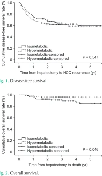
HCC recurrence
The median followup duration was 42.5 months (range, 7–103 months) in the PETnegative group and 36.4 months (range, 5–70 months) in the PETpositive group (P = 0.008). The disease
free survival rate at 1, 3, and 5 years was 76.3%, 64.4% and 60.3%
in the PETnegative group, respectively, and 70.7%, 62.2%, and 58.9% in the PETpositive group (Fig. 1) (P = 0.547). The most common site of recurrence for both groups was an intrahepatic site (82.4% in the PETnegative group compared with 78.4%
in the PETpositive group; P = 0.904). Multivariate analysis showed that serosal involvement and intrahepatic metastasis were closely associated with HCC recurrence (Table 2).
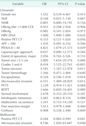
Patient survival
The overall survival rate at 1, 3, and 5 years was 96.8%, 91.1%, and 85.1% in the PETnegative group, respectively, and 98.5%, 97.0%, and 97.0% in the PETpositive group (Fig. 2). The overall survival curve for the PETpositive group was higher than that
for the PETnegative group (P = 0.046). Multivariate analysis showed that tumor size > 3.5 cm and positive preoperative PET
CT were closely associated with patient survival (Table 3).
DISCUSSION
FDG PETCT is not used in the early evaluation of HCC pa
tients because of high costs and low sensitivity. As metabolic acti vity in tumor cells can be higher than that in normal hepa
to cytes, PETCT can be used for malignant characterization of tumors by assessing metabolic activity. Many studies have suggested that a positive FDG PETCT finding is a powerful predictor of prognosis in HCC patients [5,15,16]. One of the major goals in the surgical treatment of HCC is minimizing the risk of tumor recurrence, which is strongly associated with patient survival.
However, the present study failed to prove a correlation between PET results and patient outcomes after liver resection.
Our data showed that baseline, perioperative, and pathologic characteristics in the PETnegative group were not different Table 2. Risk factors of hepatocellular carcinoma recurrence
Variable OR 95% CI Pvalue
Univariate
Female sex 0.884 0.526–1.489 0.644
NLR 0.673 0.320–1.414 0.295
NMR 0.405 0.008–20.275 0.651
HBsAg titer >1,000 COI 0.957 0.548–1.671 0.878
HBeAg 1.491 0.961–2.313 0.075
HBV DNA 1.000 1.000–1.000 0.445
Positive PET CT 1.143 0.740–1.766 0.547
AFP >200 1.004 0.612–1.647 0.987
PIVKAII >40 1.953 1.223–3.119 0.005 Laparoscopic approach 0.642 0.332–1.242 0.188 Extent of operation, major 1.149 0.742–1.780 0.534 Tumor size >3.5 cm 2.398 1.550–3.710 <0.001 Grade 3 and 4 2.741 1.656–4.214 <0.001
Tumor necrosis 1.811 1.179–2.782 0.007
Tumor hemorrhage 0.829 0.539–1.274 0.391
Encapsulation 0.502 0.278–0.907 0.022
Microvascular invasion 2.314 1.471–3.641 <0.001
PVTT 3.659 1.979–6.763 <0.001
BDTT 1.144 0.361–3.628 0.819
Serosal involvement 3.372 1.364–8.336 0.008 Intrahepatic metastasis 3.012 1.743–5.203 <0.001 Multicentric occurrence 2.392 1.103–5.190 0.027 Free resection margin 0.989 0.972–1.007 0.244
Cirrhosis 1.137 0.743–1.741 0.553
Multivariate
Serosal involvement 5.067 1.213–21.159 0.026 Intrahepatic metastasis 3.265 1.270–8.394 0.014 OR, odds ratio; CI, confidence interval; LNR, lymphocyteneutro
phil ratio; MNR, monocyteneutrophil ratio; PIVKAII, proteins induced by vitamin K antagonistII; PVTT, portal vein tumor thrombosis; BDTT, bile duct tumor thrombi.
Cumulativedisease-freesurvivalrate(%)
0
Time from hepatectomy to HCC recurrence (yr) 0
1.0
0.8
0.6
0.4
0.2
P = 0.547 Isometabolic
Hypermetabolic Isometabolic-censored Hypermetabolic-censored
1 2 3 4 5
Fig. 1. Diseasefree survival.
Cumulativeoverallsurvivalrate(%)
0
Time from hepatectomy to death (yr) 0
1.0
0.8
0.6
0.4
0.2
P = 0.046 Isometabolic
Hypermetabolic Isometabolic-censored Hypermetabolic-censored
1 2 3 4 5
Fig. 2. Overall survival.
from those in the PETpositive group with the exception of HBsAg titer, tumor size, and the presence of bile duct tumor thrombi. The diseasefree survival rates for the PETpositive group were not significantly different from those in the PET
negative group; however, the overall patient survival rates in the PETpositive group were better than those in the PETnegative group (P = 0.046). In addition, multivariate analysis showed that negative PETCT finding was an important predictor of poor patient survival.
A previous study showed that PETpositive HCC was signifi
cantly associated with AFP >200 ng/mL and microvascular invasion [17]. Another study reported that preoperative 18FFDG uptake was closely associated with metabolic activity and tumor grade. Therefore, there was a difference in uptake of 18FFDG according to degree of tumor differentiation of HCC [18]; the uptake of 18FFDG in well and welltomoderately differentiated HCC was similar to that in normal liver, whereas moderatetopoorly and poorly differentiated HCC demonstrated
increased uptake [19]. However, our results did not confirm this.
The present study revealed that positive PET CT was closely associated with tumor size >3.5 cm and HBsAg titer > 1,000 COI in patients with HBVrelated HCC who underwent curative hepatectomy. It has been reported that quantification of HBsAg is associated with the level of intrahepatic covalently closed circular DNA (cccDNA), which reflects the number of HBV
infected hepatocytes [20]. Intrahepatic cccDNA level in the tumor tissue was higher than that in the nontumor tissue of HBsAgpositive patients with HCC [21]. Our results suggest that a positive PETCT findings reflected cccDNA level based on the relationship between positive PETCT finding and high HBsAg titer.
A previous study reported a correlation between PETCT findings and prognosis in HCC patients [22]. Another study showed a good correlation among 18FFDG uptake, tumor volume doubling time, and prognosis [16]. Many studies reported similar results and strengthened the correlation bet
ween 18FFDG uptake and HCC prognosis regardless of tumor stage or treatment strategy [5,6,15,16,23]. However, the present study did not find similar outcomes in hepatectomy patients.
HCC recurrence was not different between PETpositive and PETnegative groups, and patient survival was better for the PETpositive group compared with the PETnegative group.
Tumor recurrences in most gastrointestinal cancers involve not only local recurrence, but also remote metastases. Most cases of recurrent HCC after hepatectomy in patients with HBVrelated HCC involved development of intrahepatic metastasis or de novo recurrence. Since tumor recurrence patterns are different between HCC and other gastrointestinal cancers, PETCT does not seem to predict the exact prognosis after curative surgical resection in HCC patients.
Underestimation of 18FFDG uptake by malignant lesions that contributes to PET falsenegative findings can occur because of physiological movements of the liver during emission scans [16]. The degree of this underestimation is variable, particularly in the case of subcentimeter lesions, and might even lead to nonvisualization of the lesion [24]. However, no information is available in the literature on prognosis after liver resection, during which PET can give falsenegative results.
In 2 studies, PETCT in liver transplantation patients predicted HCC recurrence [17,25]. However, those studies in
cluded many patients with various etiologies who received several locoregional therapies. The present study focused on pa tients with solitary HBVrelated HCC with preoperative loco
regional therapies who underwent curative hepatic resection.
PETCT scans in patients undergoing liver transplant or hepa
tectomy as treatment for HCC are considered to have different value in terms of tumor biology worsening during multiple loco regional therapies [26].
Table 3. Risk factors of mortality
Variable OR 95% CI Pvalue
Univariate
Female sex 1.552 0.539–4.467 0.415
NLR 0.168 0.025–1.130 0.067
NMR 0.001 0.000–14.741 0.156
HBsAg titer >1,000 COI 0.933 0.298–2.928 0.906
HBeAg 0.982 0.341–2.826 0.973
HBV DNA 1.000 1.000–1.000 0.626
Positive PET CT 0.353 0.121–1.026 0.056
AFP > 200 1.439 0.491–4.216 0.506
PIVKAII > 40 4.825 1.079–21.573 0.039 Laparoscopic approach 0.037 0.000–12.273 0.266 Extent of operation, major 2.056 0.765–5.526 0.153 Tumor size >3.5 cm 5.805 1.654–20.376 0.006 Grades 3 and 4 8.434 3.125–22.761 <0.001 Tumor necrosis 4.181 1.347–12.974 0.013 Tumor hemorrhage 1.266 0.471–3.404 0.640
Encapsulation 0.328 0.106–1.018 0.054
Microvascular invasion 6.551 1.489–28.829 0.013
PVTT 3.239 0.919–11.421 0.068
BDTT 3.666 0.820–16.401 0.089
Serosal involvement 2.678 0.353–20.310 0.341 Intrahepatic metastasis 2.682 0.863–8.334 0.088 Multicentric occurrence 3.241 0.733–14.330 0.121 Free resection margin 1.013 0.979–1.048 0.460
Cirrhosis 0.908 0.338–2.440 0.849
Multivariate
Positive PET CT 0.284 0.085–0.949 0.041 Microvascular invasion 8.196 1.020–65.847 0.048 OR, odds ratio; CI, confidence interval; LNR, lymphocyte
neutrophil ratio; MNR, monocyteneutrophil ratio; PIVKAII, proteins induced by vitamin K antagonistII; PVTT, portal vein tumor thrombosis; BDTT, bile duct tumor thrombi.
This study has several limitations. First, the study is retro
spective. Second, selection bias can occur due to the inclusion of hepatectomy patients with preoperative radiologically solitary tumor and wellpreserved liver function. Third, we did not measure standardized uptake value (SUV) in the tumor and nontumor lesions. SUV in the liver, including normal liver and tumor, was heterogeneous and therefore dependent on the arbitrary selected points. In addition, the SUV ratio varies according to the examiner. We compared PET CTpositive and PET CTnegative groups because positive and negative findings in the PET CT were clearly seen. Fourth, intrahepatic cccDNA level in the resected liver specimen was not measured;
therefore, we could not demonstrate the relationship between serum HBsAg level and intrahepatic cccDNA level in HBVrelated HCC. In addition, regular assessment of HBsAg level was not performed during the followup after resection. Consequently, the correlation between HBsAg level and PETCT findings was not investigated when HCC recurrence was detected in patients
undergoing resection.
In conclusion, the present study showed that large tumor size and increased HBsAg titer are associated with positive PET
CT findings in patients with HBVrelated HCC. Preoperative 18FFDG PET CT is not an independent prognostic factor for survival in these patients undergoing curative treatment;
however, positive PETCT appears to be associated with better patient survival. These results suggest that 18FFDG PETCT scans do not have a dominant predictive role in HCC recurrence of HBVrelated HCC patients. Our study emphasizes that further prospective studies are needed in order to assess the potential prognostic role of 18FFDG PETCT in patients undergoing hepatectomy.
CONFLICTS OF INTEREST
No potential conflict of interest relevant to this article was reported.
REFERENCES
1. Kim JM, Kwon CH, Joh JW, Park JB, Ko JS, Lee JH, et al. The effect of alkaline phosphatase and intrahepatic metastases in large hepatocellular carcinoma. World J Surg Oncol 2013;11:40.
2. Kim JM, Kwon CH, Joh JW, Ko JS, Park JB, Lee JH, et al. Creactive protein may be a prognostic factor in hepatocellular carcinoma with malignant portal vein invasion. World J Surg Oncol 2013;11:92.
3. Kim JM, Hyuck C, Kwon D, Joh JW, Lee JH, Paik SW, et al. Protein induced by vitamin K antagonistII (PIVKAII) is a reliable prognostic factor in small hepatocellular carcinoma. World J Surg 2013;37:13718.
4. Kim JM, Kwon CH, Joh JW, Park JB, Lee JH, Kim SJ, et al. Differences between hepatocellular carcinoma and hepatitis B virus infection in patients with and with
out cirrhosis. Ann Surg Oncol 2014;21:458
65.
5. Asman Y, Evenson AR, EvenSapir E, Shibolet O. [18F]fludeoxyglucose positron emission tomography and computed tomography as a prognostic tool before liver transplantation, resection, and loco
ablative therapies for hepatocellular carci
noma. Liver Transpl 2015;21:57280.
6. Gauthe M, RichardMolard M, Cacheux W, Michel P, Jouve JL, Mitry E, et al. Role of fluorine 18 fluorodeoxyglucose posi tron emission tomography/computed tomo
graphy in gastrointestinal cancers. Dig Liver Dis 2015;47:44354.
7. Lee JW, Oh JK, Chung YA, Na SJ, Hyun SH, Hong IK, et al. Prognostic significance of 18FFDG uptake in hepatocellular car ci noma treated with transarterial chemo em bolization or concurrent chemo
radio therapy: a multicenter retrospective cohort study. J Nucl Med 2016;57:50916.
8. Chen YK, Hsieh DS, Liao CS, Bai CH, Su CT, Shen YY, et al. Utility of FDGPET for investi gating unexplained serum AFP eleva tion in patients with suspected hepa tocellular carcinoma recurrence.
Anticancer Res 2005;25(6C):471925.
9. Sun L, Wu H, Guan YS. Positron emission tomography/computer tomography: chal
lenge to conventional imaging moda lities in evaluating primary and metastatic liver malignancies. World J Gastroenterol 2007;
13:277583.
10. Seo S, Hatano E, Higashi T, Hara T,
Tada M, Tamaki N, et al. Fluorine18 fluoro deoxyglucose positron emis sion tomography predicts tumor dif fer en
tia tion, Pglycoprotein expression, and out come after resection in hepatocellular car cinoma. Clin Cancer Res 2007;13(2 Pt 1):42733.
11. Kim JM, Kwon CH, Joh JW, Park JB, Lee JH, Kim SJ, et al. Outcomes after curative hepa tectomy in patients with nonB nonC hepatocellular carcinoma and hepa titis B virus hepatocellular carcinoma from noncirrhotic liver. J Surg Oncol 2014;110:97681.
12. Liver Cancer Study Group of Japan.
General rules for the clinical and patholo
gical study of primary liver cancer. 2nd ed. Tokyo: Kanehara & Co.; 2003.
13. Edmondson HA, Steiner PE. Primary carcinoma of the liver: a study of 100 cases among 48,900 necropsies. Cancer 1954;7:462503.
14. Hyun SH, Eo JS, Lee JW, Choi JY, Lee KH, Na SJ, et al. Prognostic value of (18) Ffluorodeoxyglucose positron emission tomo graphy/computed tomography in pa
tients with Barcelona Clinic Liver Cancer
stages 0 and A hepatocellular carcinomas:
a multicenter retrospective cohort study.
Eur J Nucl Med Mol Imaging 2016;43:1638
45.
15. Sun DW, An L, Wei F, Mu L, Shi XJ, Wang CL, et al. Prognostic significance of para
meters from pretreatment (18)FFDG PET in hepatocellular carcinoma: a meta
analy sis. Abdom Radiol (NY) 2016;41:33
41.
16. Tsurusaki M, Okada M, Kuroda H, Matsuki M, Ishii K, Murakami T. Clinical application of 18Ffluorodeoxyglucose posi tron emission tomography for assess
ment and evaluation after therapy for malignant hepatic tumor. J Gastroenterol 2014;49:4656.
17. Yang SH, Suh KS, Lee HW, Cho EH, Cho JY, Cho YB, et al. The role of (18)FFDG
PET imaging for the selection of liver transplantation candidates among hepa to
cellular carcinoma patients. Liver Transpl 2006;12:165560.
18. Kornberg A, Freesmeyer M, Bärthel E, Jandt K, Katenkamp K, Steenbeck J, et al.
18FFDGuptake of hepatocellular carci
noma on PET predicts microvascular tu
mor invasion in liver transplant pa tients.
Am J Transplant 2009;9:592600.
19. Ho CL, Yu SC, Yeung DW. 11Cacetate PET imaging in hepatocellular carcinoma and other liver masses. J Nucl Med 2003;44:
21321.
20. MartinotPeignoux M, Lapalus M, Asselah T, Marcellin P. The role of HBsAg quanti
fi cation for monitoring natural history and treatment outcome. Liver Int 2013;33 Suppl 1:12532.
21. Wong DK, Yuen MF, Poon RT, Yuen JC, Fung J, Lai CL. Quantification of hepatitis B virus covalently closed circular DNA in patients with epatocellular carcinoma. J Hepatol 2006;45:5539.
22. Shiomi S, Nishiguchi S, Ishizu H, Iwata Y, Sasaki N, Tamori A, et al. Usefulness of positron emission tomography with fluorine18fluorodeoxyglucose for predicting outcome in patients with hepatocellular carcinoma. Am J Gastroenterol 2001;96:187780.
23. Pant V, Sen IB, Soin AS. Role of 18FFDG PET CT as an independent prognostic
indicator in patients with hepatocellular car ci noma. Nucl Med Commun 2013;34:
74957.
24. Rohren EM, Paulson EK, Hagge R, Wong TZ, Killius J, Clavien PA, et al. The role of F18 FDG positron emission tomography in preoperative assessment of the liver in patients being considered for cura tive re sec tion of hepatic metastases from colorectal cancer. Clin Nucl Med 2002;27:
5505.
25. Hong G, Suh KS, Suh SW, Yoo T, Kim H, Park MS, et al. Alphafetoprotein and (18) FFDG positron emission tomography predict tumor recurrence better than Milan criteria in living donor liver trans
plan tation. J Hepatol 2016;64:8529.
26. Agopian VG, HarlanderLocke MP, Ruiz RM, Klintmalm GB, Senguttuvan S, Florman SS, et al. Impact of pretransplant bridging locoregional therapy for pa tients with hepatocellular carcinoma within milan criteria undergoing liver trans plan
tation: analysis of 3601 patients from the US multicenter HCC transplant con sor
tium. Ann Surg 2017;266:52535.