Letter to the Editor
Vol. 27, No. 5, 2015 639
Received November 18, 2014, Revised January 8, 2015, Accepted for publication January 16, 2015
Corresponding author: Bong Seok Shin, Department of Dermatology, Chosun University Hospital, 365 Pilmun-daero, Dong-gu, Gwangju 61453, Korea. Tel: 82-62-220-3130, Fax: 82-62-222-3215, E-mail: [email protected]
This is an Open Access article distributed under the terms of the Creative Commons Attribution Non-Commercial License (http://
creativecommons.org/licenses/by-nc/4.0) which permits unrestricted non-commercial use, distribution, and reproduction in any medium, pro- vided the original work is properly cited.
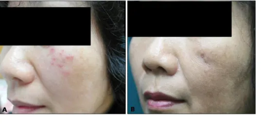
Fig. 1. (A) Multiple, erythematous, various sized, scaly papules on the left cheek. (B) After 4 months, the lesions improved with postinflam- matory hyperpigmentation and atro- phic scar.
http://dx.doi.org/10.5021/ad.2015.27.5.639
Mycobacterium marinum Infection on the Face Diagnosed by Polymerase Chain Reaction Amplification and Direct Sequencing
Hyung Woo Oh, Sang Ho Youn
1, Min Sung Kim
1, Chan Ho Na
1, Sook Jin Jang
2, Chang Ki Kim
3, Bong Seok Shin
1Departments of Radiology, 1Dermatology, and 2Laboratory Medicine, Chosun University School of Medicine, Gwangju, 3Korean Institute of Tuberculosis, Cheongju, Korea
Dear Editor:
A 56 years old Korean woman presented with asympto- matic multiple rice-sized erythematous papules on left cheek (Fig. 1A). The scaly papules were noted 2 months ago. She had no history of trauma or exposure to fish. She had been learning to swim for 3 months. Culture examina- tion of the smear for bacteria and fungus revealed no growth. The nested polymerase chain reaction (PCR) for Mycobacterium tuberculosis showed negative results.
A biopsy specimen taken from the lesion showed gran- ulomatous inflammation with granulation tissue in the deep dermis. The granulomatous infiltration was composed
of multiple histiocytes, mononuclear cells, and giant cells (Fig. 2A). Acid-fast bacilli (AFB) staining produced neg- ative findings (Fig. 2B). Hence, the diagnosis was as non- specific inflammation of the face, and the patient was treat- ed with triamcinolone injection, three times for 6 weeks.
After the injection, the lesions improved overall; however, a solitary lesion showed pus formation. Thus, in addition, mycobacterial culture including the pus was performed, which showed growth of yellow pigment-producing My- cobacterium at 32oC to 33oC in mycobacterium growth in- dicator tube (Fig. 2C). Zeihl-Neelsen staining of culture materials showed innumerable AFB (Fig. 2D), and the or-
Letter to the Editor
640 Ann Dermatol
Fig. 2. (A) Granulomatous infiltration in the deep dermis was composed of multiple histiocytes, mononuclear cells, and giant cells (H&E, ×200). (B) Negative findings (acid-fast bacillus, ×100). (C) Growth of yellow pigment producing mycobacterium on 32oC to 33oC mycobacterium growth indicator tube after 45 days. (D) Presence of innumerable acid-fast bacilli (Ziehl-Neelsen, ×1,000).
ganism was identified as Mycobacterium. Hence, PCR-re- verse blot hybridization assay was performed to identify atypical mycobacterial species, which were confirmed as M. marinum or M. ulcerans.
Furthermore, PCR amplification and direct sequencing of 16SrRNA, tuf, rpoB, hsp65 genes were performed for the rapid and accurate identification of the organism. In 16SrRNA and tuf gene analysis, both M. marium and M.
ulcerans showed >99% homology; thus, it was not possi- ble to differentiate between the two species. Finally, in the rpoB and hsp65 gene analysis, M. marium showed homol- ogy of 100% and 86%, respectively, whereas M. ulcerans showed homology of 99% and 85%, respectively. On the basis of this result, the organism was identified as M. mar- ium and the patient was treated with 200 mg minocycline and 500 mg clarithromycin for 2 months. After the treat- ment, the lesions improved, showing postinflammatory hyperpigmentation (Fig. 1B).
M. marium is a nontuberculous photochromogenic myco- bacterium1. The optimal temperature for its growth is 30oC to 32oC, and it rarely grows at 37oC. Thus, M. marium in- fection rarely occurs on the face and most commonly af- fects the cooler extremities2. Thus far, only seven cases of infection on the face have been reported worldwide3. The conventional microbiological methods used for M.
marium diagnosis are slow and solely rely on phenotypic characteristics. As delayed diagnosis is the main cause of adverse effects, rapid and accurate molecular diagnosis methods are necessary. Rapid detection of mycobacteria by using conventional broad-range PCR has previously been described in the literature4.
Nowadays, M. marinum infection due to the use of pools rarely occurs because of chlorination disinfection of pool water. Thus, we report a rare and interesting case of M.
marium infection showing an unusual location in a patient with a history of exposure to pool water. Hence, clinicians
Letter to the Editor
Vol. 27, No. 5, 2015 641
Received August 9, 2014, Revised November 10, 2014, Accepted for publication February 4, 2015
Corresponding author: Eun Joo Park, Department of Dermatology, Hallym University Sacred Heart Hospital, 22 Gwanpyeong-ro 170 beon-gil, Dongan-gu, Anyang 14068, Korea. Tel: 82-31-380-3765, Fax:
82-31-386-3761, E-mail: [email protected]
This is an Open Access article distributed under the terms of the Creative Commons Attribution Non-Commercial License (http://
creativecommons.org/licenses/by-nc/4.0) which permits unrestricted non-commercial use, distribution, and reproduction in any medium, provided the original work is properly cited.
should consider the possibility of M. marium infection even if the history and location are atypical.
ACKNOWLEDGMENT
This study was supported by the Laboratory Equipment and Research Fund of Chosun University, in 2015.
REFERENCES
1. Runyon EH. Anonymous mycobacteria in pulmonary disease.
Med Clin North Am 1959;43:273-290.
2. Adhikesavan LG, Harrington TM. Local and disseminated infections caused by Mycobacterium marinum: an unusual cause of subcutaneous nodules. J Clin Rheumatol 2008;14:
156-160.
3. Ko DY, Song KH. Mycobacterium marinum infection occur- ring on the face. J Dermatol 2013;40:773-774.
4. Chia JH, Wu TL, Su LH, Kuo AJ, Lai HC. Direct identification of mycobacteria from smear-positive sputum samples using an improved multiplex polymerase chain reaction assay.
Diagn Microbiol Infect Dis 2012;72:340-349.
http://dx.doi.org/10.5021/ad.2015.27.5.641
Imatinib Mesylate-Induced Erythema Multiforme:
Recurrence after Rechallenge with 200 mg/day Imatinib
Min Kyung Lee, Won Joo Kwon, Eun Byul Cho, Eun Joo Park, Kwang Ho Kim, Kwang Joong Kim
Department of Dermatology, Hallym University Sacred Heart Hospital, Anyang, Korea
Dear Editor:
Imatinib mesylate (Gleevec; Novartis AG, Basel, Switzer- land), a selective tyrosine receptor kinase inhibitor, is in- creasingly used for treating chronic myeloid leukemia, Philadelphia chromosome-positive acute lymphoblastic leukemia, and high-grade gastrointestinal stromal tumors (GISTs)1. Several cases of cutaneous reactions after imatinib use have been reported1. We report a case of EM after im- atinib administration for the treatment of a GIST.
A 66-year-old woman was referred for pruritus from the department of oncology. She received a diagnosis of a
GIST, for which she received adjuvant imatinib therapy af- ter gastric wedge resection. She noticed a pruritic rash on her trunk after 5 weeks of 400 mg/day imatinib therapy.
Physical examination revealed generalized variable-sized erythematous wheal-like patches with some targetoid le- sions on the trunk, face, and extremities (Fig. 1). Immuno- globulin (Ig) G and IgM antibodies to the herpes simplex virus were not detected. A skin biopsy from the trunk revealed vacuolar degeneration, tagging of lymphocytes along the dermal-epidermal junction, and perivascular lym- phocytic and some eosinophilic infiltrations in the upper dermis (Fig. 2A). Some dyskeratotic and necrotic keratino- cytes were obvious in the epidermis (Fig. 2B); therefore, EM was diagnosed. As imatinib was the only medication administered to the patient, it was considered the most probable cause. Imatinib was discontinued, and oral ste- roid and antihistamine were prescribed. For 2 weeks, 30 mg/day steroid, tapered to 5 mg/day, was administered.
One month after the discontinuation of imatinib therapy, the rash was fully cured. Imatinib treatment was restarted at a lower dose of 100 mg/day without steroids; no skin le- sion developed for 2 months. However, when the dose