Korean J Hepatobiliary Pancreat Surg 2014;18:118-121
http://dx.doi.org/10.14701/kjhbps.2014.18.4.118
Original Article
Feasibility of laparoscopic liver resection for giant hemangioma of greater than 6 cm in diameter
In Sung Kim, and Choon Hyuck David Kwon
Department of Surgery, Samsung Medical Center, Sungkyunkwan University School of Medicine, Seoul, Korea
Backgrounds/Aims: Liver hemangioma, the most common benign liver tumor, can be safely managed by clinical observation. However, surgical treatment should be considered in a subset of patients with giant hemangioma with abdominal symptoms. We reviewed the feasibility of total laparoscopic liver resection for giant hemangioma of >6 cm in diameter. Methods: Nine consecutive patients who underwent total laparoscopic liver resection for giant he- mangioma between August 2008 to December 2012 were included in this study. Medical records were retrospectively reviewed for demographic data, laboratory findings, and perioperative results. Results: The median age of patients was 36 yrs (range, 31-63). Eight females and 1 male were included in the study. The median size of hemangioma was 11 cm in diameter (range, 6-18) and 5 patients had a hemangioma >10 cm. Indications for surgical treatments were abdominal symptoms in 4 patients, increased size in 5 patients, and uncertain diagnosis in 1 patient. The median operation time was 522 minutes for right hepatectomy, 220 minutes for left lateral sectionectomy, and 90 minutes for wedge resection. The median estimated blood loss was 400 ml (range, 50-900). There was no postoperative morbid- ity, including Clanvien-Dindo grade I. Conclusions: The resection of giant hemangioma demands meticulous surgical technique due to high vascularity and the concomitant risk of intraoperative hemorrhage. Laparoscopic liver resection is feasible with minimal operative complication. Therefore, laparoscopic liver resection can be considered as an option for surgical treatment for giant hemangioma. (Korean J Hepatobiliary Pancreat Surg 2014;18:118-121)
Key Words: Giant hemangioma; Laparoscopic liver resection; Liver hemangioma
Received: October 29, 2014; Revised: November 11, 2014; Accepted: November 20, 2014 Corresponding author: Choon Hyuck David Kwon
Department of Surgery, Samsung Medical Center, 81, Irwon-ro, Gangnam-gu, Seoul 135-710, Korea Tel: +82-2-3410-0926, Fax: +82-2-3140-6982, E-mail: [email protected]
Copyright Ⓒ 2014 by The Korean Association of Hepato-Biliary-Pancreatic Surgery
This is an Open Access article distributed under the terms of the Creative Commons Attribution Non-Commercial License (http://creativecommons.org/
licenses/by-nc/3.0) which permits unrestricted non-commercial use, distribution, and reproduction in any medium, provided the original work is properly cited.
Korean Journal of Hepato-Biliary-Pancreatic Surgery ∙ pISSN: 1738-6349ㆍeISSN: 2288-9213
INTRODUCTION
Liver hemangioma, the most common benign liver tumor, can usually be managed safely by clinical observation.1,2 However, in a subset of patients with giant hemangioma of diameter >4 or 5 cm,3 surgical treatment is justified in high-volume centers if clinical symptoms are present.4,5
The resection of hemangioma demands meticulous sur- gical technique due to high vascularity and the con- comitant risk of intra-operative hemorrhage. Compared to open surgical resection, laparoscopic resection offers many advantages, such as decreased blood loss, shorter hospital stay, improved cosmetic results, and early return to normal life.6 The currently acceptable indications of laparoscopic liver resection are patients with solitary le- sions of ≤5 cm diameter located in liver segments 2 to 6. According to The Louisville Statement, giant liver he-
mangioma (>5 cm diameter) is relatively contraindicated for laparoscopic liver resection.7
Laparoscopic resection of giant hemangioma has been reported previously. However, it has only been reported anecdotally.8 Hemangiomas are more frequent in female patients as benign tumors. Patients requiring surgical treatment would benefit greatly from laparoscopic re- section of giant hemangioma if feasible. The aim of this study was to evaluate the feasibility of laparoscopic liver resection for giant hemangioma >6 cm in diameter.
MATERIALS AND METHODS
Nine patients (8 females and 1 male) with giant liver hemangioma were referred for liver resection between August 2008 and December 2012. Medical records of pa- tients were retrospectively reviewed for demographic data,
In Sung Kim and Choon Hyuck David Kwon. Laparoscopic liver resection for giant hemangioma 119
Table 1. Characteristics of patients
n=9 Median (range) Gender (n)
Male Female Age (years)
Tumor diameter (cm) BMI (kg/m2) History of cancer Yes*
No
Serum AST (U/L) Serum ALT (U/L) Indication of operation**
Abdominal symptoms Tumor enlargement Uncertainty of diagnosis
1 8
2 7
4 5 1
36 (31-63) 11 (6-18) 21.7 (16.2-25.2)
16 (10-35) 13 (9-34)
AST, aspartate aminotransferase; ALT, alanine aminotransferase.
*Lung cancer (n=1) and gastric cancer (n=1). **One patient had abdominal symptom and tumor enlargement
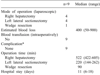
Table 2. Operative and postoperative results
n=9 Median (range) Mode of operation (laparoscopic)
Right hepatectomy Left lateral sectionectomy Wedge resection
Estimated blood loss
Blood transfusion (intraoperatively) No
Complication*
None
Operation time (min) Right hepatectomy Left lateral sectionectomy Wedge resection
Hospital stay (days)
4 4 1
9
9
400 (50-900)
522 (422-605) 220 (144-262)
90 11 (6-18)
*Including Clavien-Dindo classification of surgical complica- tion grade I
laboratory findings, hemangioma size, operation type, op- eration time, estimated blood loss and perioperative trans- fusion, complications, and hospitalization duration. Peri- operative complications and mortality were assessed over a postoperative period of 6 months.
All but 1 patient (wedge resection) underwent anatomi- cally oriented liver resection. Inflow control of the ‘to be resected section’ was performed (except for the wedge re- section) to reduce the size of the tumor and prevent mas- sive bleeding in case of accidental tearing of the hemangioma. Routine Pringle maneuver was not performed. Such a maneuver was only needed to mini- mize intraoperative blood loss and ease the operative procedures. Decompression of the hemangioma after re- section and prior to retrieval was performed in the last 2 cases to reduce the size of the incision and ease the retrieval of the mass. Total laparoscopic method was the approach in every operation.
RESULTS
The preoperative characteristics of the patients were summarized in Table 1. The median size of hemangioma was 11 cm in diameter (range, 6 to 18 cm) and included hemangioma >10 cm in 5 patients. Indications for surgi- cal treatment in patients were abdominal symptoms in 4 (44.4%), which included dyspepsia, abdominal discomfort,
epigastric pain, nausea, and vomiting. Surgical treatment was indicated in 5 (55.5%) patients due to the increasing size of the hemangioma. Diagnosis was uncertain in 1 (11.1%) patient. Hemangiomas with increasing size were 11-18 cm, with a mean increment of 1.67 cm per year.
Laparoscopic right hepatectomy was performed in 4 patients. Four patients received left lateral sectionectomy.
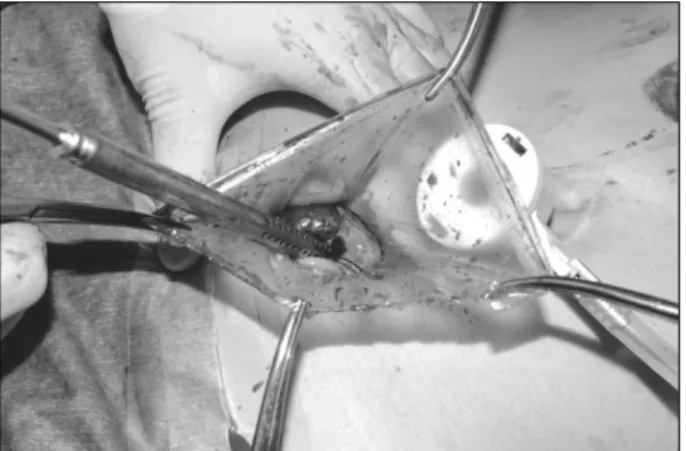
One patient underwent wedge resection. The median oper- ation time was 522 minutes (range, 422 to 605 minutes) for right hepatectomy, 220 minutes (range, 144 to 271 mi- nutes) for left lateral sectionectomy, and 90 minutes for wedge resection. The operative and postoperative results were described in Table 2. The median estimated blood loss was 400 ml (range, 50 to 900 ml). No patient re- quired blood transfusion. Incision type for specimen re- trieval was individually determined based on the previous operation scar and hemangioma size. Pfannenstiel in- cision, umbilical camera port incision extension, and pre- vious cesarean-section incision were performed on 4, 3, and 2 patients, respectively (Fig. 1). Decompression of he- mangiomas during retrieval was performed to decrease the size of incision (Fig. 2). The last 2 cases were performed with decompression. An incision of 8 cm was made on a 13 cm sized giant hemangioma. The incision size was 10 cm for an 18 cm hemangioma. There was no post- operative complication or perioperative mortality, includ- ing a Clavien-Dindo classification of surgical complica- tion grade I. The median hospitalization duration was 11 days (range, 6-18 days). There was no correlation between
120 Korean J Hepatobiliary Pancreat Surg Vol. 18, No. 4, November 2014
Fig. 1. Postoperative wound status of patient abdomen.
Fig. 2. Decompression of hemangiomas during retrieval to de- crease the size of incision. An 8-cm-long incision was made on a 13-cm-sized giant hemangioma.
hospitalization duration and the type of operation.
DISCUSSION
Over the last 2 decades, laparoscopic surgery has evolved to become the preferred approach for many ab- dominal procedures. Compared to open surgical resection, laparoscopic surgery has several advantages, such as de- creased blood loss, shorter hospital stay, improved cos- metic results, and early return to normal life.6,8 Multiple series have reported on the safety and efficacy of laparo- scopic liver surgery. Small and medium sized procedures have become commonplace in many centers. Major lapa- roscopic liver resections have been performed in highly specialized centers with the same efficacy and safety as open surgery.8
Liver hemangioma is a benign tumor. Many studies
have demonstrated that clinical observation of patients with giant hemangioma is the standard treatment option in most patients.2 Nevertheless, surgical resection is in- dicated for patients who have acceptable surgical risk, who have lesions for which a diagnosis is equivocal de- spite appropriate preoperative evaluation, and who have rapidly increasing sized lesions that cause Kasabach–
Merritt syndrome.9 Open enucleation is preferred as a cu- rative treatment by most surgeons, because in open enu- cleation, maximum amount of normal liver parenchyma is preserved, blood loss is limited, and the risk of bile leak is decreased.10 However, massive blood loss remains a problem for large hemangiomas of >10 cm diameter and for centrally located liver hemangiomas.11
Laparoscopic resection of hemangioma was not recom- mended as a surgical treatment due to high vascularity and the concomitant risk of intra-operative hemorrhage.
Therefore, anatomic liver resection was the chosen meth- od of liver resection in our series with the exception of 1 case in which wedge resection could be easily performed. Safety and efficacy were well demonstrated by median estimated blood loss of 400 ml, no requirement of perioperative blood transfusion and no postoperative complication. Minor surgical complications such as wound infection of grade I or II are documented in a ser- ies of resection. However, we had no such minor compli- cations, supporting the minimally invasive nature of the laparoscopic approach.
Two essential operative techniques enhance the feasi- bility and the outcome. First, it is essential to control the inflow to the section of the liver that has the hemangioma, in the early period of the operation. This step, not only decreases the potential for massive bleeding from the he- mangioma, but also increases surgical visibility by de- creasing the giant hemangioma, thus making the surgical procedure much easier. The Pringle maneuver should also be performed in a flexible manner to further decrease the amount of bleeding. The second technique is the decom- pression of hemangioma during retrieval. Hemangioma is a benign disease. Its size can be greatly reduced by ex- traction of blood inside the tumor and the size of incision was also reduced.
In conclusion, the resection of giant hemangioma de- mands meticulous surgical technique due to their high vascularity and the concomitant risk of intra-operative
In Sung Kim and Choon Hyuck David Kwon. Laparoscopic liver resection for giant hemangioma 121
hemorrhage. Although laparoscopic approach for tumors
>5 cm has not been previously recommended, our results suggested that laparoscopic liver resection may be consid- ered as an option for surgical treatment for giant he- mangioma contingent on some surgical precautions and the experience of the surgeon.
REFERENCES
1. Choi BY, Nguyen MH. The diagnosis and management of be- nign hepatic tumors. J Clin Gastroenterol 2005;39:401-412.
2. Schnelldorfer T, Ware AL, Smoot R, Schleck CD, Harmsen WS, Nagorney DM. Management of giant hemangioma of the liver:
resection versus observation. J Am Coll Surg 2010;211:724-730.
3. Lise M, Feltrin G, Da Pian PP, Miotto D, Pilati PL, Rubaltelli L, et al. Giant cavernous hemangiomas: diagnosis and surgical strategies. World J Surg 1992;16:516-520.
4. Yedibela S, Alibek S, Müller V, Aydin U, Langheinrich M, Lohmüller C, et al. Management of hemangioma of the liver: sur- gical therapy or observation? World J Surg 2013;37:1303-1312.
5. Duxbury MS, Garden OJ. Giant haemangioma of the liver: ob- servation or resection? Dig Surg 2010;27:7-11.
6. Katkhouda N, Mason RJ, Towfigh S, Gevorgyan A, Essani R.
Laparoscopic versus open appendectomy: a prospective random- ized double-blind study. Ann Surg 2005;242:439-448.
7. Buell JF, Cherqui D, Geller DA, O'Rourke N, Iannitti D, Dagher I, et al; World Consensus Conference on Laparoscopic Surgery.
The international position on laparoscopic liver surgery: the Louisville Statement, 2008. Ann Surg 2009;250:825-830.
8. Acharya M, Panagiotopoulos N, Bhaskaran P, Kyriakides C, Pai M, Habib N. Laparoscopic resection of a giant exophytic liver haemangioma with the laparoscopic Habib 4× radiofrequency device. World J Gastrointest Surg 2012;4:199-202.
9. Erdogan D, Busch OR, van Delden OM, Bennink RJ, ten Kate FJ, Gouma DJ, et al. Management of liver hemangiomas accord- ing to size and symptoms. J Gastroenterol Hepatol 2007;22:
1953-1958.
10. Lerner SM, Hiatt JR, Salamandra J, Chen PW, Farmer DG, Ghobrial RM, et al. Giant cavernous liver hemangiomas: effect of operative approach on outcome. Arch Surg 2004;139:818-821.
11. Hamaloglu E, Altun H, Ozdemir A, Ozenc A. Giant liver he- mangioma: therapy by enucleation or liver resection. World J Surg 2005;29:890-893.