Copyright ⓒ 2019 by Korean Society for Surgery of the Hand, Korean Society for Microsurgery, and Korean Society for Surgery of the Peripheral Nerve. All Rights reserved.
This is an Open Access article distributed under the terms of the Creative Commons Attribution Non-Commercial License (http://creativecommons.org/licenses/by-nc/4.0/) which permits unrestricted non-commercial use, distribution, and reproduction in any medium, provided the original work is properly cited.
Hand and Microsurgery
가변각 수장측 잠김 금속판을 이용한 원위 요골 골절의 치료 결과
유성림
1ㆍ이호재
2ㆍ한수홍
3ㆍ김재광
41가톨릭대학교 의과대학 정형외과학교실, 2CHA 의과대학 분당차병원 정형외과, 3CHA 의과대학 구미차병원 정형외과,
4울산대학교 의과대학 서울아산병원 정형외과학교실
A Prospective, Multicenter, Observational Study to Assess Distal Radius Fracture Treatment Outcomes Using the Variable-Angle Locking Compression Plate
Sung-Lim Yoo
1, Ho-Jae Lee
2, Soo-Hong Han
3, Jae Kwang Kim
41Department of Orthopedic Surgery, College of Medicine, The Catholic University of Korea, Seoul, Korea
2Department of Orthopaedic Surgery, CHA Bundang Medical Center, CHA University, Seongnam, Korea
3Department of Orthopaedic Surgery, CHA Gumi Medical Center, CHA University, Gumi, Korea
4Department of Orthopedic Surgery, Asan Medical Center, University of Ulsan College of Medicine, Seoul, Korea
Purpose: The 2.4 mm variable-angle locking compression plate (VA-LCP) is designed to treat a variety of distal radius fracture patterns. The purpose of this study was to evaluate the efficacy of the 2.4 mm VA-LCP in treating unstable distal radius fractures.
Methods: We recruited eligible patients treated with the device at two Korean sites. In total, we studied 61 enrolled pa- tients. We assessed clinical outcomes and radiographic union, as well as the types of plates and the number of variable- angle locking screws used in the treatment of each fracture type. Radiographic parameters were evaluated at the immedi- ate postoperative period and at 1 year after operation.
Results: A total of five complications occurred in 61 patients (8.2%). Radiographic union was achieved in 46/51 (90.2%), 50/51 (98.0%), and 51/51 (100%) patients at 3, 6, and 12 months, respectively. Radiographic parameters evaluated at the immediate postoperative period were 22.4±4.2 degrees (radial inclination), 4.7±0.4 degrees (volar tilt) and 0.8±0.2 de- grees (ulnar variance). Radiographic parameters evaluated at 1 year after operation were 21.3±3.4 degrees (radial inclina- tion), 4.1±0.4 degrees (volar tilt) and 1.2±0.3 degrees (ulnar variance). Wrist range of motion and grip strength measure- ments, as well as self-administered patient questionnaires showed continuous improvements at all follow-up time points.
The mean number of screws that were inserted through variable-angle screw guide was 3.2±0.9, while that of screws inserted through fixed-angle screw guide was 3.5±0.9. The majority of patients (85.3%) were treated with a narrow plate.
Conclusion: The findings of this study show promising results for the treatment of unstable distal radius fractures using the VA-LCP System.
Key Words: Radius, Radius fracture, Variable-angle locking compression plate
Received February 27, 2019, Revised [1] April 10, 2019, [2] May 15, 2019, Accepted May 29, 2019 Corresponding author: Jae Kwang Kim
Department of Orthopedic Surgery, Asan Medical Center, University of Ulsan College of Medicine, 88 Olympic-ro 43-gil, Songpa-gu, Seoul 05505, Korea
TEL: +82-2-3010-3523, FAX: +82-2-3010-8555, E-mail: [email protected], ORCID: https://orcid.org/0000-0001-5104-4634
Original Article
INTRODUCTION
Distal radius fractures are common, representing 10%-25% of all fractures1,2. The treatment of choice for displaced, unstable fractures is anatomic reduction with stable fixation3. Furthermore, the main treatment goals include reconstructing the congruity of the articular sur- faces as well as restoring radial height, radial inclination, and palmar tilt4. Various surgical options are available based on a number of external and internal fixation meth- ods, but the most common external fixation technique is percutaneous pinning with K-wires. Internal fixation involves the application of several types of plates, par- ticularly those with a stable angle design, which has in- creased in popularity over the last decade5. The choice of treatment method depends on the fracture type, surgeon preference, and local clinic policy.
The 2.4 mm variable-angle locking compression plate Two-Column Distal Radius (VA-LCP DR) Plate (Syn- thes, Oberdorf, Switzerland) is designed to treat a variety of distal radius fracture patterns. The VA-LCP DR Plate is the first implant to combine the freedom of variable- angle technology for fragment-specific fixation with the convenience of predefined fixed angles. Moreover, the surgeon is able to address fragments individually for each fracture, applying this plate that was conceptually de- signed up to 15 degrees off-axis screw angulation. Also, due to its anatomic shape, there is no need to contour the plates according to the bone anatomy. Other advantages of this design are the low profile of the plate and screw, polished surface, and rounded edges, which minimize potential tendon adhesions and soft tissue irritation. The VA-LCP DR Plate is the latest component of the LCP Distal Radius Plate System and has been approved for marketing in South Korea by the ministry of Food and Drug Safety in 2009. It is indicated for the fixation of intra- and extra-articular fractures and osteotomies of the distal radius.
The primary objective of this study was to observe functional and radiological improvements following sur- gery for distal radial fractures using the 2.4 mm VA-LCP DR Plate and screws system.
MATERIALS AND METHODS
This was a prospective multicenter observational study conducted from November 2010 to July 2013. Two medical centers participated in this study. This study was approved by the Institutional Review Board of Ewha Womans University Mokdong Hospital and CHA Bun- dang Medical Center (IRB No. EMCT026-1 and 2010- 08-010).
Patients were eligible for the study if they met all the following inclusion criteria: (1) male and female subjects aged over 20 years; (2) they had a fracture of the distal radius with dorsal angulation greater than 10 degrees, vo- lar angulation greater than 20 degrees, radial shortening more than 5 mm, radial inclination less than 10 degrees, or articular step-off more than 2 mm on radiographic findings after closed reduction; (3) they had an operation scheduled within 2 weeks of the injury; (4) they were psychologically, mentally, and physically able to fully comply with the protocol including adhering to scheduled visits, treatment plan, completing forms, and other study procedures; and (5) they personally signed and dated informed consent documents prior to any study-related procedures.
Patients were excluded from participation if they met any of the following criteria: (1) previous surgery of the distal radius; (2) active systemic or local infection; (3) known or documented history of communicable disease, including acquired immune deficiency syndrome or hu- man immunodeficiency virus; (4) receiving medical treatment within two years for active hepatitis; (5) active rheumatoid arthritis, non-controlled diabetes mellitus, or any other medical conditions that would increase surgical risk or interfere with normal healing; (6) immunologi- cally suppressed or received systemic steroids (excluding nasal steroids) at any dose daily for more than 1 month within the last 12 months; (7) known history of Paget’s disease, osteomalacia, or any other metabolic bone dis- ease; (8) previous known allergy to the materials con- tained in the device; (9) active malignancy; (10) current or recent history (within the last 2 years) of substance abuse; (11) pregnant or planning to become pregnant dur-
ing the study period; and (12) involved in any study of another investigational product that may affect study out- comes. A total of 61 patients were enrolled in the study.
1. Outcomes
The outcomes were evaluated postoperatively at 3, 6, and 12 months. Outcome measurement consisted of physical examination, patient self-assessments, and radio- graphic evaluation. Radiographic parameters including radial inclination, volar tilt and ulnar variance were eval- uated at the immediate postoperative period and 1 year after operation. Wrist range of motion and grip strength were checked during the physical examination. Wrist range of motion in degrees included flexion/extension, pronation/supination, and radial deviation/ulnar devia- tion. Patient self-assessments included the Patient-Rated Wrist Evaluation (PRWE) questionnaire and Disabilities of the Arm, Shoulder, and Hand (DASH) questionnaire6. Evidence of articular step-off of more than 2 mm and bone union using bridging callus was assessed by the investigators at each follow-up time point. Other analy- ses included the number of screws that were inserted through variable or fixed angle screw guides. The mean number of screws that were inserted through variable or fixed angle screw guides was evaluated. The plate types used were also analyzed, including the number of plate types used among three types (Asian, narrow, or standard plates).
2. Statistical analysis
Continuous variables in different groups presented with mean values were compared using paired t-test. All statis- tical analysis was performed using IBM SPSS Statistics for Windows ver. 24.0 (IBM Corp., Armonk, NY, USA).
The significance level was set at p-value less than 0.05.
RESULTS
1. Patient demographics
Preoperative patient demographics are reported in Table 1. The average patient age was 57.7±14.35 years, and 75.4% were female. The average body mass index was 22.8±2.34 kg/m2 and patients who had underlying diseas- es were 34.4% of the subjects. C2 was the most common fracture type in thirty-one patients of the subjects. Low energy injuries were the most common injury mecha- nism in 91.8% of the subjects, followed by high energy injuries (8.2%). Thirty-two patients (52.5%) injured their right distal radius and none of the injuries were open. Ten patients were lost during follow-up at postoperative 12 months.
Table 1. Demographic and preoperative profile
Characteristic All (n=61)
Age (yr) 57.7±14.35
Sex Male 15 (24.6)
Female 46 (75.4)
Body mass index (kg/m2) 22.8±2.34 Underlying diseases
No medication 40 (65.6)
Regular intake of medication 21 (34.4) Fracture type (AO classification)
A2 6
A3 4
B1 0
B2 11
B3 2
C1 2
C2 31
C3 5
Mechanism of injury
Low energy 56 (91.8)
High energy 5 (8.2)
Fracture side
Right 32 (52.5)
Left 29 (47.5)
Values are presented as mean±standard deviation, number (%), or number only.
2. Intraoperative data
Intravenous antibiotics were perioperatively delivered for 48 hours and oral antibiotics were administered for three days after the period. No intraoperative complica- tions occurred and the mean operation time was 48.9 (range: 17-95) minutes. The majority of patients were treated with a narrow plate (n=52). Postoperatively, none of the devices were broken and there were no surgical site infections. No patient had to be reoperated during their hospital stay.
3. Complications
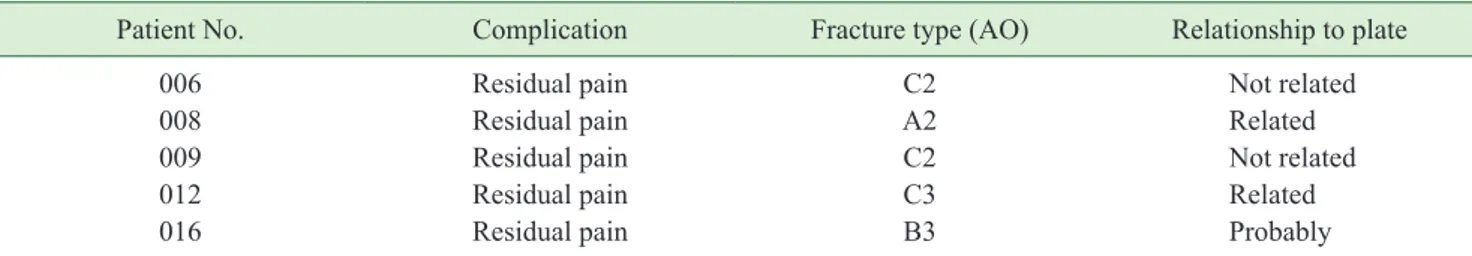
Of the 61 patients treated with a VA-LCP DR Plate, five (8.2%) experienced complications during the course of this study (Table 2). Of these complications, three experienced an implant-related complication. The com- plication reported in three patients was residual pain.
Two of these were related to the plate, and one was likely related to the plate.
4. Outcomes
Of 51 patients with postoperative radiographic data, 46 patients demonstrated union by 3 months, four pa- tients by 6 months, and one patient by 12 months. All 51 patients (100%) achieved union by 12 months. Only
one patient showed a step-off of more than 2 mm at 3 months. But this was resolved on a later follow-up, and there was no evidence of arthritic change during the course of this study. Radiographic parameters evaluated at the immediate postoperative period were 22.4±4.2 de- grees (radial inclination), 4.7±0.4 degrees (volar tilt) and 0.8±0.2 degrees (ulnar variance). Radiographic param- eters evaluated at 1 year after operation were 21.3±3.4 degrees (radial inclination), 4.1±0.4 degrees (volar tilt) and 1.2±0.3 degrees (ulnar variance). There were no significant differences between radiographic parameters evaluated at the immediate postoperative period and 1 year after operation (Table 3). Wrist range of motion in- creased continuously until the end of the study (Fig. 1).
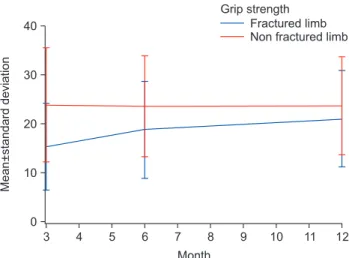
Also, the PRWE and DASH scores during the follow-up period showed continuous improvements (Table 4). The grip strength of the injured limb was compared to that of the non-injured limb at each follow-up visit (Fig. 2). The difference decreased between the 6- and 12-month time points, indicating that the grip strength normalized fol- lowing fracture union.
5. Types of screws and plates
A total of 0-5 variable-angle locking screws were used for fracture fixation in 61 patients, while 2-8 standard locking or cortex screws were placed after fracture reduc- tion (Table 5). Variable-angle screws were not used in
Table 2. Summary of all complications
Patient No. Complication Fracture type (AO) Relationship to plate
006 Residual pain C2 Not related
008 Residual pain A2 Related
009 Residual pain C2 Not related
012 Residual pain C3 Related
016 Residual pain B3 Probably
Table 3. Radiographic outcomes
Radiographic parameter The immediate postoperative period 1 year after operation p-value
Radial inclination (deg) 22.4±4.2 21.3±3.4 0.58
Volar tilt (deg) 4.7±0.4 4.1±0.4 0.26
Ulnar variance (deg) 0.8±0.2 1.2±0.3 0.31
Values are presented as mean±standard deviation.
only two patients. The mean number of screws that were inserted through variable-angle screw guide was 3.2±0.9, and that of screws inserted through fixed-angle screw guide was 3.5±0.9. There was no correlation between fracture type and mean number of variable screws.
The majority of patients were treated with a narrow plate (n=52, 85.3%). The Asian type of plate was used in six patients (n=6, 9.8%) and the standard plate was used in three patients (n=3, 4.9%).
DISCUSSION
Volar plate fixation with fixed-angle locking screws for the treatment of unstable distal radius fractures has shown generally acceptable outcomes in terms of functional and radiographic parameters7-9. The use of variable-angle locking screws has been introduced to allow increased flexibility in plate positioning and subchondral screw placement, which may possibly improve fracture pur- chase and maintain the reduction. Most patients (46/51;
90.2%) achieved union by the 3-month time point, con- sistent with other research10,11. In 98% of the fractures, an articular congruence was achieved at the 12-week
Fig. 2. Grip strength in kilograms of the fractured limb compared to the non-fractured limb at each follow-up visit.
The difference decreased between the 6- and 12-month time points, suggesting recovery of grip strength following fracture union.
Table 4. Summary of the PRWE scores and DASH scores Self-administered patient questionnaire Score
PRWE scores (mo)
3 20.61±8.06
6 14.24±8.84
12 7.56±8.25
DASH scores (mo)
3 16.14±12.21
6 10.16±12.89
12 6.34±9.30
Values are presented as mean±standard deviation.
PRWE: Patient-Rated Wrist Evaluation, DASH: Disabilities of the Arm, Shoulder, and Hand.
Table 5. Mean number and standard deviation of screw used
Screw guide used Fracture type (AO)
A2 A3 B2 B3 C1 C2 C3
Variable angle 3.3±1.6 3.5±0.6 4.1±0.3 4.0±0 3.0±1.4 2.6±1.0 4.0±0.7
Fixed-angle 3.5±1.2 3.0±0 3.0±0 3.0±0 3.5±0.7 4.0±1.1 3.0±0.5
Values are presented as mean±standard deviation.
Fig. 1. The mean wrist range of motion in degrees including flexion/extension, pronation/supination, and radial deviation/
ulnar deviation increased continuously until the end of the study. ROM: range of motion.
follow-up visit. Furthermore, sustained improvements in the range of motion and grip strength were also noted over the 12-month follow-up period. These results are in line with previous work by MacDermid12,13, who demon- strated that patients rapidly achieved most of their range of motion improvement and grip strength by 6 months, although they may continue to improve. At 6 and 12 months after surgery, patients reported low pain scores, good PRWE scores, and high levels of satisfaction. There were no cases of wound infection, tendon injury/irrita- tion, hardware failure, or loss of fracture reduction.
In this study, the mean number of variable screws used was 3.15 in 59 patients and variable-angle screws were not used in only two patients. Stanbury et al.14 reported that the variable-angle fixation exhibited a distinct me- chanical advantage over fixed-angle fixation in the setting of smaller radial styloid fragments. Hart et al.15 concluded that variable-angle screws provided 3 mm of leeway in both sagittal and coronal directions without sacrificing construct strength, which may considerably facilitate the fixation of AO C3 fractures. The recent trend toward volar plating systems with variable-angle screws has the advantage of allowing surgeons to orient screws to avoid crossing articular surfaces and allow for a more precise capture of the fragments. The flexibility of variable-angle screws can allow for fixation of the plate proximal to the watershed line while acquiring the rigid fixation of distal fragments and avoiding penetration of the subchondral bone.
In our study, we inserted screws through variable-angle guide when screws inserted through fixed angle guide
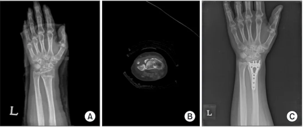
might have a risk to penetrate the articular surface, or screws inserted through variable-angle guide provided better position to stably purchase the distal fragments than screws inserted through fixed angle guide. In intra- articular fractures with sigmoid notch involvement (Fig.
3A, 3B), distal fragment that involved sigmoid notch could be fixed more stably by varying the angle of the ulnar-most screw (Fig. 3C).
Therefore, variable-angle screws have advantages in fixation of comminuted fractures and reconstruction of the complex geometry of the articular surface while avoiding potential tendon irritation and joint penetra- tion16,17.
With the exception of only three patients, the majority of patients were treated with narrow and Asian types of plates (n=58, 95.1%). According to size of plate head, there are three types of plates (Asian type: width 19.5 mm, narrow type: width 22 mm, standard type: width 25.5 mm). The standard type of plate was designed based on anatomical characteristics of Western populations.
Therefore the standard type of plate is larger than other types. Korean people have anatomical characteristics different from Western populations, such as a relatively small and short radius, especially in women18. In this study, 75% of patients were females, and as the size of the standard type of plate was too large for the small ra- dius of Korean women, narrow and Asian types of plates were used in the majority of patients. Thus, we can con- clude with confidence that distal radius fractures occur mostly in older females and therefore a relatively small plate system is useful in Korean populations.
Fig. 3. (A, B) In intra-articular fractures with sigmoid notch involvement, (C) distal fragment that involved sigmoid notch could be fixed more stably by varying the angle of the ulnar-most screw.
This study has several limitations. First, this was not a randomized controlled study and we did not compare our results with patients treated with other plate systems.
Also, because of the difference in surgeon preference and surgical technique, there was a variability in screw selec- tion (variable-angle screws versus traditional screws).
Finally, the outcomes were captured by the treating sur- geons and not independently by another researcher.
CONCLUSION
The findings of this study show promising results after volar plating of unstable distal radius fractures using the VA-LCP DR Plate System.
CONFLICTS OF INTEREST
The authors have nothing to disclose.
ACKNOWLEDGEMENTS
This study was performed by fund of Synthes Korea.
REFERENCES
1. Leung F, Chow SP. A prospective, randomized trial com- paring the limited contact dynamic compression plate with the point contact fixator for forearm fractures. J Bone Joint Surg Am. 2003;85:2343-8.
2. Letsch R, Infanger M, Schmidt J, Kock HJ. Surgical treat- ment of fractures of the distal radius with plates: a com- parison of palmar and dorsal plate position. Arch Orthop Trauma Surg. 2003;123:333-9.
3. Rozental TD, Beredjiklian PK, Bozentka DJ. Functional outcome and complications following two types of dorsal plating for unstable fractures of the distal part of the ra- dius. J Bone Joint Surg Am. 2003;85:1956-60.
4. Rein S, Schikore H, Schneiders W, Amlang M, Zwipp H.
Results of dorsal or volar plate fixation of AO type C3 distal radius fractures: a retrospective study. J Hand Surg Am. 2007;32:954-61.
5. Ring D, Prommersberger K, Jupiter JB. Combined dorsal and volar plate fixation of complex fractures of the distal
part of the radius. J Bone Joint Surg Am. 2004;86:1646- 52.
6. Gummesson C, Atroshi I, Ekdahl C. The disabilities of the arm, shoulder and hand (DASH) outcome questionnaire:
longitudinal construct validity and measuring self-rated health change after surgery. BMC Musculoskelet Disord.
2003;4:11.
7. Chung KC, Watt AJ, Kotsis SV, Margaliot Z, Haase SC, Kim HM. Treatment of unstable distal radial fractures with the volar locking plating system. J Bone Joint Surg Am. 2006;88:2687-94.
8. Osada D, Kamei S, Masuzaki K, Takai M, Kameda M, Ta- mai K. Prospective study of distal radius fractures treated with a volar locking plate system. J Hand Surg Am.
2008;33:691-700.
9. Rozental TD, Blazar PE. Functional outcome and com- plications after volar plating for dorsally displaced, unstable fractures of the distal radius. J Hand Surg Am.
2006;31:359-65.
10. Kwan K, Lau TW, Leung F. Operative treatment of distal radial fractures with locking plate system-a prospective study. Int Orthop. 2011;35:389-94.
11. Phadnis J, Trompeter A, Gallagher K, Bradshaw L, Elliott DS, Newman KJ. Mid-term functional outcome after the internal fixation of distal radius fractures. J Orthop Surg Res. 2012;7:4.
12. MacDermid JC, Richards RS, Roth JH. Distal radius frac- ture: a prospective outcome study of 275 patients. J Hand Ther. 2001;14:154-69.
13. MacDermid JC, Roth JH, Richards RS. Pain and disability reported in the year following a distal radius fracture: a cohort study. BMC Musculoskelet Disord. 2003;4:24.
14. Stanbury SJ, Salo A, Elfar JC. Biomechanical analysis of a volar variable-angle locking plate: the effect of cap- turing a distal radial styloid fragment. J Hand Surg Am.
2012;37:2488-94.
15. Hart A, Collins M, Chhatwal D, Steffen T, Harvey EJ, Martineau PA. Can the use of variable-angle volar lock- ing plates compensate for suboptimal plate positioning in unstable distal radius fractures? A biomechanical study. J Orthop Trauma. 2015;29:e1-6.
16. Fowler JR, Ilyas AM. Prospective evaluation of distal radius fractures treated with variable-angle volar locking
plates. J Hand Surg Am. 2013;38:2198-203.
17. Marlow WJ, Singhal R, Dheerendra S, Ralte P, Fischer J, Waseem M. Distal radius volar locking plates: does a variable angle locking system confer a clinical advantage?
Acta Orthop Belg. 2012;78:309-16.
18. Lim ST, Yeom JS, Lee CH, Lee YH, Chang CB, Baek GH. Development of anatomical plating system for treat- ment of distal radius fractures. J Korean Soc Surg Hand.
2007;12(3):95-104.
가변각 수장측 잠김 금속판을 이용한 원위 요골 골절의 치료 결과
유성림
1ㆍ이호재
2ㆍ한수홍
3ㆍ김재광
41가톨릭대학교 의과대학 정형외과학교실, 2CHA 의과대학 분당차병원 정형외과, 3CHA 의과대학 구미차병원 정형외과,
4울산대학교 의과대학 서울아산병원 정형외과학교실
목적: 본 연구에서는 가변각 수장측 잠김 금속판을 이용하여 원위 요골 골절을 치료한 결과를 분석하여 유효성을 평가하고자 한다.
방법: 2010년부터 2013년까지 원위 요골 골절을 진단받고 관혈적 정복술 및 가변각 수장측 잠김 금속판을 이용한 내고정술을 시행한 61명의 환자를 대상으로 관찰 연구를 시행하였다. 골절의 AO 분류에 따라 방사선적 소견, 금속 판 종류, 가변각 나사 가이드를 이용하여 삽입된 나사의 수, 임상적 결과, 합병증 발생을 분석하였다.
결과: 총 5예(8.2%)에서 합병증이 발생하였으며, 수술 후 3개월, 6개월, 12개월 추시에서 46/51 (90.2%), 50/51 (98.0%), 51/51 (100%)의 방사선적 유합을 보였다. 수술 직후 측정한 요골 경사, 수장측 경사, 척측 변위는 각각 22.4±4.2도, 4.7±0.4도, 0.8±0.2도였으며, 수술 후 1년에 측정한 요골 경사, 수장측 경사, 척측 변위는 각각 21.3±3.4도, 4.1±0.4도, 1.2±0.3도였다. 또한, 추시 기간 동안 관절의 운동 범위와 파악력은 지속적으로 증가하 였고, PRWE 및 DASH 점수는 지속적으로 감소하였다. 가변각 나사 가이드를 이용하여 삽입된 나사의 수는 평균 3.2±0.9였으며, 고정각 나사 가이드를 이용하여 삽입된 나사의 수는 평균 3.5±0.9였다. 사용된 금속판의 종류는 총 52예(85.3%)에서 narrow 금속판을 사용하였다.
결론: 원위 요골 골절의 치료로써 가변각 수장측 잠김 금속판을 이용한 내고정술은 만족할 만한 결과를 보였다.
색인단어: 요골, 요골 골절, 가변각 수장측 잠김 금속판
접수일 2019년 2월 27일 수정일 1차: 2019년 4월 10일, 2차: 2019년 5월 15일 게재확정일 2019년 5월 29일 교신저자 김재광
05505, 서울시 송파구 올림픽로 43길 88, 울산대학교 의과대학 서울아산병원 정형외과학교실 TEL 02-3010-3523 FAX 02-3010-8555 E-mail [email protected]
ORCID https://orcid.org/0000-0001-5104-4634