INTRODUCTION
A ductal adenocarcinoma is the most common tumor devel- oping from the extrahepatic bile duct (1). Benign tumors of the extrahepatic biliary duct are extremely rare, and include adenomas and tumors of the supporting structures such as leiomyomas, lipomas, carcinoids, and fibromas. These tumors are almost indistinguishable from malignant tumors and need to be resected. Schwannomas occurring in the soft tissues of the head and neck, extremities and retroperitoneum can also develop in the extrahepatic biliary duct due to the abundant anastomotic network of sympathetic and parasympathetic nerve fibers and blood vessels from branches of the hepatic and gastroduodenal arteries (2). However, there have been very few case reports in the literature.
We describe the first case of schwannomas arising in the extrahepatic bile duct in Korea, which presented as intra- and extrahepatic bile duct and gallbladder stones.
CASE REPORT
A 64-yr-old female patient was referred for a surgery for intrahepatic duct and gallbladder stones detected on a screen- ing program. She had no previous history of any other major
illness. Her vital signs were as follows: blood pressure, 140/
70 mmHg; pulse rate, 72/min; respiratory rate, 22/min; and body temperature, 36℃. There was no evidence of jaundice or abdominal pain. The laboratory studies revealed: leukocyte count, 8,230/ L; hemoglobin, 15.7 g/dL; total protein, 7.0 g/dL; total albumin, 3.8 g/dL; total bilirubin, 0.6 mg/dL;
direct bilirubin, 0.2 mg/dL; serum asparate aminotransferase, 19 IU/L; serum alanine aminotransferase 14 IU/L; serum lac- tate dehydrogenase 298 IU/L; serum alkaline phosphatase, 57 IU/L; and serum gamma-glutamyl transpeptidase, 51 IU/L.
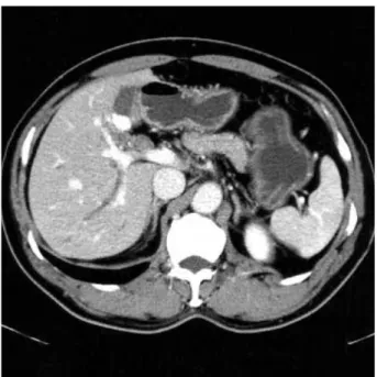
The tumor markers including CEA and CA 19-9 were within normal ranges. The abdominal computed tomography (CT) scan revealed that the intrahepatic bile ducts were slightly dilated and contained stones. The gallbladder was also filled with many small stones. However, the distal part of the com- mon bile duct and pancreas were normal (Fig. 1). Endoscop- ic retrograde cholangiography revealed a threadlike stricture at the proximal part of the common bile duct and a dilata- tion of the upstream intra- and extrahepatic bile ducts with abundant stones. In addition, the gallbladder was filled with small stones, and the cystic duct was inserted aberrantly into the right intrahepatic bile duct (Fig. 2). Surgery was planned because it was believed the intrahepatic duct stones were caused by the stricture. Furthermore, it was difficult to dif- ferentiate between a malignant or benign stricture. The pa-
Ji Heon Jung, Kwang Ro Joo, Myung Jong Chae, Jae Young Jang, Sang Gil Lee, Seok Ho Dong, Hyo Jong Kim, Byung-Ho Kim, Young Woon Chang, Joung Il Lee, Rin Chang, Youn Hwa Kim*, Sang Mock Lee�
Departments of Internal Medicine, Pathology*and Surgery�, Kyung Hee University College of Medicine, Seoul, Korea
Address for correspondence Kwang Ro Joo, M.D.
Digestive Disease Center, Kyunghee East-West Neo Medical Center, 149 Sangil-dong, Gangdong-gu, Seoul 134-090, Korea
Tel : +82.2-440-6111, Fax : +82.2-440-6295 E-mail : [email protected]
549 J Korean Med Sci 2007; 22: 549-52
ISSN 1011-8934
Copyright � The Korean Academy of Medical Sciences
Extrahepatic Biliary Schwannomas : A Case Report
Benign schwannomas arise in neural crest-derived Schwann cells. They can occur almost anywhere in the body, but their most common locations are the central ner- vous system, extremities, neck, mediastinum, and retroperitoneum. Schwannomas occurring in the biliary tract are extremely rare and mostly present with obstructive jaundice. We recently experienced a case of extrahepatic biliary schwannomas in a 64-yr-old female patient who presented with intra- and extrahepatic bile duct and gallbladder stones during a screening program. To the best of our knowledge, ex- trahepatic biliary schwannomas associated with bile duct stones have not been reported previously in the literature.
Key Words : Schwannomas; Bile Duct, Extrahepatic; Calculi, Bile Duct
Received : 14 November 2005 Accepted : 1 February 2006
550 J.H. Jung, K.R. Joo, M.J. Chae, et al.
tient underwent surgery on the bile duct. The pathology findings of the surgical specimens showed several well-demar- cated nodules in the bile ductal wall and an intact mucosal epithelium of the bile duct. The largest one measured approx- imately 1 cm in the largest dimension (Fig. 3A). Microscop- ically, the tumors were mainly composed of spindle-shaped cells arranged in short bundles or interlacing fascicles with nuclear palisading (Fig. 3B, C). Immunohistochemically, the tumor cells were stained strongly for the S-100 protein (Fig.
3D), but were negative for CD 34, CD 117, and actin. The histological diagnosis was benign multinodular schwanno- mas of the extrahepatic bile duct. The postoperative course was uneventful, and the patient has been doing well without any complications.
DISCUSSION
Schwannomas are benign neurogenic tumors that arise in the nerve sheaths of the peripheral nerves in young to mid-
dle-aged adults. They can occur almost anywhere in the body but have a predilection for the head, the neck, and the flex- ure surfaces of the upper and low extremities. They can some- times show secondary degenerative changes such as cyst for- mation, calcification, hemorrhage, and hyalinization (3). Clin- ically, these tumors are generally asymptomatic and are often discovered incidentally.
Schwannomas arising in the digestive tract are quite rare, and occur most commonly in the stomach, followed by the colon and rectum (4, 5). Schwannomas of the biliary tract can also develop because there is an abundant network of sym- pathetic and parasympathetic nerve fibers to the wall of the gallbladder and bile duct. However, schwannomas clinically arising in the biliary tract are extremely rare, and an extrahe- patic bile ductal origin is the rarest. To our knowledge, only five cases of schwannomas involving the extrahepatic biliary duct have been reported (6-10). Clinical characteristics in of these five cases are shown in Table 1. The present case differs from previously reported biliary tract schwannomas in some
Case (Ref) Age (yr) Chief complaint Site Number Characteristics Stones
1 (6) 48 F Obstructive jaundice CBD Solitary 50 mm Dumbbell, cystic GB
2 (7) 40 F Obstructive jaundice HD ligament Solitary Hen’s egg Oval, cystic -
3 (8) 62 F Abnormal liver test HD ligament Solitary 90 mm Fusiform, cystic -
4 (9) 59 F Abdominal pain Remnant CC Solitary 25 mm S/P CJ stomy� CC
5 (10) 37 M Obstructive jaundice EHB Solitary 21 mm Not cystic -
Ours 64 F Incidental finding CBD Multinodular 10 mm Not cystic GB, EHB
Table 1.Clinicopathological features of previously reported biliary schwannomas
*The largest dimension of the mass. �Choledochojejunostomy with Roux-en-Y techniques due to a choledochal cyst 15 yr ago.
F, female; M, male; CBD, common bile duct; HD ligament, hepatoduodenal ligament; CC, choledochal cyst; EHB, extrahepatic bile duct; GB, gallbladder.
Sex Size*
Fig. 1.Computed tomographic scan shows mildly dilated intrahep- atic bile duct and many small stones in the gallbladder.
Fig. 2.Endoscopic retrograde cholangiography shows a focal stric- ture of the proximal common bile duct and an upstream bile duct dilatation with stones. The cystic duct is inserted aberrantly into the right intrahepatic duct. Gallbladder stones are also observed.
Extrahepatic Biliary Schwannomas 551
clinicopathological features. First, it was found incidentally on health screenings while others mainly presented with ob- structive jaundice. Secondly, the present tumor was shown to be associated with intra- and extrahepatic bile duct stones, which were grossly pigmented stones. It seemed that bile stasis resulting from mechanical obstruction was an impor- tant antecedent to the development of pigment gallstones (11). Thirdly, our case manifested uniquely as a plexiform or multinodular growth similar to a plexiform neurofibroma.
Histologically, schwannomas originating in the digestive tract show distinct histological features that distinguish them
from conventional schwannomas. These schwannomas are S-100 protein-positive spindle cell tumors that are composed mainly of cellular (Antoni A) areas, and generally do not show a nuclear palisading pattern that is usually found in conven- tional schwannomas of the soft tissues (6, 12), Moreover, sch- wannomas of the digestive tract were recently reported to lack neurofibromatosis-2 genetic alterations, which support the theory that schwannomas of the digestive tract are unique tumors distinct from conventional schwannomas (13). In our case, the tumors showed a dominant Antoni A area, but had a nuclear palisading pattern. More cases are needed to deter-
Fig. 3.(A) The specimen shows several well-demarcated nodules in the bile duct wall. (B) A nodule shows a typical pattern of a schwan- noma consisting of spindle cells. Lymphocyte cuffing is also seen in the margin of the tumor (H&E ×100). (C) Magnified view shows inter- lacing fascicles with nuclear palisading (H&E ×200). (D) Immunochemical staining is positive for the S-100 protein (×100).
A B
C D
552 J.H. Jung, K.R. Joo, M.J. Chae, et al.
mine the pathological correlation between a biliary schwan- noma and other digestive schwannoma.
Schwannomas usually appear in CT as a homogenous mass with heterogeneous contrast enhancement and often show secondary degeneration such as a cystic change, cavity forma- tion, necrosis, or calcification. However, secondary degener- ation rarely occurs in schwannomas of the digestive tract (14).
In our case, the tumors were too small to detect these radi- ology findings. Magnetic resonance imaging (MRI) might also be useful to establish the nature of the tumor. The MRI findings of schwannomas are mainly masses with low signal intensity on the T1-weighted images and inhomogeneous high signal intensity on the T2-weighted images (15).
Schwannomas of the digestone tract have an excellent prog- nosis after a surgical resection, similar to conventional schwan- nomas, and there is no evidence to date that these tumors have a malignant potential.
In conclusion, biliary schwannomas causing a biliary stric- ture are extremely rare and a preoperative diagnosis is quite difficult. However, these potentially curable tumors should be included in the differential diagnosis of tumors arising in the biliary system.
REFERENCES
1. Malka D, Boige V, Dromain C, Debaere T, Pocard M, Ducreux M.
Biliary tract neoplasm: update 2003. Curr Opin Oncol 2004; 16:
364-71.
2. Northover JM, Terblanche J. A new look at the arterial supply of the bile duct in man and its surgical implications. Br J Surg 1979; 66:
379-84.
3. Enzinger FM, Weiss SW. Soft tissue tumors. 1nd ed. St Louis, Mo:
Mosby-Year Book 1983: 586-97.
4. Sarlomo-Rikala M. Miettinen M. Gastric schwannoma: a clinico- pathological analysis of six cases. Histopathology 1995; 27: 355-60.
5. Miettinen M, Shekitka KM, Sobin LH. Schwannomas in the colon and rectum: a clinicopathologic and immunohistochemical study of 20 cases. Am J Surg Pathol 2001; 25: 846-55.
6. Honjo Y, Kobayashi Y, Nakamura T, Kakehira Y, Kitagqwa M, Ike- matsu Y, Ozawa T, Nakamura H. Extrahepatic biliary schwannoma.
Dig Dis Sci 2003; 48: 2221-6.
7. Oden B. Neurinoma of the common bile duct: report of a case. Acta Chir Scand 1955; 108: 393-7.
8. Nagafuchi Y, Mitsuo H, Takeda S, Ohsato K, Tsuneyoshi M, Enjoji M. Benign schwannoma in the hepatoduodenal ligament: report of a case. Surg Today 1993; 23: 68-72.
9. Otani T, Shioiri T, Mishima H, Ishihara A, Maeshiro T, Matsuo A, Umekita N, Warabi M. Bile duct schwannoma developed in the rem- nant choledochal cyst-a case associated with total agenesis of the dorsal pancreas. Dig Liver Dis 2005; 37: 705-8.
10. Jakobs R, Albert J, Schilling D, Nuesse T, Riemann JF. Schwanno- ma of the common bile duct: a rare cause of obstructive jaundice.
Endoscopy 2003; 35: 695-7.
11. Shiesh SC, Chen CY, Lin XZ, Liu ZA, Tsao HC. Melatonin prevents pigment gallstone formation induced by bile duct ligation in guinea pigs. Hepatology 2000; 32: 455-60.
12. Daimaru Y, Kido H, Hashimoto H, Enjoji M. Benign schwannoma of the gastrointestinal tract: a clinicopathologic and immunohisto- chemical study. Hum Pathol 1988; 19: 257-64.
13. Lasota J, Wasag B, Dansonka-Mieszkowska A, Karcz D, Millward CL, Rys J, Stachura J, Sobin LH, Miettinen M. Evaluation of NF2 and NF1 tumor suppressor genes in distinctive gastrointestinal nerve sheath tumors traditionally diagnosed as benign schwannomas: study of 20 cases. Lab Invest 2003; 83: 1361-71.
14. Levy AD, Quiles AM, Miettinen M, Sobin LH. Gastrointestinal schwannoma: CT features with clinicopathological correlation. Am J Roentgenol 2005; 184: 797-802.
15. Rha SE, Byun JY, Jung SE, Chun HJ, Lee HG, Lee JM. Neurogenic tumors in the abdomen: tumor types and imaging characteristics.
Radiographics 2003; 23: 29-43.