대한엄상벙리사회지 : 제 16권 제 1 호 1984
례
흐 ‘ @
폐 진균증 1 예에 대한 세포소견
국럽 의료원
박선자·박노원·박태원·박효숙
Cytologic Findings of the Pulmonary Mycosis -1 case rêport-
N.찌T. Park, S.J. Park, T.W. Park, H.S. Park
Dept. ofPathology National Medical Ceη:ter
= Abstract=
The incidence of pulmonary mycotic infection is increasing these days, and it can be frequen- tly over-diagnosed as very wel1 differentiated squamous cel1 carcinoma on sputum cytology due to its simi1ar morphology.
Althouh each cell in pulmonary mycosis shows marked atypism or metaplastic changes, but we must considered the whole spectrums for differential diagnosis, ie. true tumor diathesis, volume of atypical cells, or chromatin pattern of the cell etc.
Herein we report a case of pulmonary mycosis overdiagnosed as highly suspicious squamous cel1 carcinoma on sputum cytology.
1.
서 론진균에 의한 폐철환은 근간 면역 방사선학적 치료증 례가 증가 함에 따라 임상적으로 중요한 문제를 제시 하게 되였음은 주지의 사살안 바션) Pap. 염색에 의한 객담 세포진으로서 진균을 진단하고 더 나아가 특수염 색, 생검, 면역항체검사 그러고 진균배양동청으로서 확경진단에 도달하게 되는 것이다.
객당의 Pap. 염색도말표본에서 발견 되여치는 주요
병원성 진균은 기회병원균인 Candida species, Asperg- illus spp, Mucomyces 퉁이 며 Cryptococcus neoform- ans, Cocidiodes immitis, Histoplasma capsulatum 그
리 고 Blastomyces dermatidis 등도 해 로는 발견할 수
있읍니 마. 1,6,8)
객 담도말상에 서 이 들 진균들을 발견함으로서 진균의 진단의 열쇠를 잡을 수 있다고 하겠으나 문제는 어떤 경 우의 solitary mycetoma에 서 는 펀평 상펴 암과 제 포학 척으로 너무냐 유사한 양상을 갖고있는 상펴세표가 출 현하기 써문에 암으로 위양성 보고룰 하게 되어 본의 아난 피돼릎 줄 수 있는 것이다. 이는 검도성 부위 (So
litary lesion)에 연하는 장피가 찰 분화된 훤형상피이
고 그것 의 아주 극심 한 비 정 형 상태 (atypia)를 동반한 것이기 해문이다. ',5)
그러하여 저자들은 세포학적으로 편펑상펴암과 혼돈 하였던 진균증 1 예를 말견하고 이것을 세포학적으로 분석하여 펀평상펴암과 비교한 결과를 보고하는 바이 다.
II.
증 례임상 및 X 선소견
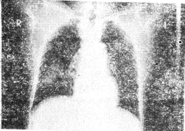
49셰의 남자로서 내원 2개월전부터 열이냐 오한없이 겸한 기침파 더불어 소량의 혈액이 객담에나왔으며 투 약을 하였£나 아무련 효파가 없었고 체중감소, 호흡 곤란 그러고 소화불량등 제증상은 또한 없었다. chest
P.A. 상에서 우측폐 하엽 부근에 경계가 비교적 뚜렷한
abnormal density가 보여 (사진 1) Bronchography를 하 였더니 하엽기관지에 폐쇄상이 관찰되고 기관지경이 환부까지 마치지 뭇하여 별다른 치관지경상 소견이 없 었 다. C.T상에 서 전문의 는 폐 외 측부의 mass와 cavity
카 있음을 보고 만성염증£로 언한 것이라고 주진단을 하였£역 펴]암을 RjO 하지는 뭇하였다(사진 2).
f’ig. 1. Chest P.A.; Showing compact homogeneous with well outlined density in R.L.L.F.
세포학척 소견
객담세포검사블 3 일 간격으로 3회 검사를 시행하였 던 바, 첫벤채의 검사에서 암세포로 생각되는 비청상 세포의 다수 출현으로 찰 분화펀 평펀상피암이라고 캉 하케 의섬하였고 나머지 2회의 검사에서는 바정상세포 의 출현없이 효모양진균이 다수로 보였다. (사진 3)
첫먼째 검사에서 나타난 세포들은 각화현상이 뚜렷 하며 농염된 핵사이로 지저분한 괴사성 물질이 보여 종양때 나나타는 백경을 연상하케 하였고, 핵파 원형 절의 심한 다행태성을 보이고(사진 4) 유첨 렌즈 상에 서 염색체는 그 분포가 블규척하며 핵막쪽으로 이몽하 여 서로 다른 두꺼l 와 오양이며 암세포와 비슷한 핵내 청정현상 (nuc1eus clearing)이 있다(사진 5,6).
Fig. 3. The fungus is seen as abudding yeast in sp.
utum(450X)
Fig. 4. Note the pleomorphism, the relatively Iow NjC ratio of the keratinized celIs, the dege.
Fig. 2. ~C.T. lung scan; Lung mass and cavity in nerate india ink nuclei and orangiohpilia of
RLL. the cytoplasm. (450X)
- 107-
Fig. 5. A heavy deposit of chromatin below the nu- clear membrane is associated \vith indenta- tions of the nuc1ear margin. (1,OOOX)
Fig. 7. Bizarre r:ucleus \vith loss of chromatin patt- ern has keratinizing cytoplasm. (450X)
합포체 (syncytium) 속에 비 정 상세 포틀의 대 소부동증 과 염색체쉬 이상이 보이고 농염된 bizzare한 핵괴- 원 형칠。1 관찰된다(사진 7). 각화형 펀평상피암의 세포 소견에서 볼 수 있는 상당히 큰 장방형 세포핵들이 농 염되여 있고 원형질의 심한 각화현상괴- 침상을 보인다 (사진 8).
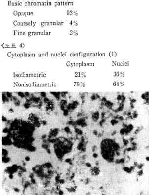
약 120여 캐 의 비 정 상세 포들중에 거 의 가 eosinophilic 하거나 orangiophilic한 염색성파 흩어진 세포배열을 보。l 고(도표 1,2) 농염, 붙투명한 핵들이 다수인 것에
비하여 굵거나 미세한 염섹체를 볼 수 있는 투명한 핵 듣응 소수에 불과했다(도표 3). 세포나 핵의 요양들은 펀펑상펴암의 세포상에 비하여 첨상형의 것들이 증가 한 형태와 함께 심한 다형태성 (pleomorphism) 을 보였
Fig. 6. A detai1ed study of the nuc1eus reveals coa- rse c1umpiJ1g of the chromatin, irregular di-
stributioη aπd nuclear c1eariηg. (1,OOOX)
Fig. 8. Fibre-like cells. The opaque, structurelessc spindle-shaped nuc1eus fills the entire width of the cel1. (40X)
다(도표 4).
〈도표 1)
Cytologic analysis of C3206j84
Number of the abnormal cell in this case 119
Nucleoli 7%
Back ground; moderately necrotic
Arrangement; Syncytia 1 % Sheet 0% Isolated 99%,、
〈도표 2)
Cytoplasmic staining reaction
(%
of 119 cells)Indeterminate 4 %
Cyanophilic 6%
Eosinophilic & Orangiophilic 90%
〈도표 3)
Basic chromatin pattern
Opaque 93%
Coarsely granular 4%
Fine granular 3 %
〈도표 4)
Cytoplasm and nuclei configuration (1) Cytoplasm Nuclei
Isodiametric 21 % 36%
Nonisodiametric 79% 64:%
Fig. 9. The right loer lobe shows grayish yel10w consolidated area with demarcation.
조직학적 소견
육얀적으로 우폐하엽의 외측부에 직경이 약 4, 5c111의 r.02ular단 연회 색 내 지 는 황색 괄 띄 운 solid 하띤서 7;3 처가 마교적 뚜렷한 부위에 부분적안 괴사와 검은 aD.- thraco~ic pigment 가 았었고 주거판지와의 떤결은 얹였
다(斗깐 g). E깐마 경 상우로는 granuloma를 동란단 댐 층 휴씌 _.~으 C냥 곰팡이가 다수 출현되여 '::;j 진균등;임녁눈 착 인다눴다子우}의 기관지는 염증과 확장이 있였고 ly- ffilJh 갚 01] 비정렌적 염중소 7단이 ;간찰 되였다(사진 10).
Fig. 10. The macrophges include many 5 micron di- ametered spores of candida with thick cap- sule. (P AS. 450X)
써1 균학적 소견
조직검사가 폐잔균증£로 잔단됨에 짜라 수술후 。1 건 따냐 진균과 돼 양등정 갈 추천 하였 던 바, B. P상어I 서 흘껴잔 균집악을 행성하녔고 Sabroud agar 상에서 균사블 형 성 하고 urea 음성 , lactose 음성 , gluc05e, sucrose maltose 등이 분폐 상을 보여 Candida species 임 을 직 감하고 API 20 Ca'1dida System 을 이 용하여 동 청한 결과 Candida tropicalis 로 진단하였다.
m.
고 찰본 혀1 의 solitary lesion 에 연하는 분화된 펀펑상피의 atypia는 괴사성만응 때문안지는 모르나 조직에서 판 찰하지는 못했다.
세포학적 측면에서 위의 세포소견을 토대로 각화형 펀평상펴암세표 소견의 통계와2, 5,긴 9) 본예블 비교하연 써1 포의 크기, 핵크기 그러고 핵과 세포질의 비율이 암 세포보다도 작고 낮았고 주로 타원행, 침상형。l 였오며,
함포체배 열 (syncytium arrangment) 은 거의 없는 훤。l 었다(도표 5). 염색체들은 눈£로 핵의 내용을 폴 수 없는 불투명하고 균칠적인 것플이 농염되여 돼부분을 차지하고 상대적오로 과련헝염색체 나 미세한 염색체 튼은 암세포와 바교함 해 매우 적은 펀이었다.
〈도표 5)
Comparative cytologic features (1)
l\1ycetoma Sq. Ca. K. type Cel1 area Smaller than Sq. Ca. 275
:t
l07 nuclear are1 Smaller than Sq. Ca. 77土 28relative nucleClr Less than Sq. Ca.
area Caudate
&
e10-ngate cell Syncytial Coarse granular
chromatin Opaque Finely granul1r
chromatin
26%
1%
4ε/ 10
93%
3 끽a
Back ground necr05is w i th fungi
29• 610
X5%
13%
59%
19%
22%
nεc 1'051s "\VI t- hout fungi 용여]에서 건띔잔 바와 같이 세포 착냐 하나과 모양 만을 중시할 것이 아니라 안_,:-jìÆ상에 닌1 딩} 갈투명험을 가진 세포가 양다먼지 서}포염석상이 어느 특경 란 곳~
로 기윷던지 또는 굵거나 당처진 엮색채릅 가잔 핵플
- 109-
2-) 수척증감등 천체적안 세포상의 흐릎도헤 B) 중을 두 어 신중허 캠토 하여야 겠다. 또한 세포학적오로 암。l 적삼펀다 할지라도 진균의 출현은 임진단의 채고롤 요 하는데 아우리 종종 나타나는 기회병원성 곰팡이라 할 지라도 정곱허 요염균으로 간주하는 것은 상한진단 (over diagnosis)으로 오진하게 되는 주요 요인이다.
어쩨든 폐진균증이 각화형 펀형상파암으로 오진할수 있는 함정임을6) 상기하고 신중히 검토 되여야 겠음을 본여l 를 통한 경험으로 보고하는 바이다.
N.
결 론1. 본예에서 보여진 형태학적연 세포상은 세포개별 적£로 볼때 찰 분화된 펀평상파암파 매우 유사하였다.
2. 천체적언 세포양상A로 보아 펀펑상펴암에 비해 a, 본예가 세포크기, 핵크기가 다소 작았으며 침상형 태의 핵 및 써I) Æ_질이 많았오며, b, 본예가 과럽형 염색 체를 가진 세폭가 현저히 캄소했고 opaque한 핵을 가 진 세포틀이 월등히 많다. c, 본예의 세포배경은 단순 한 괴사흔적이였 A며 진균이 보였다.
References
1. Compendium on Diagnostic Cytology. TutoriaJ procedings of the inetrnational Academy of Cyt-
이ogy, Chicago, ed. 5, pp.282"'292, 1983.
2. Foot N.C. The ide ntification of types of pulm- onary cancer in cytologic smear. Amer. J. Path‘
28 : 963
,
1952.3. Gompel1, Atlas of diagnostic cytology. John Wi- ley & sons 144"'146
,
1978.4. Johnston W.W.: The cytopathoogy of mycotic infections. Lab. Med. 2: 34"'40
,
1971.5. Koss L.G. Diagnostic cytology. Vo1. II
,
ed. 3,
pp.580"'584
,
1979.6. Koss L.G., Richardson H.L. Some pitfal1s of cy- tological diagnosis of lung cancer. 8: 937
,
1955.7. Lange E., and Hoeg, K.: Cytologic typing of lung cancer. Acta Cyto1., 16: 327"'330, 1972.
8. Naib Z.M. Exfoliative cytology in fungus disease- of the lung. Acta Cyto1. 6 : 413
,
1962.9. Patten S.F., Diagnostic cytology of uterine cer- vix. ed, 1, pp. 189, 1969.