제 12 장 기형학
설현주
기형유발물질의 확인
① The defect must be completely characterized.
② The agent must cross the placenta.
③ Exposure must occur during a critical developmental period.
④ There must be a biologically plausible association.
⑤ Epidemiological findings must be consistent.
⑥ The suspected teratogen causes a defect in an
animal.
Criteria for proof of human teratogenicity
① Careful delineation of clinical cases
② Rare environmental exposure associated with rare defect, with at least three reported cases—easiest if defect is severe
③ Proof that the agent acts on the embryo or fetus, directly or indirectly
④ Proven exposure to agent at critical time(s) in prenatal development
⑤ The association must be biologically plausible
⑥ Consistent findings by two or more epidemiological studies of high quality:
(a) Control of confounding factors (b) Sufficient numbers
(c) Exclusion of positive and negative bias factors (d) Prospective studies, if possible
(e) Relative risk of three or more
⑦ Teratogenicity in experimental animals, especially primates
Exposure during a critical developmental period.
① Preimplantation period Fertilization ~ 2 weeks “all or none period”
② Embryonic period.
GA 4 ~ 10 weeks, organogenesis “critical developmental period”
③ Fetal period.
GA 10 weeks ~
ex, brain
Table 14-3. Food and Drug Administration Categories for Drugs and Medications
Category A: Studies in pregnant women have not shown an increased risk for fetal abnormalities if administered during the first (second, third, or all) trimester(s) of pregnancy, and the possibility of fetal harm appears remote.
Fewer than 1 percent of all medications are in this category. Examples include levothyroxine, potassium supplementation, and prenatal vitamins, when taken at recommended doses.
Category B: Animal reproduction studies have been performed and have revealed no evidence of impaired fertility or harm to the fetus. Prescribing information should specify kind of animal and how dose compares with human dose.
or
Animal studies have shown an adverse effect, but adequate and well-controlled studies in pregnant women have failed to demonstrate a risk to the fetus during the first trimester of pregnancy, and there is no evidence of a risk in later trimesters.
Examples include many antibiotics, such as penicillins, macrolides, and most cephalosporins.
Category C: Animal reproduction studies have shown that this medication is teratogenic (or embryocidal or has other adverse effect), and there are no adequate and well-controlled studies in pregnant women. Prescribing information should specify kind of animal and how dose compares with human dose.
or
There are no animal reproduction studies and no adequate and well-controlled studies in humans.
Approximately two thirds of all medications are in this category. It contains medications commonly used to treat potentially life-threatening medical conditions, such as albuterol for asthma, zidovudine and lamivudine for human immunodeficiency viral infection, and many antihypertensives, including -blockers and calcium-channel blockers.
Category D: This medication can cause fetal harm when administered to a pregnant woman. If this drug is used during pregnancy or if a woman becomes pregnant while taking this medication, she should be apprised of the potential hazard to the fetus.
This category also contains medications used to treat potentially life-threatening medical conditions, for example:
systemic corticosteroids, azathioprine, phenytoin, carbamazepine, valproic acid, and lithium.
Category X: This medication is contraindicated in women who are or may become pregnant. It may cause fetal harm. If this drug is used during pregnancy or if a woman becomes pregnant while taking this medication, she should be apprised of the potential hazard to the fetus.
There are a few medications in this category that have never been shown to cause fetal harm but should be avoided nonetheless such as the rubella vaccine.
Counseling for teratogen exposure
women given negative information —such as a 1- to 3- percent chance of having a malformed newborn—are
more likely to perceive an exaggerated risk than women given positive information, that is, the 97- to 99-
percent chance of having a child without a malformation.
With a few notable exceptions, most commonly prescribed drugs and medications can be used with relative safety during pregnancy.
For the few drugs believed to be teratogenic, counseling should emphasize relative risk.
The concept of risk versus benefit also should be
introduced.
약물과 태아기형
Alcohol-fetal alcohol syndrome
Fetal Alcohol Syndrome and Alcohol-Related Birth Defects
Fetal Alcohol Syndrome Diagnostic Criteria—all required I. Dysmorphic facial features
a. Small palpebral fissures b. Thin vermilion border c. Smooth philtrum
II. Prenatal and/or postnatal growth impairment III. Central nervous system abnormalities
a. Structural: Head size < 10th percentile, significant brain abnormality on imaging b. Neurological
c. Functional: Global cognitive or intellectual deficits, functional deficits in at least three domains
Alcohol-Related Birth Defects
I. Cardiac: atrial or ventricular septal defect, aberrant great vessels, conotruncal heart defects II. Skeletal: radioulnar synostosis, vertebral segmentation defects, joint contractures, scoliosis III. Renal: aplastic or hypoplastic kidneys, dysplastic kidneys, horseshoe kidney, ureteral
duplication
IV. Eyes: strabismus, ptosis, retinal vascular abnormalities, optic nerve hypoplasia V. Ears: conductive or neurosensory hearing loss
VI. Minor: hypoplastic nails, clinodactyly, pectus carinatum or excavatum, camptodactyly,
"hockey stick" palmar creases, refractive errors, "railroad track" ears
약물과 태아기형
Anticonvulsant-fetal hydantoin syndrome
약물과 태아기형
Vitamin A
임신 초기 과량 섭취시 brith defect 와 연관
하루 5000 IU 이하로 섭취할 것을 권장
약물과 태아기형
Vitamin A –Isotretinoin embryopathy
약물과 태아기형
Warfarin embryopathy
임신 중 영상진단
① 이온화 방사선 (ionizing radiation):
마이크로파 (microwaves), 초음파 , 전기투열 (diathermy), 라디오파
X- 선 , 감마선
② 자기공명 단층촬영 (magnetic resonance imaging)
③ 초음파검사 (fetal ultrasonography)
방사선량 측정
① Type of study
② Type and age of equipment
③ Distance of target organ from radiation source
④ Thickness of the body part penetrated
⑤ Method or technique used for the study.
각 신체부위와 검사 에 따른 태아방사선 피폭량
각 신체부위와 검사 에 따른 태아방사선 피폭량
각 신체부위와 검사 에 따른 태아방사선 피폭량
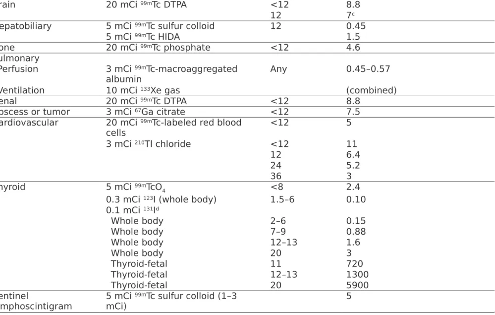
aTo convert to mrad multiply x 100.
bExposures are generally greater prior to 12 weeks compared with increasing gestational ages.
cSome measurements account for placental transfer.
dThe uptake and exposure of 131 I increases with gestational age.
DPTA = diethylenetriaminepentaacetic acid; Ga = gallium; HIDA = hepatobiliary iminodiacetic acid; I = iodine; mCi = millicurie; mSv = millisievert;
Tc = technetium; TcO4 = pertechnetate; Tl = thallium.
Table 41-7. Radiopharmaceuticals Used in Nuclear Medicine Studies Study Estimated Activity Administered per
Examination in Millicuries (mCi) Weeks'
Gestationa Dose to Uterus/Embryo per Pharmaceutical (mSv)b
Brain 20 mCi 99mTc DTPA <12 8.8
12 7c
Hepatobiliary 5 mCi 99mTc sulfur colloid 12 0.45
5 mCi 99mTc HIDA 1.5
Bone 20 mCi 99mTc phosphate <12 4.6
Pulmonary
Perfusion 3 mCi 99mTc-macroaggregated
albumin Any 0.45–0.57
Ventilation 10 mCi 133Xe gas (combined)
Renal 20 mCi 99mTc DTPA <12 8.8
Abscess or tumor 3 mCi 67Ga citrate <12 7.5
Cardiovascular 20 mCi 99mTc-labeled red blood
cells <12 5
3 mCi 210Tl chloride <12 11
12 6.4
24 5.2
36 3
Thyroid 5 mCi 99mTcO4 <8 2.4
0.3 mCi 123I (whole body) 1.5–6 0.10 0.1 mCi 131Id
Whole body 2–6 0.15
Whole body 7–9 0.88
Whole body 12–13 1.6
Whole body 20 3
Thyroid-fetal 11 720
Thyroid-fetal 12–13 1300
Thyroid-fetal 20 5900
Sentinel
lymphoscintigram 5 mCi 99mTc sulfur colloid (1–3
mCi) 5
자기공명 단층촬영 (MRI)
Maternal indication Fetal indication
임신 중 조영제는 꼭 필요한 경우가 아니면 사용하지 않을 것을
권장 .
초음파
고온 (hyperthermia) – thermal index 동공 (cavitation) – mechanical index
진단 목적의 저강도범위의 그레이 스케일의 실시간 초음파 영상
은 태아위험이 없다 .
임신 중 영상진단의 가이드라인