저작자표시-비영리-변경금지 2.0 대한민국 이용자는 아래의 조건을 따르는 경우에 한하여 자유롭게 l 이 저작물을 복제, 배포, 전송, 전시, 공연 및 방송할 수 있습니다. 다음과 같은 조건을 따라야 합니다: l 귀하는, 이 저작물의 재이용이나 배포의 경우, 이 저작물에 적용된 이용허락조건 을 명확하게 나타내어야 합니다. l 저작권자로부터 별도의 허가를 받으면 이러한 조건들은 적용되지 않습니다. 저작권법에 따른 이용자의 권리는 위의 내용에 의하여 영향을 받지 않습니다. 이것은 이용허락규약(Legal Code)을 이해하기 쉽게 요약한 것입니다. Disclaimer 저작자표시. 귀하는 원저작자를 표시하여야 합니다. 비영리. 귀하는 이 저작물을 영리 목적으로 이용할 수 없습니다. 변경금지. 귀하는 이 저작물을 개작, 변형 또는 가공할 수 없습니다.
Regenerative Effect of Recombinant Human Bone
Morphogenetic Protein-2/Absorbable Collagen
Sponge(rhBMP-2/ACS)
after Sequestrectomy of Medication-Related
Osteonecrosis of the Jaw(MRONJ)
by
Song Hee Min
Major in Oral and Maxillofacial Surgery
Department of Clinical Dentistry
Graduate School of Clinical Dentistry
Ajou University
Regenerative Effect of Recombinant Human Bone
Morphogenetic Protein-2/Absorbable Collagen
Sponge(rhBMP-2/ACS)
after Sequestrectomy of Medication-Related
Osteonecrosis of the Jaw(MRONJ)
by
Song Hee Min
A Dissertation Submitted to Graduate School of Clinical Dentistry,
Ajou University in Partial Fulfillment of the Requirements for the
Degree of Master of Science in Dentistry
Supervised by
Jeong-Keun Lee, DDS, Ph.D.
Major in Oral and Maxillofacial Surgery
Department of Clinical Dentistry
Graduate School of Clinical Dentistry
Ajou University
This certifies that the dissertation
of
Song Hee Min
is approved.
SUPERVISORY COMMITTEE
Lee Jeong Keun
Song Seung Il
Ji Suk
Graduate School of Clinical Dentistry
Ajou University
i – ABSTRACT –
Regenerative Effect of Recombinant Human Bone Morphogenetic
Protein-2/Absorbable Collagen Sponge(rhBMP-2/ACS)
after Sequestrectomy of Medication-Related Osteonecrosis
of the Jaw(MRONJ)
Objectives : Beyond the original application approved by the U.S. Food and Drug
Administration,recombinant human bone morphogenetic protein-2(rhBMP-2) is known to
be used for medication-related osteonecrosis of the jaw(MRONJ) treatment by having a bone remodeling enhancement. The purpose of the study was to investigate the bone formation effect of rhBMP-2/ACS in patients with MRONJ.
Materials and Methods: In this retrospective study, 26 female patients diagnosed as MRONJ and underwent mandibular sequestrectomy at Ajou University Dental Hospital from 2010 to 2018 were included. Experimental group was comprised of 18 patients who were received rhBMP-2/ACS after sequestrectomy, and the control group was comprised of 8 patients who were omitted this procedure after sequestrectomy. Total dose of 0.5mg of rhBMP was used in experimental group in the concentration of 0.5mg/ml. Follow up panoramic X-rays were taken immediately after the surgery, and more than 6 months after the surgery. Using those X-rays, radiographic index of bone defect area was calculated using modified Ihan Hren’s method which measuring the radiographic density of the normal bone and the defect site. Paired t-test test was used for analyzed effectiveness in
ii
each group, and Independent t-test was used for analyzed effectiveness between the experimental group and the control group.
Results: Average radiographic index in immediately after surgery and more than 6 months after the surgery for experimental group was 68.4% and 79.8%, respectively(Paired t-test, P<0.05). Average radiographic index in immediately after surgery and more than 6 months after the surgery for control group was 73.4% and 76.7%, respectively (Paired t-test, P>0.05). Average radiographic index increase of 11.4% in the experimental group and an average increase of 3.27% in the control group, respectively (Independent t-test, P>0.05) . Conclusion: According to the result, it could be a successful strategy in treatment of MRONJ patient using rhBMP-2/ACS on bone defect site after sequestrectomy.
Key words: MRONJ, rhBMP-2, BRONJ, Sequestrectomy
iii
TABLE OF CONTENTS
ABSTRACT ··· i
TABLE OF CONTENTS ··· iii
LIST OF FIGURES ··· iv
LIST OF TABLES ··· v
Ⅰ. INTRODUCTION ··· 1
Ⅱ. MATERIALS AND METHODS ··· 4
A. Study sample ··· 4
B. Surgical Procedure ··· 5
B. Bone Density Analysis ··· 7
C. Statistical analysis ··· 9 Ⅲ. RESULTS ··· 10 Ⅳ. DISCUSSION ··· 14 Ⅴ. CONCLUSION··· 16 REFERENCES ··· 17 국문요약 ··· 20
iv
LIST OF FIGURES
Fig. 1. Surgical procedure ··· 6
v
LIST OF TABLES
Table 1. Patient information of Experimental group ··· 11
Table 2. Patient information of Control group ··· 12
Table 3. Radiographic Index ··· 13
- 1 -
Ⅰ. INTRODUCTION
Osteonecrosis of the jaw (ONJ) has been a rare form of diseases until the advent of anti-osteoporotic drugs made it common form of osteonecrosis of the jaw nowadays, medication-related osteonecrosis of the jaw (MRONJ). It is the most common form of any osteonecrosis of the jaws, showing 0.04% of its incidence in Korea(Lee et al. 2013). Moreover, the incidence is much higher in patients undergoing surgical dental treatments, which makes it a significant concern of most dentists.
Diagnostic criteria for MRONJ is as follows: Current or previous treatment with antiresorptive or antiangiogenic agents, exposed bone or bone that can be probed through an intraoral or extra-oral fistula in the maxillofacial region that has persisted for more than eight weeks, no history of radiation therapy to the jaws or obvious metastatic disease to the jaws(Ruggiero et al. 2014).
There was no standard approach for treating MRONJ in the literature. Some authors have recommended conservative approach to MRONJ(Gómez et al. 2008; Ruggiero et al. 2009). Gómez et al., recommended long-term antibiotic treatment and chlorhexidine rinses 3-4 times a day in the MRONJ updateexcluding aggressive surgical treatment(Gómez et al. 2008). Ruggiero et al. also suggest oral antimicrobial rinses combined with antibiotic therapy on stage 1, 2 patients(Ruggiero et al. 2009). In addition, the positional paper of American Association of Oral and Maxillofacial Surgeons suggested that the surgical procedure be limited to patients with stage 3. However, most MRONJ patients show
- 2 -
infection-related symptoms, which do not respond well to conservative treatment. There are some studies reporting good results with surgical management of early stages of MRONJ. Vescovi et al. reported good healing of conservative surgical treatment for stage 1 BRONJ patients(Vescovi et al. 2014). Surgical approach could be a good solution in patients not responding to conservative therapy.
Recent studies have focus on using bone morphogenetic protein 2 (BMP-2) for MRONJ treatment.(Cicciù et al. 2012; Bagan et al. 2009; Park et al. 2017) Originally, FDA approved rhBMP-2 as a bone graft material for synthetic bone used for maxillary sinus augmentation, and socket preservation using its osteoinductive property(de Freitas et al. 2015). But several previous studies have demonstrated that rhBMP-2 works in situations beyond the above applications (Carter et al. 2008; Hwang et al. 2016; Chenard et al. 2012).
For optimal effect, rhBMP-2 should be bound to the appropriate scaffolds to stabilize the release of rhBMP-2 into the lesion. These scaffolds include HA (Hydroxyapatite), TCP (Tricalcium Phosphate), DBM (Demineralized Bone Matrix), and Absorbable Collagen Sponge (ACS). Li and Wozney reported that the releasing period of the rhBMP-2 was more than twice when ACS was applied than when there were no scaffold, and ACS is a suitable scaffold for rhBMP-2 application(Li and Wozney 2001; Geiger et al. 2003)
Major pharmacological effect of bisphosphonates is postulated to be the inhibition of osteoclastic activity considering this with the hypothesis that the pathogenesis of MRONJ is oversuppression of bone remodeling(Russell et al. 2008), BMP can be expected to promote the healing of the bone in MRONJ patients by having a bone remodeling
- 3 - enhancement.
The purpose of this study was to investigate the regenerative effect of rhBMP-2/ACS in patients with MRONJ. The authors expected that rhBMP-2 could promote bone healing in MRONJ, and the study aimed an quantitative evaluation of regenerative effect of rhBMP-2/ACS after sequestrectomy in MRONJ patients.
- 4 -
Ⅱ. MATERIALS AND METHODS
A.
Study Sample
26 patients were included in this investigation. All the patients are diagnosed as MRONJ and underwent sequestrectomy at Ajou University Dental Hospital from 2010 to 2018, and followed for more than 6 months. For the reliability of the data analysis, female and patients who underwent mandible site surgery were included. Based on the MRONJ diagnostic principle announced by AAOMS, the patient was diagnosed with MRONJ when a bone is exposed or probed through the fistula for more than 8 weeks and there is no history of radiation therapy for the jaw or a history of definite metastatic disease. All the patients were divided into two groups. Patients with the application of rhBMP-2/ACS in the bone defect site after sequestrectomy classified as experimental group, and patients without applying rhBMP-2/ACS to defect site were included in control group.
This study was conducted with the approval of the Institutional Review Board of Ajou University Hospital (IRB No: AJIRB-MED-MDB-19-375).
- 5 - B. Surgical Procedure
During surgical treatment, sequestrum and affected teeth were removed. Curettage performed until fresh bone was exposed. For experimental group, ACS (Ateloplug, Bioland, Seoul, Korea) soaked with rhBMP-2 (NOVOSIS, Daewoong Pharmaceutical Company, Seoul, Korea) was inserted to defect site and primary suture was performed (Fig. 1). One kit of rhBMP-2 was used for each defect site at the concentration of 0.5mg/ml for a total dose of 0.5mg. Various number of ACS(1 to 4 piece) were used depending on the size of the defect. For control group, primary suture was performed without the above process.
- 6 -
Fig. 1. Surgical procedure. For experimental group, ACS (Ateloplug, Bioland, Seoul, Korea) soaked with rhBMP-2 (NOVOSIS, Daewoong Pharmaceutical Company, Seoul, Korea) was inserted to defect site and primary suture was performed .
- 7 - C. Bone Density Analysis
In this study, it was assumed that the radiopacity and bone density of bone defect site in X-ray were proportional. The changes in bone density was estimated using radiographic density of panoramic X-ray to analyze the degree of bone healing.
Panoramic X-ray taken immediately after the surgery and more than 6months after the surgery. Panoramic X-ray (Carestream Dental - CS 8100, 73kVp, 10mA, 10.8sec) was obtained and images were converted to 256 or 4096 gray scale.
The area of bone defect site was established using INFINITT PACS software (Infinitt Co., Seoul, Korea) based on panoramic X-ray that taken immediately after surgery. Then, to measure the radiographic density of the normal bone, the mean value was calculated by measuring each of the three random site around the bone defect area. Radiopacity index was calculated by substituting the expression below. The closer the index is to 100 percent, the closer it is to the normal bone(Hren and Miljavec 2008; Yim and Lee 2009).
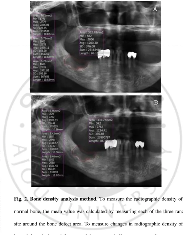
To measure the change in radiographic density, the following process was performed equally using panoramic X-ray taken after more than 6 months. The bone defect area was set to the same extent as the panoramic X-ray immediately after the surgery (Fig. 2). The above process was conducted twice by one examiner and the average of the measured values were used.
- 8 -
Fig. 2. Bone density analysis method. To measure the radiographic density of the normal bone, the mean value was calculated by measuring each of the three random site around the bone defect area. To measure changes in radiographic density of the bone defect, the bone defect area of the panoramic X-ray was set to the same extent as the panoramic X-ray immediately after surgery, and after six months of surgery. (A) Immediately after surgery (B) Post OP over six months of surgery
A
- 9 - D. Statistical Analysis
Statistical analysis was performed using SPSS version 23.0. Shapiro-Wilk test was used to determine the normality of the data. Mann-Whitney U test (non-normal distributions) used to determine difference in the area of bone defects between the experimental group and the control group with a significance level of 5%. Paired t-test (normal distributions) was used for analyzed effectiveness in each group with a significance level of 5%. Independent t-test (normal distributions) was used for analyzed effectiveness between the experimental group and the control group with a significance level of 5%.
- 10 -
Ⅲ. RESULT
Total number of patients was 26, with experimental group of 18 and control group of 8. Average age were 74.89 years and 74.88 years for experimental and control group,
respectively. Average area of bone defect were 182.90mm2 and 356.85 mm2 for
experimental and control group. As a result of Mann-Whitney U test, the P-value was 0.261, which did not have a statistically significant difference in the area of bone defects between two groups(Table 1, 2).
A. Radiographic Index
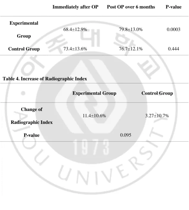
Average radiographic index for experimental group was 68.4% and 79.8% in immediately after surgery and more than 6 months after the surgery, respectively. Average radiographic index for control group was 73.4% and 76.7% in immediately after surgery and more than 6 months after the surgery, respectively. Paired t-test was done in order to verify the effects of osteogenesis immediately after surgery and after 6 months after surgery in each group. As a result, the P-value in the experimental group was 0.0003, which had a statistically significant effect, but 0.444 in the control group, which did not have a statistically significant effect (Table 3).
B. Radiographic Index Increase
Average radiographic index increase of 11.4% in the experimental group and an average increase of 3.27% in the control group, respectively. There was a difference in the amount of increase, but as a result of Independent t-test, the P-value in the experimental group was
- 11 -
- 12 - Table 1. Patient information of Experimental group
Pt. no Age Sex Bisphosphonate Reason for BP use
Follow up point Defect area(mm2 ) 1 55 F Zolendronate breast ca. (bone meta) 6 months 250.05
2 77 F Alendronate Osteoporosis 6 months 310.17
3 88 F Risedronate, Ibandronate Osteoporosis 6 months 189.49
4 65 F Alendronate Osteoporosis 10 months 179.94
5 91 F Alendronate, Ibandronate Osteoporosis 6 months 40.04
6 82 F Alendronate Osteoporosis 6 months 206.62
7 71 F Risedronate, Ibandronate Osteoporosis 12 months 76.62
8 76 F Risedronate Osteoporosis 6 months 82.09
9 76 F Ibandronate Osteoporosis 17 months 160.558
10 66 F Alendronate Osteoporosis 6 months 317.31
11 77 F Ibandronate Osteoporosis 12 months 165.44
12 79 F Unknown BP Osteoporosis 6 months 70.58
13 87 F Zolendronate Osteoporosis 6 months 153.34
14 75 F Risedronate, Ibandronate Osteoporosis 6 months 257.76
15 73 F Risedronate Osteoporosis 6 months 111.56
16 77 F Alendronate Osteoporosis 7 months 210.62
17 66 F Alendronate Osteoporosis 7 months 157.04
- 13 - Table 2. Patient information of Control group
Pt. no Age Sex Bisphosphonate Reason for BP use
Follow up
point
Defect
area(mm2)
1 77 F Unknown BP Osteoporosis 12 months 178.37
2 83 F Alendronate Osteoporosis 6 months 1073.87
3 67 F Alendronate Osteoporosis 6 months 186.16
4 76 F Alendronate Osteoporosis 6 months 336.88
5 74 F Alendronate Osteoporosis 6 months 79.79
6 63 F Unknown BP Osteoporosis 10 months 236.37
7 83 F Alendronate, Ibandronate Osteoporosis 6 months 145.72
- 14 - Table 3. Radiographic Index
Immediately after OP Post OP over 6 months P-value
Experimental Group
68.4±12.9% 79.8±13.0% 0.0003
Control Group 73.4±13.6% 76.7±12.1% 0.444
Table 4. Increase of Radiographic Index
Experimental Group Control Group
Change of Radiographic Index
11.4±10.6% 3.27±10.7%
- 15 -
IV. DISCUSSION
The purpose of this study was to investigate the bone formation effect of rhBMP-2/ACS in patients with MRONJ. This study expected that rhBMP-rhBMP-2/ACS could promote bone healing by enhancing bone remodeling in MRONJ patients, and compared the extent of bone healing to the application of rhBMP-2/ACS after sequestrectomy. In this study, bone density analysis method of bone defect area was based on the radiopacity of the panoramic X-ray. Study has shown an increase in radiographic index in the experimental group compared to the control group.
The bone density analysis method of bone defect area is based on the assumption that radiopacity of the surrounding normal bone will have the same density as normal bone. Study of Ihan Hren et al., initial radiographic index of the defect was 88%(Hren and Miljavec 2008). In this study, mean initial index of the defects was 70.3% and the index increased with time. The initial radiographic index difference from Ihan Hren's study is expected to be due to differences in the type of defect site itself or the difference in amount of radiation exposure during panoramic X-ray taking. Based on the above, bone density can be estimated using panoramic X-ray.
Cicciu et al. investigated the clinical effects of rhBMP-2 without concomitant bone grafting materials in MRONJ patients undergoing sequestrectomy and reported successful healing of the necrotic region(Cicciù et al. 2012). In this study, the experimental group identified an increase in radiographic index after 6 months of surgery (Wilcoxon signed rank
- 16 -
test, P<0.05), which may be thought as the result rhBMP-2/ACS facilitating bone healing. This result suggests that rhBMP-2/ACS contributes to new bone formation.
In Ihan Hren's study, the mean increase in radiopacity of bone defect was 27% at 6 months after cyst enucleation in healthy patients(Hren and Miljavec 2008). In this study, mean increase is 11.2% in experimental group. Despite using rhBMP-2/ACS, bone regeneration in MRONJ patients is postulated to be difficult compared to normal bone.
There were many qualitative studies on the effect of rhBMP-2 in patients with MRONJ, but few quantitatively analyzed studies. To our knowledge, the significance of this study is the demonstration of the quantitative effect of rhBMP-2 in patients with MRONJ.
This study had the following limitations: small sample sizes; difficulty in determining margin of bone defect; panoramic X-ray evaluation provide limited information in bone density evaluation; For more accurate analysis, CT-based studies are required.
In this study, rhBMP-2 was off-label used beyond FDA's suggestion, but it was expected that there would be clinical utility in the future.
- 17 -
Ⅴ. CONCLUSION
When rhBMP-2/ACS was used on bone defect sites after sequestrectomy, new bone formation rate was increased. Thus, it could be a successful strategy in treatment of MRONJ patient using rhBMP-2/ACS on bone defect site after sequestrectomy.
- 18 -
REFERENCES
1. Bagan J, Scully C, Sabater V, Jimenez Y: Osteonecrosis of the jaws in patients treated with intravenous bisphosphonates (BRONJ): A concise update. Oral
oncology 45: 551-554, 2009
2. Carter TG, Brar PS, Tolas A, Beirne OR: Off-label use of recombinant human bone
morphogenetic protein-2 (rhBMP-2) for reconstruction of mandibular bone defects in humans. Journal of Oral and Maxillofacial Surgery 66: 1417-1425, 2008
3. Chenard KE, Teven CM, He T-C, Reid RR: Bone morphogenetic proteins in
craniofacial surgery: current techniques, clinical experiences, and the future of personalized stem cell therapy. BioMed Research International 2012, 2012
4. Cicciù M, Herford AS, Juodžbalys G, Stella E: Recombinant human bone
morphogenetic protein type 2 application for a possible treatment of bisphosphonates-related osteonecrosis of the jaw. Journal of Craniofacial Surgery 23: 784-788, 2012
5. de Freitas RM, Spin‐Neto R, Junior EM, Pereira LAVD, Wikesjö UM, Susin C:
Alveolar Ridge and Maxillary Sinus Augmentation Using rh BMP‐2: A Systematic Review. Clinical implant dentistry and related research 17: e192-e201, 2015
6. Geiger M, Li R, Friess W: Collagen sponges for bone regeneration with rhBMP-2.
Advanced drug delivery reviews 55: 1613-1629, 2003
7. Gómez RF, Martínez MG, Olmos JM: Osteochemonecrosis of the jaws due to
- 19 - 13: E318-324, 2008
8. Hren NI, Miljavec M: Spontaneous bone healing of the large bone defects in the mandible. International journal of oral and maxillofacial surgery 37: 1111-1116, 2008
9. Hwang DY, On SW, Song SI: Bone regenerative effect of recombinant human bone
morphogenetic protein-2 after cyst enucleation. Maxillofacial plastic and
reconstructive surgery 38: 22, 2016
10. Lee JK, Kim K-W, Choi J-Y, Moon S-Y, Kim S-G, Kim C-H, Kim H-M, Kwon Y-D,
Kim Y-D, Lee D-K: Bisphosphonates-related osteonecrosis of the jaw in Korea: a preliminary report. Journal of the Korean Association of Oral and Maxillofacial
Surgeons 39: 9-13, 2013
11. Li RH, Wozney JM: Delivering on the promise of bone morphogenetic proteins.
Trends in biotechnology 19: 255-265, 2001
12. Park J-H, Kim J-W, Kim S-J: Does the addition of bone morphogenetic protein 2 to
platelet-rich fibrin improve healing after treatment for medication-related osteonecrosis of the jaw? Journal of Oral and Maxillofacial Surgery 75: 1176-1184, 2017
13. Ruggiero SL, Dodson TB, Assael LA, Landesberg R, Marx RE, Mehrotra B:
American Association of Oral and Maxillofacial Surgeons Position Paper on Bisphosphonate‐Related Osteonecrosis of the Jaw–2009 Update: Approved by the Board of Trustees January 2009. Australian endodontic journal 35: 119-130, 2009
- 20 -
F: American Association of Oral and Maxillofacial Surgeons position paper on medication-related osteonecrosis of the jaw—2014 update. Journal of oral and
maxillofacial surgery 72: 1938-1956, 2014
15. Russell R, Watts N, Ebetino F, Rogers M: Mechanisms of action of bisphosphonates:
similarities and differences and their potential influence on clinical efficacy.
Osteoporosis international 19: 733-759, 2008
16. Vescovi P, Merigo E, Meleti M, Manfredi M, Fornaini C, Nammour S, Mergoni G,
Sarraj A, Bagan JV: Conservative surgical management of stage I bisphosphonate-related osteonecrosis of the jaw. International journal of dentistry 2014, 2014 17. Yim J-H, Lee J-H: Panoramic analysis about spontaneous bone regeneration after
enucleation of jaw cyst. Maxillofacial Plastic and Reconstructive Surgery 31: 229-236, 2009
- 21 - - 국문요약 -
Medication-Related Osteonecrosis of the Jaw(MRONJ)
환자의 부골적출술 후 Recombinant Human Bone Morphogenetic
Protein-2/Absorbable Collagen Sponge(rhBMP-2/ACS)의
골재생 효과
아주대학교 임상치의학대학원 임상치의학과/구강악안면외과학 전공 민 송 희 (지도교수 : 이 정 근) 목적 : rhBMP-2는 골의 리모델링 향상 기능이 있어 본래 FDA가 승인한 적용 범 위를 넘어 MRONJ 치료에 사용하는 것으로 알려져 있다. 본 연구의 목적은 MRONJ 환자에 대한 rhBMP-2/ACS의 골재생 효과를 조사하는 것이었다. 재료 및 방법 : 본 후향적 코호트 연구는 2010년부터 2018년까지 아주대 치과병 원에서 MRONJ로 진단되어 하악의 부골적출술을 받은 여성 환자 26명을 대상으 로 하였다. 실험군은 부골적출술 후 rhBMP-2/ACS를 적용한 18명의 환자로 구성 되었으며, 대조군은 부골적출술 후 이 절차를 생략한 8명의 환자로 구성되었다. 실험군에서 0.5mg/ml의 농도로 총 0.5mg의 rhBMP-2를 사용하였다. 후속 파노라 마 방사선사진은 수술 직후, 수술 후 6개월이상 경과 후에 촬영하였다. 위 방사- 22 - 선사진을 이용하여, 정상골과 결손부위의 흑화도를 이용하여 골 결손 면적의 방 사선지수를 계산하는 수정된 Ihan Hren의 방법을 이용하여 각 시기별 방사선지수 를 측정하였다. 각 그룹의 효과에 검정에는 대응 t-검정을 사용했으며 실험군과 대조군 사이의 효과 비교에는 독립 t-검정을 사용하였다. 결과 : 실험군의 수술 직후와 술 후 6개월 이후의 평균 방사선지수는 각각 68.4%, 79.8%로 나타났다(Paired t-test, P<0.05). 대조군의 수술 직후와 술 후 6개 월 이후의 평균 방사선 지수는 각각 73.4%, 76.7%로 나타났다(Paired t-test, P>0.05). 또한, 평균 방사선 지수는 실험군에서 11.4%, 대조군에서 평균 3.27% 상승하였다(Independent t-test, P>0.05). 결론 : 결과에 따르면, 부골적출술 후 골 결손 부위에 rhBMP-2/ACS를 적용하는 방법은 MRONJ 환자 치료의 성공적인 전략이 될 수 있다.