대사증후군 여성에서 요가운동이 심박동변이에 미치는 영향
김하나, 서진아, 송상욱
가톨릭대학교 의과대학 성빈센트병원 가정의학과
Effect of Yoga on Heart Rate Variability in Women with Metabolic Syndrome
Ha-Na Kim, Jin-A Seo, Sang-Wook Song
Department of Family Medicine, St. Vincent’s Hospital, College of Medicine, The Catholic University of Korea, Suwon, Korea
Background: The purpose of this study was to investigate the correlation between yoga and autonomic nervous system in women with metabolic syndrome.
Methods: A randomized controlled study was performed in participants recruited from a public center for man- aging chronic diseases located in Gyeong-gi Province. The 39 women participants diagnosed with metabolic syndrome were randomly assigned to the 12-week yoga exercise group (n=22) or the wait-listed control group (n=17). Biochemical laboratory tests and heart rate variability were measured before and after the 12-week program.
Results: In post-menopausal women, the low frequency power of frequency domain significantly decreased in the yoga exercise group compared to the control group (P=0.07). On the time domain of heart rate variability and metabolic syndrome components, there were no significant differences between the groups.
Conclusions: Sympathetic activity was decreased in post-menopausal women with metabolic syndrome who practiced yoga. Our results suggest that yoga might be beneficial in improving the autonomic nervous system in post-menopausal women with metabolic syndrome.
Korean J Health Promot 2014;14(4):147-154
Keywords: Metabolic syndrome, Yoga, Autonomic nervous system, Heart rate variability
■ Received:May 9, 2014 ■ Accepted:October 6, 2014
■Corresponding author:Sang-Wook Song, MD, PhD
Department of Family Medicine, St. Vincent’s Hospital, College of Medicine, The Catholic University of Korea, 93 Jungbudaero, Paldal-gu, Suwon 442-723, Korea
Tel: +82-31-249-8246, Fax: +82-31-249-8253 E-mail: [email protected]
서 론
대사증후군은 복부비만, 고혈압, 이상지질혈증 및 인슐 린 저항성 등이 복합적으로 나타나는 질환군으로, 제2형 당뇨병 및 심혈관 질환의 발병위험을 높인다.1) 대사증후 군이 전 세계적으로 증가하면서 심혈관계 질환과 그와 관 련된 사망률 또한 증가하는 추세로2007년 국민건강영양 조사에 따르면 20세 이상의 성인에서 대사증후군 유병률 은 31.3%로 추후 국내 심혈관 질환의 증가가 우려된다.2,3)
규칙적인 운동은 제2형 당뇨병 발생과 인슐린 저항성을 낮추고, 대사를 촉진시키며, 자율신경계의 균형을 돕고, 심혈관 질환의 사망률을 줄이므로2,4-6) 대사증후군의 개선 을 위한 방법 중 하나이다. 또한 최근 요가(yoga), 태극권 (tai chi), 기공(qiqong) 등의 심신의학이 대사증후군의 치 료 방법으로 소개되고 있다.7) 특히 세계적으로 대중화된 운동인 요가는 고대의 철학으로부터 기원하며 20세기 초 부터 치료적인 목적으로 사용되기 시작하였다.8,9) 요가는 신체의 유연성과 조화, 근력을 증가시키며, 호흡 운동과 명상을 통해 평온함과 집중력을 높이고 불안, 스트레스 및 혈압을 감소시키며, 회복력 및 대사 조절을 증가시킨
다.10,11) 여러 연구에서 당뇨병, 고혈압, 고지혈증, 관상동맥
질환 및 대사증후군에서 요가 치료의 효과가 있음을 보였
다.11,12) 이는 요가운동이 항진되어 있는 교감 신경을 안정
시키고, 부교감 신경의 기능을 향상시켜 자율 신경계의 조
화를 이룸으로써,대사 상태 및 염증 반응의 조절을 유도 하는 것으로 설명될 수 있겠다.13)
자율 신경계 기능을 평가할 수 있는 방법으로 심박동변 이 검사(heart rate variability test)가 있다. 심박동변이는 1965년 Hon과 Lee에 의해 태아절박가사 전에 심박수 변 이가 먼저 발생한 것이 보고된 이후로,14 1980년대부터 급 성 심근경색에 후 돌연사를 예측하는 지표로 이용되어 왔 다.15) 최근에는 비만, 당뇨병, 고혈압, 관상동맥경화증, 뇌 졸중 및 대사증후군 환자에서 심박동변이의 감소가 보고 되고 있다.15-18) 특히, 대사증후군에서 교감 신경 활성화와 부교감 신경 불활성화로 인한 자율 신경계의 불안정성은 심근 및 심혈관 비대, 동맥의 재형성 과정과 혈관 내피의 기능저하를 유발하여 심혈관 질환으로의 진행을 촉진시킬 수 있다.19) 이에 본 연구에서는 대사증후군 여성에서 요가 운동 전후 심박동변이를 확인하여, 요가 운동이 자율신경 계 기능에 미치는 효과를 밝혀보고자 한다.
방 법
1. 연구 대상
본 연구는 경기도 소재 만성질환관리센터에 등록되어 있는 30-60세의 기혼 여성 중 대사증후군을 가진 자를 대 상으로 하였다. 제외 기준은 조절되지 않는 고혈압, 부정 맥, 울혈성 심부전, 중증의 심장 판막 질환을 가진 자, 관 상동맥질환으로 지난 6개월 내에 입원했던 자, 임신, 수유, 약물(알코올 포함) 남용자, 규칙적으로 요가 운동을 하고 있는 자, 시험을 수행하지 못할 정도의 내과적 또는 정신 과적 질환을 가진 자로 하였다. 시험 참가에 동의한 78명 의 지원자 중, 대사증후군에 해당되지 않는 자(n=32)와 제 외기준에 해당하는 자(n=7)를 제외한 39명이 본 연구에 등록되었다. 본 연구는 모든 지원자에게 연구에 대한 설명 및 고지된 시험참가 동의서를 받은 후 진행되었으며, 동일 대학병원의 기관심의위원회(Institutional Review Board, IRB)의 승인을 받아 윤리 및 안전지침을 준수하면서 시행 되었다(IRB 승인번호: VC12QISI0039).
2. 연구 방법
39명의 여성 대사증후군 환자를 대상으로 무작위 통제 연구(randomized controlled study)를 시행하였으며, 대상 자는 12주 간의 요가운동군(n=22)과 대기자 대조군(n=17) 으로 각각 할당되었다. 전체 대상자에게는 대사증후군과 관련된 생활습관 교정을 위한 기본적인 교육을 실시하였 다. 연구 시작 전 나이, 주소, 직업, 학력, 과거력, 흡연력,
음주력, 가족력, 사회경제적 상태, 운동 상태 등을 조사하 였고, 이학적 검사, 혈액검사 및 심박동변이 측정을 시행 하였다. 운동군에게 12주 간 주 2회, 회당 60분의 요가운 동 프로그램을 적용하였으며, 3개월 후 운동군 및 대조군 에 혈액검사 및 심박동변이 측정을 실시하였다.
1) 대사증후군의 진단기준
본 연구에서 대사증후군의 정의는 2005년 National Cholesterol Education Program Adult Treatment Panel
Ⅲ 가이드라인을 기본으로 하였고20) 복부비만을 정의하기 위해서는 2005년 대한비만학회에서 제시한 한국인에 적합 한 허리둘레 값(남 ≥90 cm, 여 ≥85 cm)을 사용하여21) 다음 다섯 가지 항목 중 세 가지 이상을 만족하는 경우 대사증 후군으로 분류하였다.
(1) 허리둘레 ≥90 cm (남), ≥85 cm (여)
(2) 중성지방 ≥150 mg/dL (1.69 mmol/L) 또는 약물치 료 중임
(3) 고밀도 지단백 콜레스테롤 <40 mg/dL (1.04 mmol/L) (남), <50 mg/dL (1.29 mmol/L) (여)
또는 약물치료 중임
(4) 혈압 ≥130/85 mmHg 또는 약물치료 중임
(5) 혈당 ≥100 mg/dL (6.1 mmol/L) 또는 약물치료 중임
2) 신체계측 및 혈액검사
Body Composition Analysis (Inbody 720, Biospace, Seoul, Korea)를 이용하여 키, 몸무게 및 체질량지수(body mass index) 값을 얻었다. 허리둘레는 직립자세에서 상의 를 올리고 호기 상태에서 12번째 늑골 하단부와 장골능 최상단부의 중간부위를 측정하였고, 측정은 숙련된 1인에 의해 줄자를 이용하여 0.1 cm 단위까지 시행되었다. 수축 기 및 이완기 혈압은 5분 이상 앉은 자세로 충분히 안정을 취한 후 자동 계측기로 1회 측정하였다. 혈액 검사는 12시 간 공복상태에서 상완의 정맥에서 채혈하였고 총콜레스테 롤, 중성지방, 고밀도 지단백 콜레스테롤, 공복혈당, total bilirubin, aspartate transaminase (AST), alanine trans- aminase (ALT), creatinine을 측정하였다.
3) 요가 프로그램
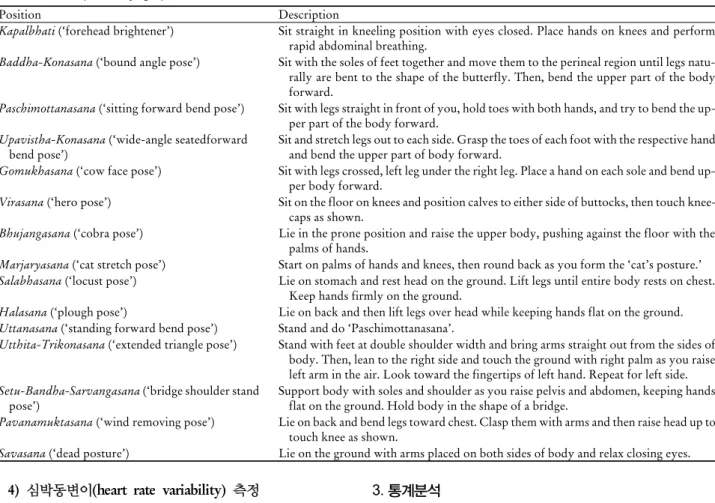
요가 프로그램은 운동군을 대상으로 12주간 주 2회씩 회당 60분간 진행하였다. 사용된 요가 프로그램은 전문가 에게 의뢰하여 개발하였으며, 프로그램 진행은 자격증을 소유한 경험 있는 전문 요가 강사가 담당하였다. 요가 프 로그램은 각각 5분 이내로 약 50분간 실시된 14가지의 자 세(asana)와 약 10분 간의 명상 이완(shavasana)으로 구성하 였다(Table 1).
Table 1. Description of yoga positions
Position Description
Kapalbhati (‘forehead brightener’) Sit straight in kneeling position with eyes closed. Place hands on knees and perform rapid abdominal breathing.
Baddha-Konasana (‘bound angle pose’) Sit with the soles of feet together and move them to the perineal region until legs natu- rally are bent to the shape of the butterfly. Then, bend the upper part of the body forward.
Paschimottanasana (‘sitting forward bend pose’) Sit with legs straight in front of you, hold toes with both hands, and try to bend the up- per part of the body forward.
Upavistha-Konasana (‘wide-angle seatedforward
bend pose’) Sit and stretch legs out to each side. Grasp the toes of each foot with the respective hand and bend the upper part of body forward.
Gomukhasana (‘cow face pose’) Sit with legs crossed, left leg under the right leg. Place a hand on each sole and bend up- per body forward.
Virasana (‘hero pose’) Sit on the floor on knees and position calves to either side of buttocks, then touch knee- caps as shown.
Bhujangasana (‘cobra pose’) Lie in the prone position and raise the upper body, pushing against the floor with the palms of hands.
Marjaryasana (‘cat stretch pose’) Start on palms of hands and knees, then round back as you form the ‘cat’s posture.’
Salabhasana (‘locust pose’) Lie on stomach and rest head on the ground. Lift legs until entire body rests on chest.
Keep hands firmly on the ground.
Halasana (‘plough pose’) Lie on back and then lift legs over head while keeping hands flat on the ground.
Uttanasana (‘standing forward bend pose’) Stand and do ‘Paschimottanasana’.
Utthita-Trikonasana (‘extended triangle pose’) Stand with feet at double shoulder width and bring arms straight out from the sides of body. Then, lean to the right side and touch the ground with right palm as you raise left arm in the air. Look toward the fingertips of left hand. Repeat for left side.
Setu-Bandha-Sarvangasana (‘bridge shoulder stand
pose’) Support body with soles and shoulder as you raise pelvis and abdomen, keeping hands flat on the ground. Hold body in the shape of a bridge.
Pavanamuktasana (‘wind removing pose’) Lie on back and bend legs toward chest. Clasp them with arms and then raise head up to touch knee as shown.
Savasana (‘dead posture’) Lie on the ground with arms placed on both sides of body and relax closing eyes.
4) 심박동변이(heart rate variability) 측정
심박동변이는 일중변동을 보이므로 오전에 측정하였으 며, 12시간 이상의 금식상태에서 검사 2시간 전까지 격렬 한 운동, 음주, 흡연 및 카페인 섭취를 금한 뒤 충분히 안 정을 취한 후 의자에 착석한 자세에서 심박동변이 측정 장 치(SA-2000EⓇ, Medicore, Seoul, Korea)를 사용하여 측정 하였으며, 대상자의 양측 손목과 좌측 발목부분 각각에 전 극을 부착시킨 후 5분간 측정하였다. 시간 범주 분석은 교 감 및 부교감 신경과 다른 신체적인 영향을 함께 반영하는 R-R 간격의 표준편차(the standard deviation of nor- mal-to-normal intervals, in milliseconds, SDNN)와 주로 부교감 신경에 의한 변화를 반영하는, 인접한 R-R 간격의 차이를 제곱한 값의 평균의 제곱근(the square root of the mean squared differences of successive normal-to-normal intervals, in milliseconds, RMSSD)을 측정하였다. 주파수 범주 분석으로 전체 주파수 강도(total power: VLF, LF, HF를 포함한 5분 동안의 전체 power를 의미, TP), 저주 파 영역(low frequency: 0.04-0.15 Hz에 해당하는 주파수 대역의 강도, LF), 고주파 영역(high frequency: 0.15-0.4 Hz에 해당하는 주파수 대역의 강도, HF)을 측정하였으 며, 저주파영역과 고주파영역의 비(LF/HF ratio)를 구하 여 분석을 시도하였다.
3. 통계분석
연구 대상자들의 일반적 특성을 특성의 비교를 위해 Student’s t-test와 chi-square test를 이용하여 분석하였고 두 군 간의 요가운동 전후의 심박동변이 지표의 비교를 위 해 ANCOVA(analysis of covariance)를 이용하였다. 심박 수변이의 주파수 범주 값은 심한 사편위를 보이므로 자연 로그변환 후 통계 분석하였다. 자료 분석은 SPSS ver.12.0 (SPSS Inc., Chicago, IL, USA)을 이용하였으며 각 분석에 서 유의 수준 5% 이하를 통계적으로 유의한 것으로 간주 하였다.
결 과
1. 연구 대상자들의 일반적 특성
연구 대상자는 총 39명으로 요가운동군은 22명, 대조군 은 17명이었다. 연령, 키, 체중, 총콜레스테롤, total bilir- ubin, AST, ALT, creatinine 및 고혈압, 당뇨병 및 뇌혈관질 환의 가족력 유무 모두 두 군 간에 통계적으로 유의한 차 이를 보이지 않았다(Table 2).
Table 2. Baseline characteristics of participantsa
Characteristics Yoga exercise (n=22) Control (n=17) Pb
Age, y 51.05±8.26 48.24±7.22 0.273
Height, cm 160.55±0.05 158.47±0.05 0.214
Weight, kg 64.8±10.03 66.29±7.06 0.612
BMI, kg/m2 25.12±3.88 26.35±2.32 0.258
TChol, mg/dL 196.91±32.46 197.88±26.72 0.920
Tbil, mg/dL 0.51±0.15 0.53±0.23 0.673
AST, mg/dL 19.82±3.51 20.29±6.03 0.784
ALT, mg/dL 17.00±6.44 22.47±14.37 0.161
Cr, mg/dL 0.73±0.08 0.73±0.10 0.941
Family history
Hypertension 14 (63.6) 12 (70.6) 0.747
Diabetes 6 (27.3) 8 (47.1) 0.310
CVA 9 (40.9) 11 (45.8) 0.754
Abbreviations: BMI, body mass index; TChol, total cholesterol; Tbil, total bilirubin; AST, aspartate transaminase; ALT, alanine transaminase;
Cr, creatinine; CVA, Cerebro-vascular accident.
aData are presented as means ± standard deviations or N (%).
bP values were obtained by t-test or chi-square test.
Table 3. Changes in metabolic syndrome components at baseline and post-yoga exercisea
Yoga exercise (n=22) Control (n=17) Pb
Baseline Post Baseline Post
WC, cm 90.14±7.98 90.41±8.73 91.12±6.60 90.03±5.17 0.201
SBP, mm Hg 120.91±11.09 121.36±13.20 118.24±10.74 114.71±8.74 0.108
DBP, mm Hg 79.09±8.11 78.64±8.34 77.65±7.52 7 7.06±5.88 0.533
FBS, mg/dL 105.82±15.88 95.45±15.23 115.76±33.54 99.88±24.55 0.792
HDL-C, mg/dL 44.59±13.15 47.55±12.98 46.76±10.64 54.29±15.26 0.145
TG, mg/dL 186.05±99.92 154.95±85.83 176.06±84.31 129.29±52.95 0.312
Abbreviations: WC, waist circumference; SBP, systolic blood pressure; DBP, diastolic blood pressure; FBS, fasting blood sugar; HDL-C, high-density lipoprotein cholesterol; TG, triglyceride.
aValues are presented as mean ± standard deviations.
bP values correspond to between-group comparisons for the differences over time for each variable, and are obtained by analysis of covariance.
Table 4. Changes in heart rate variability indices at baseline and post-yoga exercisea
Yoga exercise (n=22) Control (n=17) Pb
Baseline Post Baseline Post
SDNN, ms 33.96±16.10 32.03±11.47 27.63±11.66 25.56±9.27 0.213
RMSSD, ms 25.02±12.93 19.80±9.53 20.63±12.36 18.59±9.05 0.662
cln TP, ms2 6.41±0.98 6.23±0.89 5.87±0.91 5.86±1.02 0.927
cln LF, ms2 4.74±1.16 4.40±1.26 4.09±0.83 4.50±0.98 0.061
cln HF, ms2 4.74±1.20 4.13±1.12 4.20±1.35 4.00±1.13 0.585
LF/HF ratio 1.52±1.29 2.43±4.25 1.91±3.00 2.27±2.10 0.819
Abbreviations: SDNN, standard deviation of normal to normal intervals; RMSSD, square root of the mean squared differences of successive normal to normal intervals; TP, total power for the 5-minute cycle (1.15×10⁻⁵-0.40Hz); LF, low frequency power (0.004-0.15 Hz); HF, high frequency power (0.15-0.40Hz); LF/HF ratio, ratio of power in LF/HF.
aValues are presented as mean ± standard deviations.
bP values correspond to between-group comparisons for the differences over time for each variable, and are obtained by analysis of covariance.
cln: natural logarithm. (The spectral power data were log transformed).
2. 요가운동 전후의 대사증후군 지표 비교
요가운동 전후의 대사증후군 지표를 비교해 보았을 때 허리둘레, 수축기 및 이완기 혈압, 공복혈당, 저밀도 콜레 스테롤, 중성 지방 모두 두 군 간에 통계적으로 유의한 차 이를 보이지 않았다(Table 3).
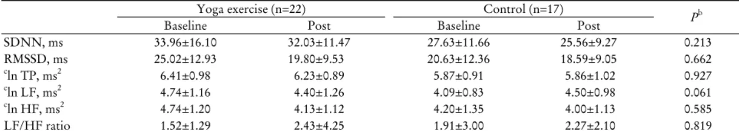
3. 요가운동 전후의 심박동변이의 비교
요가운동 시행 전후의 심박동변이 지표를 비교해 보았을 때 ln LF은 요가운동 후 감소되는 경향을 보였으나(P=0.061), SDNN, RMSSD, ln TP, ln HF, LF/HF ratio는 두 군 간 에 통계적으로 유의한 차이를 보이지 않았다(Table 4).
Table 5. Changes in heart rate variability indices at baseline and post-yoga exercise in pre-menopausal womena
Yoga exercise (n=8) Control (n=10) Pb
Baseline Post Baseline Post
SDNN, ms 32.57±17.56 33.76±13.35 23.57±12.40 24.54±9.42 0.210
RMSSD, ms 23.35±15.93 20.93±10.76 19.45±14.91 16.44±7.12 0.373
cln TP, ms2 6.15±1.03 6.56±0.87 5.65±0.95 5.54±1.18 0.098
cln LF, ms2 4.67±1.42 4.98±1.36 4.19±0.95 4.36±1.20 0.590
cln HF, ms2 4.35±1.53 4.21±1.46 4.03±1.58 3.97±1.22 0.895
LF/HF ratio 1.80±1.38 4.21±6.78 2.22±3.46 1.87±1.44 0.312
Abbreviations: SDNN, standard deviation of normal to normal intervals; RMSSD, square root of the mean squared differences of successive normal to normal intervals; TP, total power for the 5-minute cycle (1.15×10⁻⁵-0.40Hz); LF, low frequency power (0.004-0.15 Hz); HF, high frequency power (0.15-0.40Hz); LF/HF ratio, ratio of power in LF/HF.
aValues are presented as mean ± standard deviations.
bP values correspond to between-group comparisons for the differences over time for each variable, and are obtained by analysis of covariance.
cln: natural logarithm (The spectral power data were log transformed).
Table 6. Changes in heart rate variability indices at baseline and post-yoga exercise in post-menopausal womena
Yoga exercise (n=14) Control (n=7) Pb
Baseline Post Baseline Post
SDNN, ms 34.75±15.83 31.05±10.66 33.43±8.06 28.43±8.94 0.613
RMSSD, ms 25.97±11.44 19.15±9.11 22.32±8.27 21.66±11.13 0.153
cln TP, ms2 6.57±0.76 6.05±0.87 6.19±0.79 6.31±0.49 0.062
cln LF, ms2 4.79±1.04 4.08±1.12 3.94±0.66 4.70±0.55 0.007
cln HF, ms2 4.95±0.97 4.09±0.92 4.42±1.02 4.05±1.07 0.429
LF/HF ratio 1.35±1.26 1.41±1.16 1.46±2.39 2.83±2.83 0.064
Abbreviations: SDNN, standard deviation of normal to normal intervals; RMSSD, square root of the mean squared differences of successive normal to normal intervals; TP, total power for the 5-minute cycle (1.15×10⁻⁵-0.40Hz); LF, low frequency power (0.004-0.15 Hz); HF, high frequency power (0.15-0.40Hz); LF/HF ratio, ratio of power in LF/HF.
aValues are presented as mean ± standard deviations.
bP values correspond to between-group comparisons for the differences over time for each variable, and are obtained by analysis of covariance.
cln: natural logarithm (The spectral power data were log transformed).
4. 폐경 유무에 따른 요가운동 전후의 심박동변이의 비교
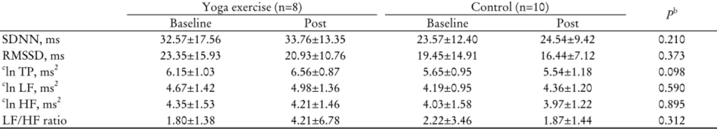
폐경 전 여성에서 요가운동 시행 전후의 심박동변이 지 표를 비교해 보았을 때 ln TP은 요가운동 후 증가하는 경 향을 보였으며(P=0.090), SDNN, RMSSD, ln LF, ln HF, LF/HF ratio는 두 군 간에 통계적으로 유의한 차이를 보 이지 않았다(Table 5).
폐경 후 여성에서 요가운동 시행 전후의 심박동변이 지 표를 비교해 보았을 때 ln LF은 요가운동 시행 후 요가운 동군에서 대조군에 비해 유의하게 감소하였다(P=0.007).
LF/HF ratio는 요가운동 시행 후 대조군에서 요가운동군 보다 더 증가하는 경향을 보였으며(P=0.064), ln TP은 요가 운동 후 요가운동군에서 감소하고 대조군에서는 증가하는 경향을 보였다(P=0.062). 그 외 SDNN, RMSSD, ln HF 값 은 두 군 간에 유의한 차이를 보이지 않았다(Table 6).
고 찰
본 연구는 대사증후군 여성에서 요가운동 시행 후 심박 동변이 및 대사증후군 지표 변화를 살펴보았다. 전체 환자
를 대상으로 요가운동 후 심박동변이를 분석하였을 때 시 간 범주 분석값은 통계적으로 유의하지 않았으며, 주파수 범주 분석에서는 요가운동 후 LF 값이 요가운동군에서는 감소하였고, 대조군에서는 증가하는 경향을 보였다. 심박 동변이는 연령 및 에스트로겐의 영향을 받으므로 전체 환 자를 폐경 전후 여성으로 나누어 분석하였다.22,23) 폐경 전 후 여성으로 나누어 분석한 결과에서도 시간 범주 분석 값 은 통계적으로 유의한 차이를 보이지 않았다. 폐경 후 여 성에서, 주파수 범주 분석값 중 LF값은 요가운동 후 대조 군에서는 증가한 반면, 요가운동군에서는 감소하였으며 이는 통계적으로 유의하였다. 또한 폐경 후 여성에서, 요 가운동 후 LF/HF ratio는 대조군에서 요가운동군보다 더 증가하는 경향을 보였다. TP값은 폐경 전 여성 중, 요가운 동군에서는 증가 그리고 대조군에서는 감소하는 경향을 보였으나, 폐경 후 여성의 요가운동군에서 감소한 반면 대 조군에서 증가하는 경향을 보였다. 요가 운동 전후 대사증 후군 지표는 두 군 간의 유의한 변화는 없었다.
정상인을 대상으로 요가운동 후 심박동변이를 측정한 Muralikrishnan 등의 연구23)에서 요가운동군이 대조군에 비해 낮은 LF값을 보인 것은 본 연구 결과와 일치하였다.
Muralikrishnan 등24)은 LF값의 감소를 요가운동 후 교감 신경 활성의 저하로 설명하고 있으며 요가운동이 심혈관 계에 좋은 영향을 미칠 것이라고 기대하였다. 또 다른 연 구에서는 노인여성의 요가운동 전후 심박동변이를 측정하 였는데 요가운동군에서 LF값의 변화는 관찰되지 않았으 나 HF값의 유의한 증가를 보고하였다.25) 이러한 결과는 이미 여러 연구들에서 증명되었듯이, 요가운동이 교감 신 경 활성을 저하시키고 부교감 신경의 활성은 증가시켜 자 율 신경계가 교감 신경 위주에서 부교감 신경 위주로 조화 를 이루게 하는 것으로 설명할 수 있다.26)
LF값은 교감 및 부교감 신경계의 활동을 동시에 반영하 나 대부분 교감 신경 활동의 지표로 활용된다.15) 본 연구 에서 폐경 후 여성의 요가운동 후 LF값은 요가운동군에서 감소한 반면 대조군에서 증가하였고 이는 통계적으로 유 의하였다. 이는 폐경 후 여성에서 요가운동 후 요가운동군 의 교감 신경계 활성이 감소하였다는 것을 의미한다. 폐경 은 그 자체로 신체적, 물리적, 신경 내분비학적인 노화를 촉진시키며 심혈관 질환의 위험을 증가시키는 위험 인자 로, 교감신경 활성을 증가시키고 부교감신경 활성을 저하 시켜 심혈관 질환의 발생을 증가시킬 수 있다.27) 그러므로 요가운동 후 교감 신경계 활성 감소를 통한 자율 신경계의 조화는 폐경 후 여성에서 심혈관계 질환의 감소에 긍정적 영향을 미치는 근거가 될 수 있다.
고주파 영역에 대한 저주파 영역의 비인 LF/HF ratio는 주로 교감 신경 활성을 반영하며 자율 신경계의 조화를 나 타내는 지표로 사용되어 교감 신경의 활성이 증가하거나 부교감 신경의 활성이 감소할 때 이 비는 높아지게 된 다.15) 본 연구에서 폐경 후 여성의 LF/HF ratio값은 통계 적으로 유의하지는 않았으나 요가운동 후 대조군에서 요 가운동군보다 더 증가하는 경향을 보였다. 이는 12주 간의 요가 운동 후 요가운동군에서 대조군보다 교감신경계의 활성이 감소한 결과로 요가운동군에서 자율신경계가 더 균형을 이루는 경향을 보인 것으로 볼 수 있다. 노인여성 의 요가운동 전후 심박동변이를 측정한 연구에서도 요가 운동군에서 LF/HF ratio값이 유의하게 감소하는 결과를 보고하였는데 이를 요가운동 후에 교감 신경 활성도저하 로 해석하였다.25)
TP값은 자율 신경계의 전체적인 활성 정도를 반영하는 값으로 자율 신경계 조절능력을 의미하며 시간 범주의 SDNN과 높은 상관성을 보이지만, 본 연구에서는 TP값 과 SDNN값의 연관성은 관찰되지 않았다.28) 폐경 후 여성 에서의 TP값은 요가운동군에서는 감소하는 경향을 보인 반면, 대조군에서는 증가하는 경향을 보였다. TP값은 운 동의 강도가 증가함에 따라 감소하는데, 이는 점차적으로 부교감 신경계 활성도가 감소하는 것을 의미한다.29) 따라
서 폐경 후 여성에서의 TP값의 감소는 요가 운동의 영향 으로 인해 요가운동군에서 감소한 것으로 설명할 수 있다.
그러나 폐경 전 여성에서의 TP값은 폐경 후 여성에서와는 달리 요가운동 후 요가운동군에서는 증가하였고 대조군에 서는 감소한 경향을 보였다. 이를 폐경 전 여성에서 요가 운동 후 자율신경계 조절능력이 향상된 결과라고 설명하 기에는 SDNN, RMSSD, LF, HF, LF/HF ratio값 모두 두 군 간에 통계적으로 유의한 차이를 보이지 않았다.
요가운동 시행 전후 요가운동군과 대조군 간에 대사증 후군 지표를 비교하였을 때 두 군 간에 통계적으로 유의한 차이를 보이지 않았다. 그러나 비만 소년을 대상으로 한 Seo 등의 연구30)에서는 요가운동군이 대조군에 비해 체중, 신체 비만지수, 지방제외체중, 기초대사율, 총 콜레스테롤 수치가 두 군 간에 유의하게 호전되었다. 또한 비만한 폐 경 여성을 대상으로 한 16주 간의 요가운동 후 대사증후 군 지표 변화에 대한 연구에서, 요가운동 후 허리둘레, 고 밀도 지단백 콜레스테롤, 이완기 혈압이 호전되었다.31) 그 러나 본 연구에서는 요가운동 전후 대사증후군 지표에서 두 군 간에 유의한 차이를 보이지 않았는데 이는 대사증후 군 진단 시 요가운동군 및 대조군을 포함한 전체 참여자를 대상으로 시행한 생활습관 개선 교육에서 기인한 것으로 생각된다.
본 연구는 몇 가지 제한점을 가지고 있다. 첫째, 표본크 기가 작아 통계적 유의성이 감소되었을 가능성이 있다. 둘 째, 연구대상자가 일개 만성질환관리센터의 등록자 내에 서 모집되어 대표성을 갖기 어려웠다. 셋째, 심박동변이는 여성 호르몬의 영향을 받으므로 본 연구에서 폐경 전후 여 성으로 나누어서 분석하였으나, 폐경 전 여성의 생리주기 를 고려하지 않았다. 정상 생리 주기 동안 심박동변이를 관찰한 결과 황체기 때 교감신경계 활성도가 높다는 결과 를 보고한 연구가 있다.23)
이러한 제한점에도 불구하고 이번 연구를 통해 요가운 동은 대사증후군 여성, 특히 폐경 후 여성에서 교감 신경 계를 안정화시킬 수 있는 효과를 가지며, 나아가 심혈관 질환 발생 예방에 영향을 미칠 수 있다고 기대해 볼 수 있 겠다. 추후 대사증후군 여성환자를 대상으로 요가 운동을 통한 자율 신경계의 안정화와 심혈관계질환 예방의 연관 성에 대한 추가적 연구가 필요하다.
요 약
연구배경: 제2형 당뇨병 및 심혈관 질환의 위험과 관련 이 있는 대사증후군의 개선 방법의 하나로 요가 등의 심신 의학이 소개되고 있다. 본 연구에서는 대사증후군 여성에 서 요가 운동이 자율 신경계의 기능에 미치는 효과를 심박
동변이 측정을 통해 알아보고자 한다.
방법: 경기도 소재 만성질환관리센터에 등록되어 있는 30-60세의 39명의 여성 대사증후군 환자를 대상으로 무작 위 통제 연구를 시행하였으며, 대상자는 요가운동군 (n=22)과 대기자 대조군(n=17)으로 할당되었다. 연구 시작 전 혈액검사 및 심박동변이를 시행하였으며 운동군에게 12주 간 주 2회, 회당 60분의 요가 프로그램을 시행하였 고, 3개월 후 대조군과 운동군에게 혈액검사 및 심박동변 이를 측정하였다.
결과: 전체 환자를 대상으로 요가운동 시행 전후의 심박 동변이 지표를 비교해 보았을 때 LF값은 요가운동 후 감 소되는 경향을 보였다(P=0.061). 폐경 전후 여성으로 나누 어 분석하였을 때, 폐경 전 여성에서 요가운동 후 TP값이 증가하는 경향을 보였다(P=0.090). 폐경 후 여성에서는 요 가운동 시행 후 LF값은 요가운동군에서 대조군에 비해 통 계적으로 유의하게 감소하였으며(P=0.007), LF/HF ratio 는 대조군에서 요가운동군보다 더 증가하는 경향을 보였 고, TP 값은 요가운동군에서 감소하고 대조군에서는 증가 하는 경향을 보였다(P=0.062). 요가운동 후의 대사증후군 지 표는 두 군 간에 통계적으로 유의한 차이를 보이지 않았다.
결론: 요가운동은 대사증후군을 가진 폐경 후 여성에서 교감 신경계를 안정화시킬 수 있는 효과를 나타냈다. 추후 대사증후군 여성환자를 대상으로 요가 운동을 통한 자율 신경계의 안정화와 심혈관계질환 예방의 연관성에 대한 추가적 연구가 필요하다.
중심단어: 대사증후군, 요가, 자율신경계, 심박동변이
REFERENCES
1. Kassi E, Pervanidou P, Kaltsas G, Chrousos G. Metabolic syn- drome: definitions and controversies. BMC Med 2011;9:48.
2. Gallagher EJ, Leroith D, Karnieli E. The metabolic syndrome- from insulin resistance to obesity and diabetes. Med Clin North Am 2011;95(5):855-73.
3. Lim S, Shin H, Song JH, Kwak SH, Kang SM, Won Yoon J, et al.
Increasing prevalence of metabolic syndrome in Korea: the Korean national health and nutrition examination survey for 1998-2007. Diabetes Care 2011;34(6):1323-8.
4. Derouich M, Boutayeb A. The effect of physical exercise on the dynamics of glucose and insulin. J Biomech 2002;35(7):911-7.
5. Furlan R, Piazza S, Dell'Orto S, Gentile E, Cerutti S, Pagani M, et al. Early and late effects of exercise and athletic training on neural mechanisms controlling heart rate. Cardiovasc Res 1993;
27(3):482-8.
6. O'Connor GT, Buring JE, Yusuf S, Goldhaber SZ, Olmstead EM, Paffenbarger RS Jr, et al. An overview of randomized trials of rehabilitation with exercise after myocardial infarction.
Circulation 1989;80(2):234-44.
7. Anderson JG, Taylor AG. The metabolic syndrome and mind-body therapies: a systematic review. J Nutr Metab 2011;
2011:276419.
8. Büssing A, Michalsen A, Khalsa SB, Telles S, Sherman KJ.
Effects of yoga on mental and physical health: a short summary of reviews. Evid Based Complement Alternat Med 2012;2012:
165410.
9. Kirkwood G, Rampes H, Tuffrey V, Richardson J, Pilkington K. Yoga for anxiety: a systematic review of the research evidence. Br J Sports Med 2005;39(12):884-91.
10. Yang K. A review of yoga programs for four leading risk factors of chronic diseases. Evid Based Complement Alternat Med 2007;4(4):487-91.
11. Sahay BK. Role of yoga in diabetes. J Assoc Physicians India 2007;55:121-6.
12. Singh S, Malhotra V, Singh KP, Madhu SV, Tandon OP. Role of yoga in modifying certain cardiovascular functions in type 2 di- abetic patients. J Assoc Physicians India 2004;52:203-6.
13. Whang W, Bigger JT Jr. Comparison of the prognostic value of RR-interval variability after acute myocardial infarction in pa- tients with versus those without diabetes mellitus. Am J Cardiol 2003;92(3):247-51.
14. Lee ST, Hon EH. The fetal electrocardiogram. IV. unusual variations in the qrs complex during labor. Am J Obstet Gynecol 1965;91:56-60.
15. Task Force of The European Society of Cardiology and The North American Society of Pacing and Electrophysiology.
Heart rate variability. Standards of measurement, physiological interpretation, and clinical use. Eur Heart J 1996;17(3):354-81 16. Wijngaarden MA, Pijl H, van Dijk KW, Klaassen ES, Burggraaf
J. Obesity is associated with an altered autonomic nervous sys- tem response to nutrient restriction. Clin Endocrinol (Oxf) 2013;79(5):648-51.
17. Vinik AI, Maser RE, Mitchell BD, Freeman R. Diabetic auto- nomic neuropathy. Diabetes Care 2003;26(5):1553-79.
18. Singh JP, Larson MG, Tsuji H, Evans JC, O'Donnell CJ, Levy D. Reduced heart rate variability and new-onset hypertension:
insights into pathogenesis of hypertension: the Framingham Heart Study. Hypertension 1998;32(2):293-7.
19. Grassi G. Sympathetic overdrive and cardiovascular risk in the metabolic syndrome. Hypertens Res 2006;29(11):839-47.
20. Grundy SM, Cleeman JI, Daniels SR, Donato KA, Eckel RH, Franklin BA, et al. Diagnosis and management of the metabolic syndrome: an American Heart Association/National Heart, Lung, and Blood Institute scientific statement: executive summary.
Crit Pathw Cardiol 2005;4(4):198-203.
21. Lee SY, Park HS, Kim DJ, Han JH, Kim SM, Cho GJ, et al.
Appropriate waist circumference cutoff points for central obe- sity in Korean adults. Diabetes Res Clin Pract 2007;75(1):72-80.
22. Parati G, Di Rienzo M. Determinants of heart rate and heart rate variability. J Hypertens 2003;21(3):477-80.
23. Yildirir A, Kabakci G, Akgul E, Tokgozoglu L, Oto A. Effects of menstrual cycle on cardiac autonomic innervation as assessed by heart rate variability. Ann Noninvasive Electrocardiol 2002;
7(1):60-3.
24. Muralikrishnan K, Balakrishnan B, Balasubramanian K, Visnegarawla F. Measurement of the effect of Isha yoga on cardiac autonomic nervous system using short-term heart rate variability. J Ayurveda Integr Med 2012;3(2):91-6.
25. Choi YS, Joo KC, Park JH. Is yoga beneficial for improving
physical fitness, autonomic nervous functions, and psychosocial health in older female adults? Korean J Health Promot 2012;
12(4):211-7.
26. Innes KE, Bourguignon C, Taylor AG. Risk indices associated with the insulin resistance syndrome, cardiovascular disease, and possible protection with yoga: a systematic review. J Am Board Fam Pract 2005;18(6):491-519.
27. Innes KE, Selfe TK, Taylor AG. Menopause, the metabolic syn- drome, and mind-body therapies. Menopause 2008;15(5):1005-13.
28. Lombardi F. Clinical implications of present physiological un- derstanding of HRV components. Card Electrophysiol Rev
2002;6(3):245-9.
29. Perini R, Veicsteinas A. Heart rate variability and autonomic ac- tivity at rest and during exercise in various physiological conditions. Eur J Appl Physiol 2003; 90(3-4):317-25.
30. Seo DY, Lee S, Figueroa A, Kim HK, Baek YH, Kwak YS, et al.
Yoga training improves metabolic parameters in obese boys.
Korean J Physiol Pharmacol 2012;16(3):175-80.
31. Lee JA, Kim JW, Kim DY. Effects of yoga exercise on serum adiponectin and metabolic syndrome factors in obese post- menopausal women. Menopause 2012;19(3):296-301.