Congenital muscular torticollis (CMT) consists of fi- brous contracture of the unilateral sternocleidomastoid
muscle (SCM muscle). The incidence of CMT varies from 0.4% to 1.3%, and it is the third most common ab- normality of the musculoskeletal system, followed by dislocation of the hip and club foot (1-3).
Contracted muscle fibers of the affected SCM muscle may force infants to tilt their heads toward the affected side and this may result in limited rotation of the neck.
Persistent head tilting and limited rotation of the neck may cause asymmetric pressure in the growing cranium and so this can affect the growth and development of
The Sonographic Correlation between The Sternocleidomastoid Muscle Thickness and the
Prognosis of Congenital Muscular Torticollis
1Daekeon Lim, M.D., Woocheol Kwon, M.D., Seung-Whan Cha, M.D., Hoseok Yoo, M.D., Sanghyeok Lim, M.D., Jeong Mee Park, M.D.2, Myung Soon Kim, M.D.
1Department of Radiology, Wonju Christian Hospital, Yonsei University Wonju College of Medicine
2Department of Rehabilitation Medicine, Wonju Christian Hospital, Yonsei University Wonju College of Medicine
Received September 3, 2008 ; Accepted October 20, 2008
Address reprint requests to : Woocheol Kwon, M.D., Ph.D., Department of Radiology, Yonsei University Wonju College of Medicine, Wonju Christian Hospital, 162 Ilsan-dong, Wonju, Gangwondo 220-701, Korea.
Tel. 82-33-741-1467 Fax. 82-33-732-8281 E-mail: [email protected]
Purpose: We wanted to predict the prognosis of patients with CMT by the A/N ratio of the thickness and the circumference of the SCM muscle on ultrasonography, and we wanted to correlate the echogenecity of the affected muscle and the prognosis.
Materials and Methods: Ultrasonography was performed on 24 patients from June 2004 to March 2007. We measured the thickness and the cross sectional circumference of the SCM muscle at three levels; below the mastoid process, at the level of the carotid artery bifurcation and at the level of the sternum and clavicle. The ratio of the affected side to the normal side (the A/N ratio) of the SCM muscle was calculated. We performed fol- lowed up ultrasonography at 2 months intervals until the end of treatment. The Wilcoxon signed-rank test was used to correlate the A/N ratio before and after the treat- ment. Spearman’s rank test was used to correlate the A/N ratio and the total treatment duration. Paired T-tests were used to correlate the echogenecity of the SCM muscle and the treatment duration divided by less than or greater than 12 months.
Results: With measuring the thickness of the SCM muscle, the A/N ratio after treat- ment (1.36) was decreased compared with the initial A/N ratio (2.31) (p<0.05). The correlation between the A/N ratio of the thickness with the total treatment duration was statistically significant (p<0.05). The echogenecity of the affected SCM muscle was not correlated with the duration of treatment.
Conclusion: The A/N ratio of the thickness of the SCM muscle is useful to predict the prognosis of patients with CMT.
Index words :Torticollis Ultrasonography Neck muscle
the spine, causing progressive deformity of the skull (plagiocephaly) and facial hemihypoplasia (4).
CMT is diagnosed when the clinician palpates a firm mass within the SCM muscle and the patients tilt their heads to the affected side while their chin is pointing to the opposite site (1). Ultrasonography has been used to evaluate and differentiate the mass within the SCM muscle in CMT patients. Real-time high resolution ultra- sonography has many advantages such as relatively low cost, a short study time, it provides patient comfort and there is no anesthesia and no exposure to radiation. In spite of these advantages, ultrasonography has had only a small role in evaluating the prognosis of CMT (4).
Although several studies have used ultrasonography for the evaluation of CMT, they used only simple cross sectional or longitudinal information or the ultrasono- graphic features. To the best of our knowledge, there has been no study about predicting the prognosis of CMT with using ultrasonography (4).
The aim of our study is to predict the prognosis of CMT with using ultrasonography and by new method, that is, the A/N ratio, and to correlate the echogenecity of the affected SCM muscle and the prognosis.
Materials and Methods
Twenty four CMT patients who were clinically diag- nosed were followed up from June 2004 to March 2007.
The presenting clinical features, including head tilting in the upright position, the facial asymmetry and the pas- sive range of motion of the neck, were evaluated before performing an ultrasonographic examination. All the pa- tients underwent regular follow-up in the rehabilitation clinic during the treatment at regular intervals of two months until the final assessment. All the patients con- tinuously received physiotherapy, including range of motion exercises, postural training and gentle stretch- ing, during the follow up period. The patients who were enrolled in our study had no other anomalies such as a craniocervical junction anomaly or ophthalmologic ab- normalities.
Serial ultrasonographic examinations of the bilateral SCM muscle were done by two radiologists. A 6~11 MHz linear-array transducer (LOGIQ 700, General Electric Company, Milwaukee, WI) was used. The pa- tients were examined in the supine position with their head slightly rotated to the opposite side while they were sedated. We measured the thickness and the cross sectional circumference of the SCM muscle on both
sides at the three levels: below the mastoid process, at the level of the carotid artery bifurcation and at the level of the sternum and clavicle. The thickness of the SCM muscle was measured as the largest dimension on the longitudinal view, and then the cross sectional circum- ference of the SCM muscle was measured on the axial view at the same area. But the thickness and the cross sectional circumference of the SCM muscle at the level of the sternum and clavicle were measured only on the axial view because the bony structure of the clavicle and sternum blocked the longitudinal view. All the parame- ters were measured using an ultrasound machine.
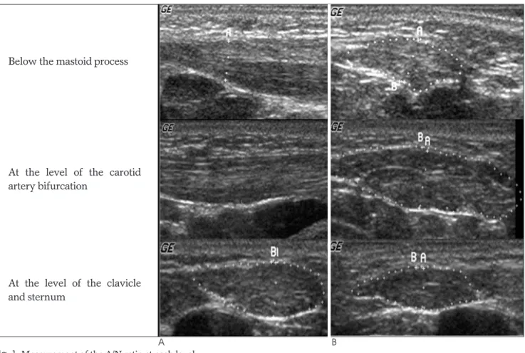
The firm, hard palpable mass detected during the physical examination was defined as “the affected mus- cle”. The ultrasonographic views of the affected muscle were compared with those views of the contra lateral normal side, and the ratio of the affected side to the nor- mal side (the A/N ratio) was calculated (Fig. 1). We fol- lowed up the ultrasonographic results from the begin- ning of physiotherapy at each two months interval until the final assessment was done. All the patients had their ultrasonographic neck procedure done every 2 months.
After every neck ultrasonographic procedure was per- formed, informed consent was obtained from all the parents of the patients before performing the follow up ultrasonographic procedure. Because the ultrasono- grams were retrospectively rechecked by a radiologist, no IRB approval was required. The study was finished when the clinical symptoms along with the palpable masses disappeared. We classified the ultrasonographic echogenecity of the affected SCM muscle into the het- erogeneous or homogenous groups, as compared to those of the contralateral unaffected side, before the treatment, and we followed up the ultrasonographic echogenecity after the treatment.
The data was analyzed with SPSS 12.0KO for Windows (SPSS Inc., Chicago, Illinois, U.S.A.). The Wilcoxon signed-rank test was used to correlate the A/N ratios obtained before and after the treatment.
Spearman’s rank test was used to correlate the A/N ratio and the duration of the total treatment. Using paired T- tests, we tried to correlate the A/N ratio with the treat- ment duration as divided into less than or greater than 12 months. Using paired T-tests, we tried to correlate the echogenecity of the SCM muscle and the treatment duration as divided into less than or greater than 12 months. The Wilcoxon signed-rank test was used to cor- relate the changes in echogenecity of the SCM muscle before and after the treatment. A p value less than 0.05
was regarded as statically significant.
Results
Twenty four unilaterally affected CMT patients (15 boys and 9 girls) with ages ranging from 5 days to 12 months (mean age: 2 months) were analyzed. The right SCM muscle (15 patients) was more frequently affected than the left SCM muscle (9 patients). Six patients were treated for a time longer than 12 months and 18 patients were treated for a time shorter than 12 months. The to- tal number of calculated A/N ratios, with each derived from its thickness and cross sectional circumference) was 96. The A/N ratio of the affected SCM muscle was chosen as 24. We calculated each mean value of the thickness and the cross sectional circumference, and we then compared these values before and after the treat- ment. The most frequently affected site was at the level of the carotid artery bifurcation for 10 of 24 sites.
A significant change in the mean A/N ratio was found for both the thickness and the cross sectional circumfer- ence when the palpable mass disappeared from the neck and the head tilting was normalized. The mean A/N ratio
Fig. 2. Significant change of the A/N ratio was found for both the thickness and the cross sectional circumference of the SCM muscle after the treatment.
A B
Fig. 1. Measurement of the A/N ratio at each level.
A. Longitudinal views of the SCM muscle show the muscle thickness.
B. Axial views of the SCM muscle show the cross sectional circumference.
At the level of the clavicle and sternum, only the axial view was obtained due to obscuring by the bony structure.
The A/N ratio = Affected side SCM muscle (The thickness, circumference) Normal side SCM muscle (The thickness, circumference) Below the mastoid process
At the level of the carotid artery bifurcation
At the level of the clavicle and sternum
of the thickness decreased from 2.31±0.47 to 1.36±
0.39. The mean A/N ratio of the cross sectional circum- ference also decreased from 1.28±0.29 to 1.12±0.24, and this was statistically significant (p<0.05) (Fig. 2).
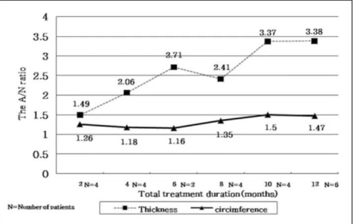
The A/N ratio was proportional to the total treatment duration, and the correlation between the A/N ratio of the thickness and the total treatment duration was statis- tically significant (p<0.05) (Fig. 3). When the treatment duration was longer than 12 months, the mean A/N ra- tio of the thickness was calculated to be 3.38 (range:
2.45-4.31) and when the duration was shorter than 12 months the mean value was calculated to be 2.04 (range:
1.14-2.94). This shows that as the A/N ratio is increased, the treatment duration is also significantly prolonged (p<0.05). Yet the A/N ratio of the cross sectional circum- ference was not correlated with the treatment duration.
The A/N ratio of the thickness between 2.45 and 2.94 before the treatment is the borderline value for deter- mining whether the treatment duration will be longer or shorter than 12 months (Table 1).
There was no significant correlation between the ul- trasonographic echogenecity of the affected SCM mus- cle before the treatment and the treatment duration as divided into less than or greater than 12 months (Table 2). All the patients with homogenous echogenecity be- fore the treatment kept the same echogenecity after the treatment. Most of the patients’ heterogeneous echo- genecity before the treatment was changed into ho- mogenous echogenecity, and this was statistically signif- icant (p < 0.05) (Table 3). So either the patients with ini- tial homogenous echogenecity or the patients with ini- tial hetrogeneous echogenecity that changed to homoge- nous echogenecity during follow up displayed a recov- ered status of the affected muscle.
Discussion
CMT has been described in the literature for many years, yet the etiology of CMT remains unknown de- spite the many hypotheses concerned with its origin (5, 7). Many cases of CMT have been detected at birth or shortly after birth and these cases are related to a history of trauma, a difficult delivery or a breech delivery.
Both light and electron microscopy have consistently shown replacement of the SCM muscle by dense fibrous tissue. This has led to the hypothesis that this condition represents the sequelae of an intrauterine or perinatal compartment syndrome of injury through the birth canal (5).
About 80% of the cases of CMT resolve spontaneously without treatment, but Canale et al found that persistent CMT beyond age of 12 months did not resolve sponta- neously (4). The treatment of CMT consists of physio- therapy, including range of motion exercises, postural training and gentle stretching. Craniofacial asymmetry may persist and scoliosis can develop in children with severe torticollis and who have been treated inadequate-
Table 1. The Mean (±standard deviation) A/N Ratio and the Treatment Duration Divided by Less than or Greater than 12 months
The mean The mean A/N ratio A/N ratio (Cross sectional (Thickness) Circumference) Shorter than 12 months 2.04±0.90 1.40±0.32
n=18
Longer than 12 months 3.38±0.93 1.47±0.28 n=6
p < 0.05 p = 0.064
Table 2. Echogenecity of the Affected SCM Muscle before the Treatment and the Total Treatment Duration
Treatment duration Heterogeneous Homogenous
Shorter than 12 months 7 11
Longer than 12 months 4 2
p = 0.27 p = 0.56
Table 3. Change of the Echogenecity of the SCM Muscle after the Treatment
Echogenecity Before the After the
Treatment treatment p
Homogenous 13 20 p<0.05
Heterogeneous 11 4 p<0.05
Fig. 3. The correlation between the A/N ratio and the total treatment duration was statistically significant for the thick- ness.
ly or for whom surgery has been delayed. For these pa- tients, early surgical correction is crucial for improving not only the head tilting, neck bending and loss of the neck contour, but also to prevent craniofacial asymme- try. Surgery is considered when the symptoms persist after 12 months of physiotherapy or if severe complica- tions are present (4, 8). Thus, predicting the treatment duration is clinically important for determining the need for surgery or continuing the physiotherapy (1-5).
One of the hypotheses suggests that CMT results from the replacement of SCM muscle by the dense fibrous tis- sue that produces either a fibrous mass or a tumor that leads to shortening of the affected muscle (5, 6).
Replacement with fibrous tissue in the SCM muscle may result in the change of the morphological features and ultrasonographic echogenecity of the affected SCM muscle. Yet the morphologically changed SCM muscle is not uniformly outlined. Thus, for this indeterminate form of the affected SCM muscle, we tried to use a new method: the A/N ratio of the thickness and the cross sec- tional circumference.
A significant decrease the A/N ratio of the thickness and the cross sectional circumference after the treat- ment implied that the A/N ratio can use for assessing the treatment effect (Fig. 2). The A/N ratio was proportional to the total treatment period, and so the A/N ratio can help predict the total treatment period and determine the need for surgery (Fig. 3) (Table 1). Particularly, the A/N ratios greater than the mean value in the group with a treatment duration longer than 12 months (greater than 3.38 in the thickness) means they have the possibility of a long treatment duration and they proba- bly need surgical treatment.
Ultrasonographic imaging of the SCM muscle pro- vides information about the arrangement of muscle fibers. Skeletal muscle fibers are grouped in fascicles that are separated from one another by connective tis- sue called perimysium, and the whole muscle is covered by the epimysium. On the axial scans of the normal SCM muscle, the perimysium appears in cross- section as fine dotted echoes and short scattered lines that deter- mine the ultrasonographic echogenecity. A combination of the perimysium and the fibrous component deter- mine the ultrasonographic echogenecity, be it heteroge- neous or homogenous. Microscopically, there is replace- ment of the muscle fiber and the various stages of de- generation of the muscle fibers result in various degrees of ultrasonographic echogenecity. The affected SCM muscles in the patients with CMT have various stages of
fibrotic components, and this may result in various de- grees of ultrasonographic echogenecity (4, 5).
Bias can arrive from the indeterminate morphologic outline of the affected SCM muscle and the morphologic changes in concordance with the variable degree of muscle stretching. In addition to assessing the A/N ratio of the thickness and the cross sectional circumference of the affected SCM muscle, evaluating the thickness seems to be useful for predicting the treatment effect, the treatment duration and to determine the need for surgery. This is because measuring the A/N ratio of the thickness is easy and simple, and there is less alteration of the A/N ratio in proportion to the degree of muscle stretching. In contrast, it is difficult to measure the cross sectional circumference as a constant value. The cross sectional circumference is largely affected by this bias, and so the A/N ratio of the cross sectional circumference has a limited role in predicting the prognosis. Thus, the A/N ratio of the thickness of the SCM muscle has more value in daily practice to predict the prognosis of CMT than does the A/N ratio of the cross sectional circumfer- ence because of these problems.
Our study has some limitations. First, the sample size was too small to define an accurate threshold value for determining the need for surgery. No follow up studies were done after the patients completed treatment.
Second, there was no severe complicated CMT case that required surgery. Although six CMT patients had treat- ment durations longer than 12 months, all of them fully recovered without surgical correction. Therefore, we could not evaluate the ultrasonographic echogenecity and the A/N ratios in the patients who required surgery.
A future study that will include a larger study group of severely complicated cases will help to more accurately determine the prognostic factors and values of patients with CMT.
In conclusion, the A/N ratio of the thickness of the SCM muscle is useful to predict the prognosis of CMT.
References
1. Tatli B, Aydinli N, Caliskan M, Ozmen M, Bilir F, Acar G.
Congenital muscular torticollis: evaluation and classification.
Pediatr Neurol 2006;34:41-44
2. Dudkiewicz I, Ganel A, Blankstein A. Congenital muscular torti- collis in infants: ultrasound-assisted diagnosis and evaluation. J Pediatr Orthop 2005;25:812-814
3. Tang SF, Hsu KH, Wong AM, Hsu CC, Chang CH. Longitudinal follow up study of ultrasonography in congenital muscular torti- collis. Clin Orthop Relat Res 2002; 403:179-185
4. Hsu TC, Wang CL, Wong MK, Hsu KH, Tang FT, Chen HT.
Correlation of clinical and ultrasonographic features in congenital muscular torticollis. Arch Phys Med Rehabil 1999;80:637-641 5. Lin JN, Chou ML. Ultrasonographic study of the sternocleidomas-
toid muscle in the management of congenital muscular torticollis. J Pediatr Surg 1997;32:1648-1651
6. Sonmez K, Tu¨rkyilmaz Z, Demiroqullari B, Ozen IO, Karabulut R,
Baqbanci B, et al. Congenital muscular torticollis in children. ORL J Otorhinolaryngol Relat Spec 2005;67:344-347
7. Cheng JC, Metreweli C, Chen TM, Tang S. Correlation of ultra- sonographic imaging of congenital muscular torticollis with clini- cal assessment in infants. Ultrasound Med Biol 2000;26:1237-1231 8.박창일, 문재호. 재활의학 서울: 한미의학, 2007:645-646
대한영상의학회지 2009;60:133-138
선천성근육사경 환아의 초음파 검사에서 흉쇄유돌근 두께와 예후와의 관계1
1연세대학교 원주의과대학 원주기독병원 영상의학과
2연세대학교 원주의과대학 원주기독병원 재활의학과
임대건∙권우철∙차승환∙유호석∙임상혁∙박정미2∙김명순
목적: 선천성 사경 환자의 예후를 초음파를 이용하여 흉쇄유돌근의 두께와 둘레의 A/N ratio를 구하여 예측하고, 병변이 이환된 흉쇄유돌근의 메아리 양상과 예후와의 관계를 알아보고자 한다.
대상과 방법: 2004년 6월부터 2007년 3월까지 24명의 선천성 사경 환자를 대상으로 초음파를 시행하였다. 흉쇄유 돌근의 두께와 둘레를 3군데에서 측정 하였다. 유양돌기 아래부위, 경동맥 분지 부위, 흉골 부위와 쇄골 부위에서 근 육의 두께, 둘레를 각각 양측에서 측정하였다. 흉쇄유돌근의 병변의 이환부에 대한 정상부의 비율(A/N ratio)을 구 하였다. 치료가 끝날 때까지 2개월마다 정기적으로 추적 관찰하였다. Wilcoxon signed-rank test로 치료 전, 후 의 A/N ratio의 변화를 분석하였고, Spearman’s rank test로 A/N ratio와 총 치료 기간 과의 관계를 평가하였 다. Paired T-test를 사용하여 12개월의 치료기간을 기준으로 나눈 두 그룹 간의 흉쇄유돌근의 메아리 양상을 비교 하였다.
결과: 치료 전 흉쇄유돌근 두께의 A/N ratio (2.31)는 치료 후 의미 있게 감소하였으며(1.36), 통계적으로 유의하 였다(p < 0.05). 두께의 A/N ratio와 총 치료기간은 의미 있는 상관관계를 보였다(p < 0.05). 치료 전 흉쇄유돌근 의 메아리 양상은 치료기간과 상관관계가 없었다.
결론: 흉쇄유돌근 두께의 A/N ratio는 선천성 사경 환자의 예후 예측에 도움을 줄 수 있다.