후측방 접근법을 이용한 흉요추부 척추체 성형술
연세대학교 의과대학 신경외과학교실
김경현ㆍ김긍년ㆍ윤도흠ㆍ신현철ㆍ이 성ㆍ김상현
Posterolateral Approach of Percutaneous Vertebroplasty in Thoracolumbar Fractures
Kyung Hyun Kim, M.D., Keung Nyun Kim, M.D., Do Heum Yoon, M.D., Hyun Chul Shin, M.D., Seong Yi, M.D., and Sang Hyun Kim, M.D.
Department of Neurosurgery, Yonsei University, College of Medicine, Seoul, Korea
Objective: Transpedicular percutaneous vertebroplasty has been widely used for the treatment of osteoporotic vertebral com- pression fractures. The authors introduce a novel method of approach by the posterolateral route for percutaneous vertebro- plasty.
Materials and Methods: A retrospective review was conducted on 30 consecutive patients with 42 vertebrae of acute osteo- porotic thoracolumbar compression fractures treated from Mar. 2002 to May 2003 with percutaneous vertebroplasty using this approach. Pain response, volume of polymethylmethacrylate (PMMA), epidural leakage of PMMA, distribution of PMMA in vertebral body and complications were investigated.
Results: Among 30 patients, 27 had marked to complete pain relief, 2 moderate pain relief and 1 no significant change during follow up period. A mean volume of PMMA injected by this unilateral approach was 5.8cc. Epidural leakage of PMMA was noted on 28 vertebrae(66.7%) on postoperative CT scan. All vertebrae except one injected by this method showed bilateral distribution of PMMA on postoperative CT scan. Minor complication (transient radicular pain) occured in 2(6.7%) patients.
Conclusion: Unilateral posterolateral approach of percutaneous vertebroplasty was a safe and effective method for the treatment of acute osteoporotic thoracolumbar compression fractures.
Key Words: Osteoporotic compression fracture․Vertebroplasty․Posterolateral approach
Crresponding Author: Hyun Chul Shin, M.D.
Department of Neurosurgery, 134, Shinchon-dong, Seodaemun-gu, Seoul, 120-752, Korea
Tel: 82-2-2228-2150/2161, Fax: 82-2-393-9979 E-mail: [email protected]
서 론
1987년 Galibert 등이 척추 혈관종 환자에서 최초로 polyme- thylmetacrylate (PMMA)를 추체내에 주입한 이래4) 경피적 척 추 성형술은 악성 종양이나 혈관종 뿐만 아니라, 골다공증성 추체 골절에 널리 이용되고 있다5,7-9). 골다공증성 척추 골절 에서 경피적 척추 성형술은 통증의 빠른 호전과 조기 보행이
가능하고, 치료에 따른 합병증이 적어서 아주 유용한 방법으 로 보고 되고 있다. 그러나 비교적 드물지만 여러 가지 크고 작은 합병증이 보고 되고있다6,15). 이러한 합병증은 대개 척추 에 주입하는 골시멘트가 추체 외로 유출되거나 잘못된 천자침 의 위치, 방향 때문에 발생한다. 또한 증상의 호전은 주입한 PMMA의 양이나 분포와는 무관하다고 발표되고 있으나2,9), 척 추체 성형술의 생체역학적 측면에서는 적당한 양이 들어가고 추체에 균일하게 분포할 수록 안정성이 더 잘 유지된다고 보 고되고 있다1,11). 현재까지 추체 접근법으로는 추경을 통한 양 측성 또는 일측성 접근법이 주로 시행되어 왔다. 저자들은 흉 요추부 추체 골절에 대해서는 추경을 통하지 않고, 후측방으 로 추체에 직접 접근하여 일측성으로 골성형술을 시행하였 다. 이에 흉요추 추체골절에 대한 골성형술에서 후측방 접근
Fig. 2. Entry point on posterior inferior corner of vertebral body.
Fig. 1. Entry point is 7~8 cm lateral from midline and the angle of needle is approximately 45~60 degree horizontally.
Fig. 4. Vertebral venogram shows paravertebral and epi- dural drainage.
Fig. 3. Needle is directed towards the anterior-superior margin.
법을 소개하고, 그 임상 결과를 보고 하고자 한다.
대상 및 방법
2002년 3월부터 2003년 5월까지 골다공증성 추체 골절로 진단되어 경피적 추체 성형술을 시행한 80명의 환자 중 후측 방 접근법을 시행한 환자 30명(42 척추체)을 대상으로 임상 자료와 방사선 자료를 후향적으로 조사하였다.
1. 수술 전 검사 및 적응증
심한 흉요배부 통증을 호소하면서 단순 X-선 촬영상 압박 골절로 인한 척추체의 변형이 있는 환자를 대상으로 골 밀도 검사, 전신 골 주사 검사, 척추 자기공명영상 검사를 하였다.
시술의 적응증은 진통제 투여 물리치료 등 고식적 치료에 반 응이 없고, 신경근이나 척수 압박 소견이 없는 환자를 대상 으로 하였다. 이들 환자 중 압통부위와 자기공명영상에서 T1 강조 영상상 저음영을 보이며, 골 주사 검사에서 강한 섭취 를 보인 부위가 일치하는 환자를 시술하였다.
2. 수술 방법
시술은 혈관 촬영실에서 시술대 위에 환자를 복와위로 눕 히고 혈압, 맥박, 심전도를 감시할 수 있게 하였다. 일반적인 방법으로 시술할 부위를 소독한 후에 척추 정중선에서 7~8
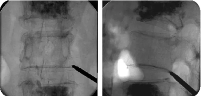
cm 외측에 Biplane C-arm fluoroscope 하에서 1% lidocaine으 로 국소 마취 후 수평면에서 45~60도 각도로 11G 천자침을 넣는다(Fig. 1). 천자침의 방향은 추체의 posterior-inferior cor- ner에서 anterior-superior 방향으로 넣는다(Fig. 2).
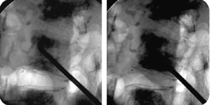
천자침의 위치는 측방에서 추체의 2/3 정도 앞쪽에 전후 방에서는 반대편 추경 부위에 위치한다(Fig. 3) 조영제를 주 입하여 척추체 정맥조영술을 시행한다(Fig. 4). 이때 추체 주
위의 정맥이나 척추강내로 조영제가 유출되는 지를 관찰하 여 정확한 천자침의 위치를 정하고 PMMA의 점도를 결정한 다. 적당한 점도의 PMMA를 5 cc syringe 두개에 나누어 넣은 후 천자침을 서서히 빼면서 2 cc씩 3회 주입한다(Fig. 5). 이 때 전과정은 투시 검사 장치로 감시하면서 PMMA가 척추체 외 조직이나 정맥, 혹은 척추 강내로 유출되는 소견이 보이 면 즉시 주입을 중단한다.
Fig. 5. PMMA was injected by slow withdrawal method under the fluoroscopic guidance.
Fig. 6. Postoperative CT scan shows even and bilateral distribution of PMMA in the body.
Table 1. Clinical results of percutaneous vertebroplasty via posterolateral approach (postoperative 1day and 3 months)
Result postop 1 days postop 3 months Excellent
Good Fair Poor
8 18 3 1
16 11 2 1
Total 30 30
3. 자료 수집 및 정리
이들 환자의 임상자 료와 방사선 소견을 검토하여 수술 후 통증의 호전 정도(시술 1일째와 3개월째), 주입한 PMMA 의 양, PMMA의 척추강 내로의 유출 빈도, PMMA의 추체내 에 분포양상, 합병증 등을 조사하였다. 수술 후 통증의 호전 정도는 완전히 통증이 소실된 경우는 excellent, 현저히 호전 된 경우는 good, 경미한 호전을 보인 것은 fair, 호전이 없거 나, 악화된 경우는 poor로 나누어 조사하였다.
결 과
2002년 3월부터 2003년 5월까지 총 30명의 환자 중 남자가 7명 여자가 23명이었으며, 평균 나이는 62.5세였다. 모두 일측 성으로 실시하였고, 양측성으로 시행한 경우는 없었다. 한 부 위를 시행한 환자가 20명이었고, 두 부위가 8명 세 부위가 2
명으로 총 42 척추체에 대하여 시술하였다. 시술한 부위는 제 12 흉추가 16, 제1 요추가 20, 제2 요추가 3, 제3 요추가 2, 제4 요추가 1개 있었다. 수술 후 통증의 호전 정도를 조사한 결과 시술 후 1일째 excellent가 8명 good이 18명 fair가 3명, poor가 1명이었고, 3개월째 는 excellent가 16명, good이 11명 fair가 2 명, poor가 1명이었다(Table 1). 수술 후 통증의 호전이 없었던 한 환자는 척추체 외로의 유출이 심했던 경우였다. PMMA의 양은 최저가 2 cc, 최고가 8 cc를 주입하였으며 평균 5.8 cc를 주입하였다. 수술 후 1일째 전 환자에서 전산화단층촬영(CT) 을 시행하였으며, CT상 척추강 내로 PMMA의 유출이 28 척 추체(66.7%)에서 관찰되었다. 이중 2례는 골절된 척추체 후면 을 통해 유출된 것으로 사료되며, 나머지는 경막외 정맥을 통 해 유출된 것으로 사료 된다. CT 소견상 PMMA의 분포를 보 면 2 cc 만을 주입한 한 예를 제외하고는 전 예에서 시술한 반대편까지 PMMA가 분포하는 것을 확인할 수 있었다. 합병 증으로는 2례에서 일시적인 신경근 이상 소견이 있었으나, 보 존적 치료 후 모두 소실되었다.
고 찰
골다공증성 척추 골절에서 경피적 척추 성형술은 통증의 빠른 호전과 조기 보행이 가능하고, 치료에 따른 합병증이 적어서 아주 유용한 방법으로 보고되고 있다5,7-9). 척추체 성 형술에서 통증의 완화 기전은 골절된 골편의 안정화와 추체 로 전달되는 압박 압력의 감소, 추체내의 신경 말단의 파괴 로 설명되고 있다3,10,14,16). Tohmeh 등은 사체 척추에 PMMA 를 주입 후 측정한 추체의 강도를 측정하여 압력에 대한 저 항이 증가한 것을 확인하여 척추 성형술의 생체 역학적 근거 를 제시하였다17). 척추 성형술의 치료 성적을 발표한 여러 논문에서 주입한 PMMA의 양이나 분포 정도와 임상적 치료 성적은 별 관계가 없다고 주장되어 지고 있고, 오히려 적은 량을 주입할 때 추체외로의 유출을 막을 수 있어 합병증을
예방할 수 있다고 주장되기도 한다2,9). 그러나 이러한 임상 결과의 보고는 아직 장기 추적 결과가 아닌 초기의 임상 성적 이다. 오히려 최근에 발표되는 생체역학적 논문에서는 적정량 이상의 PMMA의 주입이 골저항력을 증가시키고, PMMA의 분 포도 양측성으로 된경우에만, 적정한 저항력을 유지 할 수 있다고 발표되었다1,11). 기존에 흉요추 추체 성형술에서는 대 부분 경추경적 방법으로 시도되고 있고, 극히 일부에서 후측 방 접근법이 시도되어졌다. 즉 후방기기 고정술을 시행한 부 위에 골다공증성 골절이 생긴 경우나, 종양이 추경을 침범한 경우에 제한적으로 시도되었다12). 후측방 접근법으로 시도한 경우에도 추체외로의 유출이 많은 것으로 보고되기도 하였 다13). 그러나 저자들의 경험으로는 추체외로의 유출은 그간 발표된 논문들과 비교하여 차이가 없으며, 추체외로의 유출 은 접근법의 문제 보다는 골절된 추체의 해부학적 변화, 주입 하는 PMMA의 점도, 주입시의 압력과 더 밀접한 관계가 있을 것으로 사료된다. 저자들이 경험한 후측방 접근법의 치료성적 은 경추경 접근법을 이용한 방법과 차이가 없으며, PMMA의 추체외로의 유출도 비슷한 결과를 보이고 있다. 오히려 생체 역학적 측면에서는 대부분의 경우 추체 내에 양측성으로 PMMA를 분포 시킬 수 있어 경추경법에 비해 보다 유리한 측면이 있다. 경추경법은 천자침이 추경을 통과할 때 경우 척추강과 인접해서 통과 해야 하는 부담이 있고, 일측성으로 만 되지않고, 양측성으로 해야 하는 경우가 자주 발생하므로 경제적인 부담면에서도 차이가 있다. 후측방 접근법은 추간 반 조영술이나 관절경을 이용한 추간반 제거술과 비슷한 경 로를 갖는 접근법이다. 따라서 척추 전문의사는 누구나 경험 이 많은 접근법이다. 그러나 11흉추 이상의 흉추 골절 환자에 서는 폐와 횡경막의 손상을 줄 위험이 있어 시행할 수 없는 단점이 있다. 또한 천자침이 추간공 주위를 통과해야 하므로 추간공 부근에서 신경근 손상을 주지 않도록 주의를 요한다.
본 연구에 대상이 된 환자 중 1례에서 천자침 주입시 하지통 이 발생한 경우가 있었다. 후측방 접근법을 이용한 추체 성형 술은 장기 추적 결과의 관찰이 요구되나, 초기 성적으로는 비 교적 안전하며, 높은 성공률을 보이는 방법으로 사료된다. 향 후 경추경 접근법과의 비교 연구가 필요할 것으로 사료된다.
결 론
후측방 접근법은 일측성으로 시행함에도 PMMA를 추체 의 양측에 균등하게 분포시킬 수 있는 장점이 있는 방법이
며, 통증의 호전이나 합병증의 발생에서는 기존의 경추경법 과 비슷한 결과를 보이는 방법이다. 그러나 흉추 11번 이상 의 흉추체는 폐나 횡경막 때문에 본 접근법을 시행할 수 없 으며, 천자침 삽입 시 추간공 부위에서 신경근 손상에 유의 하여야 할 것이다. 골다공증성 흉요추체 압박 골절의 골성형 술에서 일측성 후측방 접근법은 안전하며, 효과적인 시술 방 법으로 사료된다.
참 고 문 헌
1. Belkoff SM, Mathis JM, Jasper LE, Deramond H: The bio- mechanics of vertebroplasty. The effect of cement volume on mechanical behavior. Spine 26(14):1537-1541, 2001 2. Cotten A, Dewatre F, Cortet B, Assaker R, Leblond D,
Duquesnoy B, et al: Percutaneous vertebroplasty for osteoly- tic metastases and myeloma: Effects of the percentage of lesion filling and the leakage of methyl methacrylate at clinical follow-up. Radiology 200(2):525-530, 1996
3. Dean JR, Ison KT, Gishen, P: The strengthening effect of percutaneous vertebroplasty. Clin Radiol 55(6):471-476, 2000 4. Galibert P, Deramond H, Rosat P, Le Gars D: Preliminary
note on the treatment of vertebral angioma by percutaneous acrylic vertebroplasty. Neurochirurgie 33(2):166-168, 1987 5. Gangi A, Kastler BA, Dietemann JL: Percutaneous verteb-
roplasty guided by a combination of CT and fluoroscopy.
AJNR Am J Neuroradiol 15(1):83-86, 1994
6. Harrington KD: Major neurological complications following percutaneous vertebroplasty with polymethylmethacrylate: A case report. J Bone Joint Surg Am 83-A(7):1070-1073, 2001 7. Jensen ME, Dion JE: Vertebroplasty relieves osteoporosis
pain. Diagn Imaging(San Franc) 19(9):68, 71-72, 1997 8. Jensen ME, Dion JE: Percutaneous vertebroplasty in the treat-
ment of osteoporotic compression fractures. Neuroimaging Clin N Am 10(3):547-568, 2000
9. Jensen ME, Evans AJ, Mathis JM, Kallmes DF, Cloft HJ, Dion JE: Percutaneous polymethylmethacrylate vertebroplasty in the treatment of osteoporotic vertebral body compression fractures: Technical aspects. AJNR Am J Neuroradiol 18 (10):1897-1904, 1997
10. Levine SA, Perin LA, Hayes D, Hayes WS: An evidence-
based evaluation of percutaneous vertebroplasty. Manag Care 9(3):56-60, 63, 2000
11. Liebschner MA, Rosenberg WS, Keaveny TM: Effects of bone cement volume and distribution on vertebral stiffness after vertebroplasty. Spine 26(14):1547-1554, 2001
12. Martin JB, Jean B, Sugiu K, San Millan Ruiz D, Piotin M, Murphy K, et al: Vertebroplasty: Clinical experience and follow-up results. Bone 25(2 Suppl):11S-15S, 1999
13. Mathis JM, Barr JD, Belkoff SM, Barr MS, Jensen ME, Deramond H: Percutaneous vertebroplasty: A developing stand- ard of care for vertebral compression fractures. AJNR Am J Neuroradiol 22(2):373-381, 2001
14. Maynard AS, Jensen ME, Schweickert PA, Marx WF, Short JG, Kallmes DF: Value of bone scan imaging in predicting
pain relief from percutaneous vertebroplasty in osteoporotic vertebral fractures. AJNR Am J Neuroradiol 21(10):1807- 1812, 2000
15. Ratliff J, Nguyen T, Heiss J: Root and spinal cord compre- ssion from methylmethacrylate vertebroplasty. Spine 26(13):
E300-302, 2001
16. San Millan Ruiz D, Burkhardt K, Jean B, Muster M, Martin JB, Bouvier J: Pathology findings with acrylic implants. Bone 25(2 Suppl):85S-90S, 1999
17. Tohmeh AG, Mathis JM, Fenton DC, Levine AM, Belkoff SM: Biomechanical efficacy of unipedicular versus bipedi- cular vertebroplasty for the management of osteoporotic com- pression fractures. Spine 24(17):1772-1776, 1999