ISSN 0378-6471 (Print)⋅ISSN 2092-9374 (Online)
http://dx.doi.org/10.3341/jkos.2015.56.12.1906
Original Article
Dynamic Contour Tonometry와 안혈류분석계의 안구 박동 크기 비교
Comparison of Ocular Pulse Amplitude Measured Using Dynamic Contour Tonometry and Ocular Blood Flow Analyzer
김용현1⋅이마빈2⋅안재홍1
Yong Hyun Kim, MD1, Mar Vin Lee, MD2, Jae Hong Ahn, MD, PhD1
아주대학교 의과대학 안과학교실1, 분당제생병원 안과2
Department of Ophthalmology, Ajou University School of Medicine1, Suwon, Korea Department of Ophthalmology, Bundang Jesaeng Hospital2, Seongnam, Korea
Purpose: To compare ocular pulse amplitude (OPA) measured using dynamic contour tonometry (DCT) and ocular blood flow analyzer (BFA).
Methods: Thirty-five eyes of 35 patients were enrolled in this cross-sectional and retrospective study. OPA was measured using DCT. Pulse amplitude (PA) and pulsatile ocular blood flow were measured using BFA.
Results: OPA measured using DCT (2.79 ± 0.89 mm Hg) was not significantly different from PA measured with BFA (3.02 ± 0.90 mm Hg; p = 0.082) and both were significantly correlated (r = 0.663, p < 0.001). Mean difference ± limit of agreement was -0.22 ± 1.44 mm Hg between OPA and PA. OPA correlated significantly with intraocular pressure (IOP) measured using Goldmann applanation tonometry (r = 0.330, p = 0.047) but not PA (r = 0.057, p = 0.745). Both PA and OPA did not show sig- nificant correlation with the spherical equivalent of refractive error and central corneal thickness.
Conclusions: Although both OPA and PA measure IOP fluctuation and are not significantly different, they showed different rela- tionships with IOP.
J Korean Ophthalmol Soc 2015;56(12):1906-1912
Key Words: Intraocular pressure, Ocular blood flow, Ocular pulse amplitude
■Received: 2015. 2. 6. ■ Revised: 2015. 7. 24.
■Accepted: 2015. 9. 25.
■Address reprint requests to Jae Hong Ahn, MD, PhD
Department of Ophthalmology, Ajou University Hospital, #164 World cup-ro, Yeongtong-gu, Suwon 16499, Korea
Tel: 82-31-219-5261, Fax: 82-31-219-5909 E-mail: chrisahn@ajou.ac.kr
ⓒ2015 The Korean Ophthalmological Society
This is an Open Access article distributed under the terms of the Creative Commons Attribution Non-Commercial License (http://creativecommons.org/licenses/by-nc/3.0/) which permits unrestricted non-commercial use, distribution, and reproduction in any medium, provided the original work is properly cited.
박동 안구 혈류(Pulsatile ocular blood flow, POBF)는 공 기 압평 안압계를 통해 측정되는 값으로, 녹내장의 병인과 연관이 있다고 보고된 바 있다.1-3 POBF는 심장의 수축과 이완에 따라 눈에 유입되는 혈류량의 차이로 인해 발생하 게 되며 눈주위 혈류량을 반영하는 인자로 생각되고 있는
데 과거의 연구들을 살펴보면 인위적으로 안구로의 관류압 을 떨어트린 눈에서는 POBF가 감소하며 임신 후 채액량이 증가하게 되면 POBF가 증가한다는 보고가 있다.4,5 POBF 는 안구 박동 크기(ocular pulse amplitude, OPA)라 불리는 안압의 주기적인 변화로부터 얻어진다. OPA는 심박동에 의한 수축기와 이완기의 혈압 차이에 의해 발생하는 것으 로 보이나, 전신적인 혈역학적 수치와의 연관성은 명확하 지 않다고 보고되어 왔다.6,7 이는 OPA가 안압, 혈관 저항, 맥락막 두께, 자동 조절 기능, 안구 경직 정도 등 다양한 변 수에 의해 영향을 받는 수치이기 때문으로 생각된다.8-12 또 한 OPA에 영향을 주는 이러한 다양한 변수들은 녹내장의
병인과 연관이 있을 것으로 추정되어 왔다.
Dynamic contour tonometry (DCT)는 안압을 측정할 수 있는 오목한 모양의 탐침을 각막에 접촉하여 연속적으로 안압을 측정하여 OPA를 측정하게 되며,13 다른 안압계의 측정 방식과는 다르다. 이전의 연구들에서는 OPA가 비압 평방식의 접촉 방식인 DCT, 혹은 각막에 가해지는 공기압 력과 각막의 반발력의 균형점을 찾는 공기 압평 방식의 안 구 혈류 분석계(Ocular blood flow analyzer, BFA)를 통해 서 측정되었다.8,13,14 이러한 두 가지 방식으로 측정한 안구 박동 크기의 경우 매우 짧은 시간 동안의 안압 변화를 측정 하여 얻게 되는 값이나, 서로 다른 측정 알고리즘으로 인해 안구 혹은 전신적인 혈역학적 측정값과의 상관관계에 있어 서 차이가 생길 수 있다.
본 연구에서 저자들은 DCT와 BFA를 통해 측정된 안압, 안구 박동 크기를 비교하였고, 이들 값과 안구 계측치 및 전신적인 혈역학적 측정값과의 관계를 알아보고자 하였다.
대상과 방법
본 연구는 후향적-단면 연구로, 2012년 8월부터 2012년 10월까지 아주대학교병원 녹내장 클리닉에 내원한 환자들 의 의무기록을 분석하여 진행하였다. 79명의 환자 146안에 서 30분 이내에 Pascal Dynamic Contour Tonometry (DCT, Swiss Microtechnology AG, Port, Switzerland)와 Ocular Blood-Flow Analyzer (BFA, Paradigm Medical, Salt Lake City, UT, USA)를 통해 OPA를 측정하였다. 본 연구는 아 주대학교병원 Institutional Review Board의 승인을 받았다 (AJIRB-MED-MDB-12-350).
모든 환자에서 병력 문진, 시력, 굴절이상의 구면렌즈 대 응치, 세극등 검사, 안저 검사 등 전반적인 안과 검사가 시행 되었다. Ultrasonic pachymetry (DGH-500; DGH Technology, Exton, PA, USA)를 통해 중심 각막 두께를 측정하였고, 골 드만 압평 안압계(Goldmann applanation tonometry, GAT) 를 이용하여 안압을 측정하였다. DCT를 이용하여 안압과 OPA를 측정하였고, BFA를 이용하여 평균 안압, 안압변동 의 크기인 박동 크기(pulse amplitude measured by BFA, PA), 박동 시 용적의 변화량인 박동 용적(pulse volume measured by BFA, PV), POBF를 측정하였다. 안압의 측정 은 진료실에서 GAT와 DCT의 순서대로 측정되었으며 검 사실로 이동하여 BFA 검사를 시행하였으며 모든 검사는 30분 이내에 측정이 이루어졌다.
앉은 상태에서 3분 이상 휴식을 취한 뒤 자동 혈압계를 통해 우측 상완부에서 수축기 혈압(systolic blood pressure, SBP) 및 이완기 혈압(diastolic blood pressure, DBP)을 측
정하였다. 환자들에게는 혈압 측정 전 혈압에 영향을 줄 수 있는 육체적 활동을 자제하도록 하였다. 혈압은 2번 측정하 였으며, 두 값 중 낮게 측정된 혈압을 분석에 이용하였다.
평균 동맥압(mean arterial pressure, MAP)은, MAP=[2 DBP+SBP]/3의 공식을 이용하여 계산하였다.15 평균 안구 관류압(mean ocular perfusion pressure, MOPP)은 MOPP=2/3 (DBP+1/3 [SBP-DBP])-GAT intra ocular pressure (IOP)의 공식을 이용하여 계산하였다.16
본 연구는 안질환이나 수술 및 안압하강제 사용 등이 결 과에 미치는 영향을 배제하기 위해 검사를 시행한 환자의 의무기록을 녹내장 전문의가 검토하여 녹내장을 포함한 다 른 전신질환이나 안과적 질환이 없는 정상인을 대상으로 진행하였으며, 연구에 참여한 모든 환자들의 최대교정시력 은 20/30 이상이었다. DCT와 BFA는 제조사로부터 제공된 측정 방법대로 측정하였다. DCT로 측정된 quality score (Q)가 3보다 크고 BFA 측정이 incomplete로 기록되거나 분 산 값이 크게 측정된 경우는 연구에서 제외하였다. 시행한 검사에서 녹내장으로 진단되었거나 이전에 녹내장 또는 고 안압으로 진단되어 약물치료를 시행한 환자는 대상에서 제 외하였으며, 이전에 백내장을 포함한 안내 수술을 받았거 나, 굴절 교정수술을 받은 경우, 아르곤 레이저 주변부 홍 채절개술을 포함한 레이저 시술을 받은 안과적 과거력이 있는 경우는 연구에서 제외하였다.
두 가지 장비의 기술 통계학적 통계분석을 시행하였고, 두 변수 간의 상관관계는 SPSS 프로그램(ver. 20.0; SPSS Inc., Chicago, IL, USA)을 이용해 평가하였다. 양안 모두 본 연구 대상의 포함 기준에 만족하는 경우는 SPSS 프로그 램의 난수발생기를 이용하여 양안 중 무작위로 연구의 대 상안을 선택하였다. 두 장비의 재현성에 대한 통계 분석은 MedCalc 프로그램(Version 11, MedCalc Software, Ostend, Belgium)을 이용하였다. 모든 통계 분석에 있어서 p값이 0.05보다 작은 경우를 통계적으로 의미 있다고 판단하였다.
결 과
79명 중 녹내장으로 진단되었거나 녹내장으로 진단되어 약물치료 중인 환자 총 42명이 제외되었으며 4안은 검사 결과의 신뢰도가 충족기준에 맞지 않아 제외되었다. 최종 적으로 35명의 환자 35안이 본 연구에 포함되었다. 환자들 의 평균 나이는 47.0 ± 10.4세였고, GAT, DCT, BFA로 측 정한 평균 안압은 각각 15.8 ± 3.17, 20.01 ± 4.21, 22.89 ± 5.78 mmHg였다. DCT로 측정한 OPA의 평균값은 2.79 ± 0.89 mmHg였으며, BFA로 측정한 PA의 평균값은 3.02 ± 0.90 mmHg였다(Table 1). 3가지 장비를 통해 측정한 안압
Table 1. Descriptive statistics of enrolled subjects
Parameter Mean ± SD Minimum Median value Maximum
Age (years) 47.0 ± 10.4 31 45 71
Male/female ratio 17/18 - - -
MAP (mm Hg) 94.2 ± 10.1 70.3 94.7 115.7
PP (mm Hg) 50.3 ± 10.9 33 50 75
MOPP (mm Hg) 47.0 ± 6.8 33.9 46.7 60.2
PR (beats per minute) 77.8 ± 11.4 54 76 108
CCT (μm) 545.84 ± 38.06 458.0 543.0 629.3
SE (diopters) -2.036 ± -2.790 -9.50 -0.50 1.50
GAT IOP (mm Hg) 15.8 ± 3.17 10 15 21
DCT IOP (mm Hg) 20.01 ± 4.21 11.6 20.6 29.1
OPA (mm Hg) 2.79 ± 0.89 1.3 2.7 5.3
BFA IOP (mm Hg) 22.89 ± 5.78 12.8 22.9 35.9
PA (mm Hg) 3.02 ± 0.90 1.5 3.1 5.9
PV (μL) 4.92 ± 1.75 1.8 4.9 9.5
POBF (μL/sec) 12.21 ± 3.83 6.2 12.6 21.8
Values are presented as mean ± SD unless otherwise indicated.
SD = standard deviation; MAP = mean arterial pressure; PP = pulse pressure; MOPP = mean ocular perfusion pressure; PR = pulse rate;
CCT = central corneal thickness; SE = spherical equivalent; GAT = Goldmann applanation tonometry; IOP = intraocular pressure; DCT
= dynamic contour tonometry; OPA = ocular pulse amplitude; BFA = ocular blood flow analyzer; PA = pulse amplitude measured by BFA;
PV = pulse volume measured by BFA; POBF = pulsatile ocular blood flow measured by BFA.
Table 2. Comparison of IOP differences measured by 3 different devices and correlations between IOP measurements
Compared parameters Mean difference(± standard deviation) p-value* LOA (mm Hg)
Range (mm Hg)
Correlation† between
two parameters p-value
GAT IOP-DCT IOP -4.2 ± 2.9 <0.001 -9.8, 1.4 11.2 0.733 <0.001
GAT IOP-BFA IOP -7.1 ± 3.8 <0.001 -14.5, 0.3 14.8 0.795 <0.001
DCT IOP-BFA IOP -2.9 ± 3.4 <0.001 -9.6, 3.9 13.6 0.808 <0.001
OPA-PA -0.22 ± 0.74 0.082 -1.66, 1.22 2.88 0.663 <0.001
Values are presented as mean ± SD unless otherwise indicated.
IOP = intraocular pressure; LOA = limit of agreement; GAT = Goldmann applanation tonometry; DCT = dynamic contour tonometry; BFA
= ocular blood flow analyzer; OPA = ocular pulse amplitude measured by DCT; PA = pulse amplitude measured by BFA.
*Paired t-test; †Pearson correlation coefficient.
Table 3. Correlations between IOP or OPA-derived parameters and systemic or other ocular parameters (Pearson correlation co-
efficient R and p-value in parenthesis)Age CCT SE
GAT IOP -0.211 (0.224) 0.296 (0.047) -0.278 (0.106)
DCT IOP -0.004 (0.980) 0.235 (0.174) -0.268 (0.120)
BFA IOP -0.258 (0.134) 0.144 (0.409) -0.900 (0.079)
OPA -0.031 (0.861) -0.146 (0.404) 0.154 (0.376)
PA 0.123 (0.482) -0.165 (0.344) 0.308 (0.072)
POBF 0.209 (0.228) -0.339 (0.047) 0.431 (0.010)
IOP = intraocular pressure; OPA = ocular pulse amplitude measured by DCT; CCT = central corneal thickness; SE = spherical equivalent;
GAT = Goldmann applanation tonometry; DCT = dynamic contour tonometry; BFA = ocular blood flow analyzer; PA = pulse amplitude measured by BFA; POBF = pulsatile ocular blood flow measured by BFA.
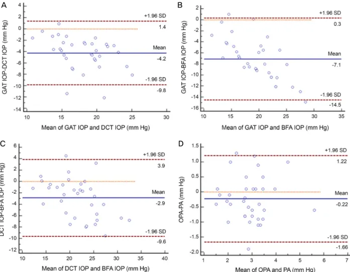
은 통계적으로 유의하게 달랐다(p<0.001). BFA로 측정한 안압이 가장 높았고, 이어서 DCT, GAT 순이었다. BFA로 측정한 PA값은 DCT로 측정한 OPA 값에 비해 높았지만 통 계적으로 유의하지는 않았다(평균값 차 0.22 ± 0.74 mmHg, p=0.082; Table 2). 장비 간 안압 측정의 일치도를 Bland- Altman plot으로 측정하였으며 Mean differences ± limit of
agreement (LOA)로 산출하였다. LOA는 95% 신뢰구간에 해당하는, 표준편차에 1.96을 곱한 값으로 하였다. GAT와 DCT에서는 -4.2 ± 5.6 mmHg, GAT와 BFA에서는 -7.1 ± 7.4 mmHg, DCT와 BFA에서는 -2.9 ± 6.7 mmHg였다.
OPA 측정의 일치도를 평가하기 위한 LOA는 DCT와 BFA 에서 -0.22 ± 1.44 mmHg였다(Table 2, Fig. 1).
Figure 1. Agreement between intraocular pressure and ocular pulse amplitude measured by different devices. (A) Bland-Altman plot
showing the inter-instrument repeatability of IOP measurements by DCT and GAT. (B) Bland-Altman plot showing the inter-instru- ment repeatability of IOP measurements by ocular BFA and GAT. (C) Bland-Altman plot showing the inter-instrument repeatability of IOP measurements by BFA and DCT. (D) Bland-Altman plot showing the inter-instrument repeatability of OPA measurements by DCT with measurements using BFA. GAT = Goldman applanation tonometry; IOP = intraocular pressure; DCT = dynamic contour tonometry; BFA = ocular blood flow analyzer; OPA = ocular pulse amplitude; PA = pulse amplitude measured by BFA;SD = standard deviation.
Table 4. Correlations between OPA-derived parameters and IOP measurements (Pearson correlation coefficient R and p-value in pa-
renthesis)GAT IOP DCT IOP BFA IOP
OPA 0.330 (0.047) 0.374 (0.027) 0.330 (0.053)
PA 0.057 (0.745) 0.100 (0.566) 0.003 (0.986)
POBF -0.328 (0.049) -0.373 (0.027) -0.373 (0.003)
OPA = ocular pulse amplitude measured by DCT; IOP = intraocular pressure; GAT = Goldmann applanation tonometry; DCT = dynamic contour tonometry; BFA = ocular blood flow analyzer; PA = pulse amplitude measured by BFA; POBF = pulsatile ocular blood flow meas- ured by BFA.
안압과 OPA, 그리고 PA 모두 평균 동맥압(MAP), 평균 안구 관류압(MOPP)과 통계적으로 유의한 상관관계를 보 이지 않았다(data not shown, p>0.05). 중심 각막 두께는 GAT 안압(r=0.296, p=0.047)과는 양의 상관관계를 보였으 나 DCT 안압(r=0.235, p=0.174), BFA 안압(r=0.144, p=0.409)
과는 상관관계를 보이지 않았다. 구면렌즈 대응치는 안압 및 OPA, 그리고 PA와는 상관관계를 보이지 않았으나 POBF (r=0.431, p=0.010)와는 양의 상관관계를 보였다(Table 3).
DCT OPA는 GAT (r=0.330, p=0.047)와 DCT (r=0.374, p=0.027)로 측정한 안압과 양의 상관관계를 보였다. 또한
A B
C D
BFA로 측정한 PA (r=0.663, p<0.001) 및 POBF (r=0.351, p=0.039)와 유의한 상관관계를 보였다. 반면에 BFA PA는 세 가지 장비로 측정한 안압과 통계적으로 유의한 상관관 계를 보이지 않았다(r=0.003-0.100, p>0.500). POBF는 모든 안압 측정치와 음의 상관관계를 보였다(r=-0.328~-0.373, p<0.05) (Table 4).
고 찰
POBF는 안구 혈류의 박동성을 평가할 수 있는 측정값이 며, 매우 짧은 시간 동안의 안압 변화인 OPA 측정을 통해 구할 수 있다. POBF는 OPA 측정을 통해 얻는 값으로, OPA와 상관관계가 높은 것으로 보고되었다.14 그동안 OPA 는 POBF를 대체할 수 있는 값으로 여겨져 왔을 뿐만 아니 라, DCT가 도입되면서부터 DCT를 통한 OPA 측정이 이전 의 공기압 안압 측정 방식을 통한 장비로 OPA를 측정하는 것에 비해 용이하고, 높은 재현성을 보여 많은 OPA 관련 논문들이 보고되었다.17 DCT로 측정된 OPA는 POBF를 측 정하는 장비로부터 결정된 박동 크기와 그 특성이 유사하 다는 개념을 바탕으로 하여 이러한 DCT OPA 관련 연구들 이 보고되어 왔다.
이전에 DCT를 통해 측정한 OPA와 BFA를 통해 측정한 PA를 비교한 연구가 없었으나, 이전 연구에서 다른 장비로 측정한 안압 측정치들은 서로 차이를 보였으며, 다른 변수 들과도 서로 다른 연관성을 보였다.17,18 본 연구에서 GAT, DCT, BFA를 통해 측정한 안압은 서로 차이가 있으며 OPA와 PA는 유의한 차이가 없으나 BFA로 측정된 PA가 DCT로 측정된 OPA에 비해 높은 경향을 보여주었다(Table 2). 이는 본 연구에서 측정된 DCT IOP보다 BFA IOP가 더 높게 측정된 것의 영향일 수도 있으나 PA가 안압 측정치에 영향을 받지 않는 것으로 보여(Table 4) 이로 인한 영향은 적을 것으로 생각된다. DCT와 GAT로 측정된 안압을 비교 한 이전의 연구에서는 두 안압계 간의 유의한 차이가 없다 는 보고도 있으나19 DCT로 측정한 안압이 GAT로 측정한 안압에 비해 유의하게 높다는 연구가 더 많았으며 이러한 차이는 각막 두께에 영향을 받는다고 보고된 바 있다.20-22 즉 각막두께가 얇은 경우 DCT 안압은 GAT 안압보다 더 높게 측정되는 경향이 있다. BFA로 측정한 안압과 다른 안 압계로 측정된 안압에 대한 직접적인 비교 연구는 아직 없 었는데 이는 BFA를 사용하는 목적이 안압보다는 POBF와 PA를 측정하기 위한 것이기 때문으로 생각된다. 본 연구에 서 BFA로 측정된 안압이 다른 안압계에 비해 높게 측정된 것은 아직 이에 대한 비교 연구가 없어 원인을 분석하기 어 려우나 대상 환자의 수가 적어 각막두께, 안축장, 굴절이상
등의 안인자와 전신적 인자의 분포가 편중되어 나타난 결 과일 수도 있으며 향후 이를 보완한 연구가 더 필요할 것이 라 생각된다.
본 연구에서 DCT OPA는 BFA PA와 유의한 상관관계를 보였으나(Pearson r=0.663, p<0.001), POBF와의 상관계수는 (r=0.351) 이보다 낮았다(Table 2, 4). DCT OPA와 BFA PA 는 다른 안과적 요소와 유사한 연관성을 보였으나(Table 3), OPA는 안압과 유의한 상관관계를 보인 반면에 PA는 그렇 지 않았다(Table 4). 따라서 DCT로 측정된 OPA의 경우 BFA로 측정된 PA에 비해 안압에 더 영향을 받는 것으로 보인다. 즉 Pascal DCT로 측정한 OPA의 감소는 POBF가 감소했음을 의미할 수도 있지만 안압이 낮아졌음을 시사한 다. Acetazolamide가 안압 및 OPA에 미치는 영향에 대한 서로 다른 결과의 두 가지 보고가 있었다. Haustein et al23 에 의해 보고된 공기압 안압 측정 방식(OBF system 3000) 을 통해 측정된 pulse amplitude와 POBF는 안압이 baseline 보다 20% 이상 감소하여도 pulse amplitude와 POBF는 유 의한 변화가 없었다고 보고하였다. 반면에 DCT를 통해 OPA를 측정한 다른 연구에서는 acetazolamide 투약 후 2시 간 뒤 OPA와 IOP가 각각 21%, 17% 감소했다고 보고하였 다.24 또한 섬유주 절제술과 백내장 수술 후 DCT OPA의 감소는 술 후 안압 하강 정도와 유의한 상관관계가 있었다 는 연구 결과도 있었다.25,26 이러한 연구 결과는 OPA는 안 압의 변화에 영향을 받고 PA는 받지 않음을 보여준 본 연 구 결과와 일치하는 결과로 생각할 수 있다. 따라서 DCT OPA는 POBF 측정의 근거가 되는 BFA PA와 좋은 상관관 계를 보이지만, 안압에 영향을 받으며 POBF와의 상관관계 도 다소 낮으므로 POBF를 반영하는 측정치로 사용하기에 는 제한점이 있다고 생각된다.
OPA와 POBF의 경우 안축장 길이, 안압 등 여러 요소들 에 의한 영향을 받을 수 있어,3,8,12,27 환자들의 안축장 길이 를 측정하지 않았다는 점은 본 연구의 제한점이 될 수 있 다. 또한 측정값들의 일중 변화를 확인하기 위하여 하루 중 다른 시기에 반복 측정을 했다면 결과는 더 신뢰할 만했을 것으로 생각된다.
결론적으로, DCT OPA는 BFA PA와 BFA PA로부터 계 산된 POBF와 유의한 상관관계를 보였으나 DCT OPA의 경우 BFA PA에 비해 안압에 더 영향을 받기 때문에 DCT 로 측정된 OPA를 POBF를 반영하는 측정치로 사용하는 데 는 한계가 있을 것으로 생각된다.
REFERENCES
1) Fontana L, Poinoosawmy D, Bunce CV, et al. Pulsatile ocular
blood flow investigation in asymmetric normal tension glaucoma and normal subjects. Br J Ophthalmol 1998;82:731-6.
2) Kerr J, Nelson P, O'Brien C. A comparison of ocular blood flow in untreated primary open-angle glaucoma and ocular hypertension.
Am J Ophthalmol 1998;126:42-51.
3) Kerr J, Nelson P, O'Brien C. Pulsatile ocular blood flow in primary open-angle glaucoma and ocular hypertension. Am J Ophthalmol 2003;136:1106-13.
4) Kergoat H. Using the POBF as an index of interocular blood flow effects during unilateral vascular stress. Vision Res 1997;37:1085-9.
5) Centofanti M, Migliardi R, Bonini S, et al. Pulsatile ocular blood flow during pregnancy. Eur J Ophthalmol 2002;12:276-80.
6) Grieshaber MC, Katamay R, Gugleta K, et al. Relationship between ocular pulse amplitude and systemic blood pressure measurements.
Acta Ophthalmol 2009;87:329-34.
7) Choi J, Lee J, Park SB, et al. Factors affecting ocular pulse ampli- tude in eyes with open angle glaucoma and glaucoma-suspect eyes.
Acta Ophthalmol 2012;90:552-8.
8) Kaufmann C, Bachmann LM, Robert YC, Thiel MA. Ocular pulse amplitude in healthy subjects as measured by dynamic contour tonometry. Arch Ophthalmol 2006;124:1104-8.
9) Stalmans I, Harris A, Fieuws S, et al. Color doppler imaging and ocular pulse amplitude in glaucomatous and healthy eyes. Eur J Ophthalmol 2009;19:580-7.
10) De Moraes CG, Reis AS, Cavalcante AF, et al. Choroidal ex- pansion during the water drinking test. Graefes Arch Clin Exp Ophthalmol 2009;247:385-9.
11) Aykan U, Erdurmus M, Yilmaz B, Bilge AH. Intraocular pressure and ocular pulse amplitude variations during the Valsalva maneuver. Graefes Arch Clin Exp Ophthalmol 2010;248:1183-6.
12) Dastiridou AI, Ginis HS, De Brouwere D, et al. Ocular rigidity, oc- ular pulse amplitude, and pulsatile ocular blood flow: the effect of intraocular pressure. Invest Ophthalmol Vis Sci 2009;50:5718-22.
13) Kanngiesser HE, Kniestedt C, Robert YC. Dynamic contour ton- ometry: presentation of a new tonometer. J Glaucoma 2005;14:344-50.
14) Silver DM, Farrell RA. Validity of pulsatile ocular blood flow measurements. Surv Ophthalmol 1994;38 Suppl:S72-80.
15) Razminia M, Trivedi A, Molnar J, et al. Validation of a new for- mula for mean arterial pressure calculation: the new formula is su- perior to the standard formula. Catheter Cardiovasc Interv 2004;
63:419-25.
16) Sehi M, Flanagan JG, Zeng L, et al. Relative change in diurnal mean ocular perfusion pressure: a risk factor for the diagnosis of primary open-angle glaucoma. Invest Ophthalmol Vis Sci 2005;
46:561-7.
17) Kaufmann C, Bachmann LM, Thiel MA. Comparison of dynamic contour tonometry with goldmann applanation tonometry. Invest Ophthalmol Vis Sci 2004;45:3118-21.
18) Hsu SY, Sheu MM, Hsu AH, et al. Comparisons of intraocular pressure measurements: Goldmann applanation tonometry, non- contact tonometry, Tono-Pen tonometry, and dynamic contour tonometry. Eye (Lond) 2009;23:1582-8.
19) Ouyang PB, Li CY, Zhu XH, Duan XC. Assessment of intraocular pressure measured by Reichert Ocular Response Analyzer, Goldmann Applanation Tonometry, and Dynamic Contour Tonometry in healthy individuals. Int J Ophthalmol 2012;5:102-7.
20) Jimenez-Roman J, Gil-Carrasco F, Martinez A, et al. Comparison of Goldmann applanation and dynamic contour tonometry in a pop- ulation of Mexican open-angle glaucoma patients. Int Ophthalmol 2013;33:221-5.
21) Lee J, Lee CH, Choi J, et al. Comparison between dynamic contour tonometry and Goldmann applanation tonometry. Korean J Ophthalmol 2009;23:27-31.
22) Realini T, Weinreb RN, Hobbs G. Correlation of intraocular pres- sure measured with goldmann and dynamic contour tonometry in normal and glaucomatous eyes. J Glaucoma 2009;18:119-23.
23) Haustein M, Spoerl E, Boehm AG. The effect of acetazolamide on different ocular vascular beds. Graefes Arch Clin Exp Ophthalmol 2013;251:1389-98.
24) Zinkernagel MS, Ebneter A. Acetazolamide influences ocular pulse amplitude. J Ocul Pharmacol Ther 2009;25:141-4.
25) Breusegem C, Fieuws S, Zeyen T, Stalmans I. The effect of trabe- culectomy on ocular pulse amplitude. Invest Ophthalmol Vis Sci 2010;51:231-5.
26) Plange N, Rennings C, Herr A, et al. Ocular pulse amplitude before and after cataract surgery. Curr Eye Res 2012;37:115-9.
27) Dastiridou AI, Ginis H, Tsilimbaris M, et al. Ocular rigidity, ocular pulse amplitude, and pulsatile ocular blood flow: the effect of axial length. Invest Ophthalmol Vis Sci 2013;54:2087-92.
= 국문초록 =
Dynamic Contour Tonometry와 안혈류분석계의 안구 박동 크기 비교
목적: Dynamic contour tonometry (DCT)와 안구 혈류 분석계(blood flow analyser, BFA)를 이용해 측정한 안구 박동 크기를 비교하 고자 한다.
대상과 방법: 본 연구는 후향적 단면 연구로 정상안 35명 35안을 대상으로 하였다. 안구 박동 크기(ocular pulse amplitude, OPA)는 DCT를 이용하여 측정하였고 박동 크기(pulse amplitude, PA) 및 박동 안구 혈류(pulsatile ocular blood flow, POBF)는 BFA를 통해 측정하였다.
결과: DCT를 이용해 측정한 OPA (2.79 0.89 mmHg)는 BFA를 이용해 측정한 PA (3.02 0.90 mmHg)와 유의한 차이는 없었고 (p=0.082) 두 측정치 간에는 유의한 상관관계를 보였다(r=0.663, p<0.001). OPA와 PA 사이의 Mean difference limit of agreement는 -0.22 1.44 mmHg였다. OPA는 골드만 안압계로 측정된 안압과 유의한 상관관계를 보였으나(r=0.330, p=0.047) PA 는 그렇지 않았다(r=0.057, p=0.745). PA와 OPA는 모두 굴절이상의 구면렌즈 대응치와 중심각막두께와는 유의한 상관관계를 보이지 않았다.
결론: OPA와 PA는 모두 안압 측정 시 안압의 변동폭을 나타내는 값이며 유의한 차이는 없었으나 각각의 값과 안압과의 상관관계는 서로 다른 경향을 보였다.
<대한안과학회지 2015;56(12):1906-1912>