ISSN 0378-6471 (Print)⋅ISSN 2092-9374 (Online)
https://doi.org/10.3341/jkos.2019.60.5.455
Original Article
녹내장 및 분지망막정맥폐쇄에서 두 가지 시야검사를 통한 망막신경절세포층 손상의 비교 연구
Comparison of Retinal Ganglion Cell Damage in Glaucoma and Retinal Vein Occlusion by Visual Field
이지영⋅전수지⋅박혜영
Jiyoung Lee, MD, Sooji Jeon, MD, Hae-Young Lopilly Park, MD, PhD
가톨릭대학교 의과대학 서울성모병원 안과학교실
Department of Ophthalmology, Seoul St. Mary’s Hospital, College of Medicine, The Catholic University of Korea, Seoul, Korea
Purpose: We analyzed and compared retinal ganglion cell damage between patients with glaucoma and those with branched retinal vein occlusion (BRVO). We performed two types of visual field examinations.
Methods: We retrospectively reviewed the medical records of 40 glaucoma eyes and 40 BRVO eyes. We compared the median deviation (MD), the pattern standard deviation (PSD), and sensitivity of damaged visual hemifield from frequency-doubling tech- nology (FDT) C24-2 and standard automated perimetry (SAP) C24-2 visual field tests evaluation. We sought correlations be- tween the MDs and retinal nerve fiber layer thickness as revealed by optical coherence tomography.
Results: MDs did not differ between the groups. PSD value was higher in glaucoma patients with FDT C24-2 test (p = 0.022), but no difference between two groups with SAP C24-2 test (p = 0.144). In terms of the sensitivity of the damaged visual hemifield, glaucoma patients had larger areas of damage in the FDT C24-2 test (p < 0.01). In regression analyses, the log R2 values of both tests were higher in glaucoma patients.
Conclusions: Glaucoma patients had a greater damaged visual field area in the FDT C24-2 test than the SAP C24-2 test. The BRVO patients exhibited similar extents of damage in both tests. Thus, the subtypes and distributions of damaged retinal gan- glion cells may differ between the conditions, facilitating differential diagnosis.
J Korean Ophthalmol Soc 2019;60(5):455-462
Keywords: Glaucoma, Retinal ganglion cell, Retinal vein occlusion, Visual field
■Received: 2019. 1. 3. ■ Revised: 2019. 2. 27.
■Accepted: 2019. 4. 25.
■Address reprint requests to Hae-Young Lopilly Park, MD, PhD Department of Ophthalmology, Seoul St. Mary’s Hospital, College of Medicine, The Catholic University of Korea, #222 Banpo-daero, Seocho-gu, Seoul 06591, Korea
Tel: 82-2-2258-6329, Fax: 82-2-599-7405 E-mail: [email protected]
*Conflicts of Interest: The authors have no conflicts to disclose.
ⓒ2019 The Korean Ophthalmological Society
This is an Open Access article distributed under the terms of the Creative Commons Attribution Non-Commercial License (http://creativecommons.org/licenses/by-nc/3.0/) which permits unrestricted non-commercial use, distribution, and reproduction in any medium, provided the original work is properly cited.
녹내장은 망막신경절세포(retinal ganglion cell)의 소실에 의한 시신경의 점진적인 퇴행을 특징으로 하는 질환이며1
망막신경절세포는 magnocellular cell (M-cell), parvocellular cell (P-cell), koniocellular cell (K-cell)의 세 종류로 나눈다.
이 가운데 약 10%를 차지하는 M-cell은 Magnocellular pathway를 담당하여 사물의 형태 및 움직임을 인식하는 것 에 관여하는데,2 녹내장에 의해 M-cell이 손상될 경우 주파 수배가시야검사(frequency-doubling Technology, FDT)를 통해 세포 손상을 선택적으로 검사함으로써 녹내장성 변화 를 조기에 확인할 수 있음이 여러 연구를 통해 입증되었 다.3-5 한편 표준자동시야검사(Standard Automated Perimetry, SAP)는 비선택적으로 망막신경절세포의 손상을 검사하는 검사법으로,6 이를 통해 녹내장성 질환뿐 아니라 허혈성 망
막 혈관 질환의 시야손상 정도를 확인할 수 있다.7,8 녹내장 환자에서 M-cell의 손상이 먼저 시작된다는 연구 결과가 있 지만9 M-cell과 P-cell이 함께 손상된다는 연구 결과도 있어 아직 논란은 있다.10,11 그러나 크기가 큰 M-cell이 담당하는 Magnocellular pathway는 안압에 의한 압박성 손상에 더 취 약한 반면 크기가 작은 P-cell이 담당하는 Parvocellular pathway는 허혈성 손상에 더 취약할 수 있다는 가설을 토 대로, 본 연구에서는 안압에 의한 망막신경절세포의 손상 및 기능 변화는 주파수배가시야검사를 통해 먼저 발견할 수 있고 허혈에 의한 망막신경절세포의 손상 및 기능 변화 는 표준자동시야검사를 통해 발견할 수 있을 것이라 생각 하였다. 따라서 망막신경절세포의 손상을 보이는 대표 질 환 중 녹내장환자와 분지망막정맥폐쇄(Branched retinal vein occlusion, BRVO)환자를 대상으로 두 가지 시야검사를 시 행하여 결과를 비교 분석하였다.
대상과 방법
본 연구는 2017년 1월부터 2017년 12월 사이 본원에서 녹내장 및 분지망막정맥폐쇄로 진단받은 환자 각 40안, 총 80안의 의무기록을 후향적으로 분석하였다. 본 연구는 헬 싱키선언에 입각한 의학연구윤리 심의위원회(Institutional Review Board, IRB)로부터 심의 승인을 받아(승인번호:
KC18RESI0852) 연구를 시행하였다.
모든 환자들은 세극등현미경검사, 골드만압평안압계검 사, 전방각경검사, 안저검사, 자동굴절검사, 안축장측정검 사, 중심각막두께측정검사를 포함한 전반적인 안과검사를 시행받았으며 기능-구조 분석을 위해 FDT C24-2 및 SAP C24-2 시야검사, 시신경유두주위와 황반부의 빛간섭단층촬 영(optical coherence tomography, OCT)을 시행하였다. 녹 내장환자와 분지정맥망막폐쇄환자들은 시신경유두주위 OCT 검사에서 평균 망막신경섬유층(retinal nerve fiber layer, RNFL)의 두께를 1:1 대응하여 선택하였다.
시력 20/40 이상, 안구길이는 27 mm 이하이며 합병증을 동반하지 않은 백내장수술 이외의 안과적 수술을 받지 않 은 환자를 대상으로 하였다. 또한 녹내장 이외의 시신경 손 상을 일으킬 만한 전신 질환이 있는 경우, 임상적으로 의미 있는 매질 혼탁이나 시야검사 및 빛간섭단층촬영 결과에 영향을 줄 수 있는 다른 시신경 및 망막 질환이 있는 경우, 시야검사 시 동공 크기가 3.0 mm 이하인 경우, 안외상의 기왕력이 있는 경우, 안내 레이저광응고술을 받은 경우, 6개 월 이상 병원에 내원하지 않은 경우는 연구 대상에서 제외 하였다.
녹내장은 전방각이 열려 있는 경우에 한하여 녹내장성
시신경 변화(시신경유두테 얇아짐, 시신경주위출혈) 또는 망막신경섬유층(RNFL) 손실을 보이면서 상응하는 부위에 비측계단이나 궁상암점이 두 번 이상의 시야검사에서 보이 는 경우 진단하였다. 이 중 정상안압녹내장은 여러 번 측정 한 안압이 21 mmHg 미만인 경우로, 원발개방각녹내장은 여러 번 측정한 안압이 21 mmHg 이상인 경우로 정의하였 다. FDT C24-2와 SAP C24-2 시야검사 모두 초진 후 3개월 이내에 시행하였으며, 두 가지 시야검사를 포함하여 필요 한 검사를 모두 시행한 후 녹내장으로 진단한 시점에 골드 만압평안압계검사로 측정한 안압 수치를 기록하였다. 치료 시작 시에는 Prostaglandin analogue 안압하강제를 우선적 으로 사용하였다.
분지망막정맥폐쇄의 진단은 안저검사에서 망막 상이측 혹은 하이측 사분면에 국한된 망막출혈 및 망막정맥의 울 혈과 사행, 삼출물이 관찰되면서 형광안저혈관조영술에서 폐쇄된 정맥의 형광충만 지연 소견을 보일 때로 진단하였 다. 황반부종은 빛간섭단층촬영에서 망막 내의 낭포성 변 화가 관찰되며 중심구역황반두께가 300 µm 이상인 경우로 정의하였는데, 이러한 소견을 보이는 환자는 연구 대상에 서 제외하였다. 첫 진단 후 3-6개월 이내에 경과 관찰 시 안 저검사에서 망막출혈이 소실된 것을 확인 후 시야검사 및 빛간섭단층촬영검사를 시행하였으며 망막출혈 또는 황반 부종이 진행하였을 경우 연구 대상에서 제외하였다.
시야검사의 경우 FDT C24-2는 The FDT Humphrey Matrix (Carl Zeiss Meditec, Dublin, CA, USA)를 이용하여 5° 크기, 0.5 cycles/deg의 spatial frequency, 18 Hz의 tem- poral frequency를 갖는 시표를 이용하였고, SAP C24-2는 Humphrey field analyzer (Carl Zeiss Meditec)를 이용하였 으며 시표의 크기는 0.43°, 골드만 사이즈 III로 하였다. 이 때 신뢰할 수 있는 검사 결과(fixation losses<20%, false positives and false-negatives<33%)만을 채택하였다.
두 시야검사의 결과를 비교 분석할 때는 Mean deviation (MD) 및 Pattern standard deviation (PSD)을 이용하였다.
또한 Pattern deviation plot에서 생리적 암점을 제외한 52 지 점을 상부 26 지점과 하부 26 지점의 hemifield로 나누었다.
모집된 녹내장과 분지망막정맥폐쇄환자들 중에서 상부 또 는 하부 중 한쪽의 hemifield에 시야결손이 있는 경우만 분 석을 하였다. 시야손상이 발생한 상부 또는 하부 hemifield 의 26 지점의 raw sensitivity의 평균으로 hemifield sensi- tivity를 계산하였다. 그리고 FDT의 hemifield sensitivity에 서 SAP의 hemifield sensitivity를 뺀 값을 구하였다. 마지막 으로 전체 26 지점에서 각 지점 중 confidence limits 5% 및 1% 이하에 해당하는 지점의 비율을 계산하여 백분율로 환 산하였고, 역시 FDT의 5% 및 1% 지점의 백분율에서 SAP
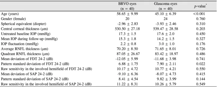
BRVO eyes (n = 40)
Glaucoma eyes
(n = 40) p-value*
Age (years) Gender (female)
Spherical equivalent (diopter) Central corneal thickness (μm) Untreated baseline IOP (mmHg) Mean IOP during follow-up (mmHg) IOP fluctuation (mmHg)
Average RNFL thickness (μm) Quadrant RNFL thickness (μm) Mean deviation of FDT 24-2 (dB)
Pattern standard deviation of FDT 24-2 (dB)
Raw sensitivity in the involved hemifield of FDT 24-2 (dB)
58.65 ± 9.99 20 -2.96 ± 2.83 530.50 ± 27.18
17.3 ± 1.5 15.3 ± 1.8 2.2 ± 0.8 70.20 ± 8.50 87.05 ± 26.67 -12.05 ± 5.99
6.88 ± 1.75 10.17 ± 4.72
45.10 ± 6.39 24 -3.93 ± 2.46 539.47 ± 28.58
17.6 ± 2.0 14.2 ± 1.5 3.0 ± 1.0 71.65 ± 8.01 83.42 ± 18.97 -11.68 ± 3.98
7.90 ± 2.11 10.77 ± 4.21
<0.001 0.760 0.310 0.203 0.450 0.327 0.176 0.726 0.486 0.741 0.022 0.550 Mean deviation of SAP 24-2 (dB)
Pattern standard deviation of SAP 24-2 (dB)
-9.10 ± 6.36 8.41 ± 4.54
-8.07 ± 4.73 9.82 ± 3.99
0.415 0.144 Raw sensitivity in the involved hemifield of SAP 24-2 (dB) 11.22 ± 8.31 10.26 ± 5.79 0.549 Values are presented as mean ± standard deviation unless otherwise indicated.
BRVO = branched retinal vein occlusion; IOP = intraocular pressure; RNFL= retinal nerve fiber layer; FDT = frequency doubling techni- que; SAP = standard automated perimetry.
*Student t-test, p < 0.05.
Table 1. Demographic of patients and calculation of visual fields parameters
의 5% 및 1% 지점의 백분율을 각각 빼 주었다.12,13 빛간섭단층촬영은 Cirrus HD OCT 4000 (Carl Zeiss Meditec)을 이용하여 황반의 14.13 mm2의 타원고리 모양에 해당하는 부위의 망막신경섬유층의 두께를 측정하였다.
본 연구의 통계 분석은 IBM SPSS ver. 22.0 software (IBM Corp., Armonk, NY, USA)를 이용하였으며 t-test를 이용하여 두 군 간 평균 안압 및 변동폭, 빛간섭단층촬영 시 망막신경섬유층의 두께, 시야검사에서 mean deviation 및 pattern deviation, raw sensitivity를 비교하였으며, 이 중 통계학적으로 유의미한 지표를 이용하여 다중선형회귀분 석을 시행하였을 때(결정계수 R2 value) p값이 0.05 미만일 경우를 통계적으로 의의가 있는 것으로 정하였다.
결 과
녹내장환자 40안 중 14안은 정상안압녹내장, 26안은 원 발개방각녹내장환자였으며 평균 연령은 45.10 ± 6.39세, 진 단 시 평균 안압은 17.6 ± 2.0 mmHg, 평균 RNFL 두께는 71.65 ± 8.01 μm였다. 분지망막정맥폐쇄로 진단받은 총 40안 의 평균 연령은 58.65 ± 9.99세, 평균 안압은 17.3 ± 1.5 mmHg, 평균 RNFL 두께는 70.20 ± 8.50 μm였다. 분지망막정맥폐 쇄환자들이 유의하게 고령이었다.
MD의 경우 FDT C24-2에서 녹내장환자는 -11.68 ± 3.98 dB, 분지망막정맥폐쇄환자는 -12.05 ± 5.99 dB였으며 SAP C24-2 에서 녹내장환자는 -8.07 ± 4.73 dB, 분지망막정맥폐쇄환자
는 -9.10 ± 6.36 dB로 모두 통계학적으로 유의한 차이는 없 었다. 반면 PSD의 경우 FDT C24-2에서 녹내장환자는 7.90 ± 2.11 dB, 분지망막정맥폐쇄환자는 6.88 ± 1.75 dB로 유의 한 차이를 보였다(p=0.022). SAP C24-2의 PSD는 녹내장환 자에서 9.82 ± 3.99 dB, 분지망막정맥폐쇄환자에서 8.41 ± 4.54 dB로 유의한 차이를 보이지는 않았다(p=0.144, Table 1).
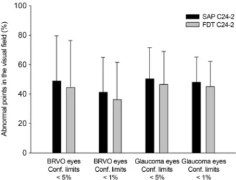
각 시야검사의 Pattern deviation plot에서 시야손상이 발 생한 해당 hemifield의 26 지점을 분석했을 때, FDT C24-2 와 SAP C24-2의 평균 hemifield sensitivity는 두 군 간 차이 가 없었다. 또한 시야손상이 발생한 hemifield에서 신뢰구 간 5% 및 1% 이하로 감도가 떨어져 있는 지점의 비율을 계산한 결과, FDT C24-2와 SAP C24-2 두 가지 모두 녹내 장 및 분지망막정맥폐쇄환자에서 유의한 차이를 보이지 않 았다(Fig. 1).
하지만 FDT C24-2의 hemifield sensitivity에서 SAP C24-2의 hemifield sensitivity를 뺀 값은 녹내장 군에서는 양의 값이었던 반면 분지망막정맥폐쇄환자에서는 음의 값 으로, 녹내장환자의 경우 FDT C24-2에서 보이는 시야손상 의 범위가 SAP C24-2보다 유의하게 컸다(p<0.01, Fig. 2).
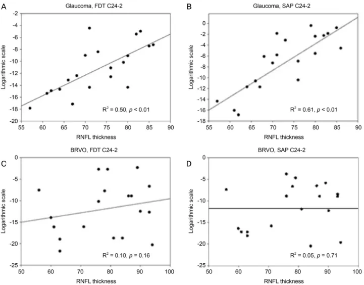
한편 구조-기능 관계를 알아보기 위해 평균 RNFL 두께와 시야검사의 MD 간 선형회귀분석을 시행한 결과, 녹내장환 자는 FDT C24-2에서 R2=0.50, SAP C24-2에서 R2=0.60으 로 분지망막정맥폐쇄환자에 비해 높은 R2값을 보였으며 모 두 통계적으로 유의하였다(p<0.01). 분지망막정맥폐쇄환자 는 전반적으로 구조-기능 관계가 약하였지만 SAP C24-2에
Figure 1. Percentage of abnormal points in the FDT C24-2 and
SAP 24-2. It is based on confidential limits of 5% and 1% on glaucoma patients and BRVO patients. FDT = frequency dou- bling technique; SAP = standard automated perimetry; BRVO= branched retinal vein occlusion; Conf. limits = confidential limits.
Figure 2. Comparison as mean difference between FDT C24-2
and SAP C24-2 on glaucoma patients and BRVO patients. The percentage of the raw sensitivity points and where the con- fidence limit is less than 5% and 1% was calculated. FDT = frequency doubling technique; SAP = standard automated perimetry; BRVO = branched retinal vein occlusion. *The dif- ference of raw sensitivity value from FDT C24-2 to SAP C24-2. In glaucoma patients, it was positive value but in BRVO patients, it was negative value.비해 FDT C24-2에서 더 큰 상관관계(R2=0.10)를 보였다 (Fig. 3). 앞서 통계학적으로 유의하게 판정된 지표들을 이 용하여 다중선형회귀분석을 시행하였으며 망막신경섬유의 두께가 얇을수록, 분지망막정맥폐쇄환자가 아닐수록 FDT C24-2에서 손상을 보이는 지점의 비율이 SAP C24-2보다 높았다(Table 2).
고 찰
녹내장환자는 FDT C24-2에서 보이는 시야손상의 범위 가 SAP C24-2에서 보이는 것보다 유의하게 컸는데, 이는 FDT C24-2가 망막신경절세포의 중첩(redundancy)을 줄여 민감도를 높일 수 있고 더 넓은 범위를 확인할 수 있기 때 문에 초기 녹내장을 좀 더 잘 발견할 수 있다는 의견에 부 합하는 결과라고 할 수 있겠다.14-16 반면 분지망막정맥폐쇄 환자에서 두 가지 시야검사를 시행하였을 때는 시야손상의 범위가 서로 유의한 차이를 보이지 않았음을 본 연구를 통 해 확인할 수 있었다. 이러한 차이는 각 질환에서 주로 손 상받은 망막신경절세포의 종류 및 그 분포가 서로 다를 수 있음을 시사한다고 볼 수 있을 것이다.
Hu et al17은 녹내장의증환자 221안에서 FDT C24-2 와 SAP C24-2를 시행한 후 분석한 결과, FDT C24-2에서 보다 조기에 혹은 비슷한 시기에 녹내장을 발견할 수 있으 나 이것이 RNFL 두께 변화로 이어지는 구조-기능 상관관 계와는 무관하다고 보고한 바 있다. 반면 Patyal et al18은
녹내장의증환자 55안과 정상안을 비교 분석한 결과 FDT C24-2에서 SAP C24-2보다 선행하는 녹내장성 변화를 발 견하지 못하였다고 주장하였으며, 이는 대상 질환군, 장비 및 추적 기간의 차이, 시야검사 기계 및 이를 분석하는 방 법의 상이함 때문인 것으로 생각해 볼 수 있을 것이다. 기 존의 여러 연구 결과에서 밝힌 바와 같이 본 연구에서도 FDT C24-2가 초기 녹내장성 변화를 확인하는 데에 그 유 용성이 입증되었다고 할 수 있으며, 이는 안압에 의한 압박 성 손상 시 망막신경절세포 중 M-cell의 손상이 조기에 일 어날 수 있음을 시사할 수 있겠다. 단, M-cell은 망막신경절 세포 중 10%에 불과하여 그 수가 적기 때문에 그중 일부가 손상되어도 시야검사상에서는 보다 조기에 잘 발견될 수도 있을 것이다.
반면 분지망막정맥폐쇄환자는 FDT C24-2와 SAP C24-2 간 시야손상 영역의 범위가 서로 유의한 차이를 보이지 않 고 비슷하였다. 이는 분지망막정맥폐쇄에서 발생하는 망막 신경절세포의 손상이 저산소성-허혈성 변화로 인한 뉴런의 퇴화 및 세포사멸사에 의한 것이기 때문에19,20 세포손상의 정도에 비례하여 시야손상이 나타나는 것으로 파악된다.21 즉 M-cell 외에도 망막신경절세포의 대부분을 차지하는 P-cell 손상이 전반적으로 함께 발생하고 있음을 시사할 수 있겠다.
구조-기능 관계 분석을 위해 RNFL 두께와 시야검사의 MD 간 상관성을 비교한 결과 분지망막정맥폐쇄환자는 녹
A B
C D
Figure 3. Correlation between average RNFL thickness and median deviation categorized by diagnosis and visual field examination.
(A) Glaucoma patients, FDT C24-2 (B) glaucoma patients, SAP C24-2 (C) BRVO patients, FDT C24-2 (D) BRVO patients, SAP C24-2. FDT = frequency doubling technique; SAP = standard automated perimetry; BRVO = branched retinal vein occlusion;
RNFL = retinal nerve fiber layer.
Variable Beta 95% CI p-value* Beta 95% CI p-value*
Age
Gender (female) Spherical equivalent Central corneal thickness Untreated baseline IOP Mean IOP during follow-up IOP fluctuation
Average RNFL thickness Quadrant RNFL thickness BRVO eyes
-0.091 0.103 -1.565 -0.008 0.063 0.130 0.905 0.243 0.110 0.505
-0.202 to 0.019 -0.080 to 0.289 -0.393 to 0.805 -0.045 to 0.029 -0.009 to 0.136 -0.049 to 0.309 -1.495 to 3.305 0.146 to 0.341 0.064 to 0.156 0.072 to 0.938
0.102 0.264 0.193 0.668 0.287 0.151 0.455
<0.001
<0.001 0.023
-0.076 -1.225
0.258 0.023 0.520
-0.208 to 0.055 -4.158 to 1.838
0.088 to 0.427 -0.050 to 0.097 0.089 to 0.952
0.253 0.380
0.003 0.529 0.019 CI = confidence Interval;IOP = intraocular pressure; RNFL= retinal nerve fiber layer; BRVO = branched retinal vein occlusion.
*Student t-test, p < 0.05.
Table 2. Multiple linear regression analysis of mean difference between frequency doubling technology perimetry C24-2 (FDT
C24-2) and Standard automatic perimetry C24-2 (SAP 24-2) for the percentage of depressed pointsA B C
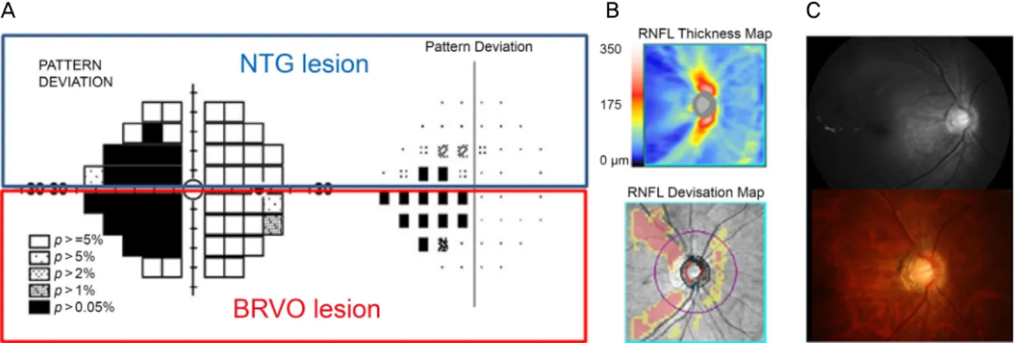
Figure 4. A 41-year-old patient with NTG at superior hemifield and BRVO at inferior hemifield. (A) FDT C24-2 and SAP C24-2.
On superior hemifield, the FDT C24-2 showed progressive visual defect than SAP C24-2, but on inferior hemifield two examination showed smiliar results. (B) OCT showed superior and inferior RNFL defect. (C) Disc and red-free photographs show glaucomatous cupping and localized RNFL defect. NTG = normal tension glaucoma; BRVO = branched retinal vein occlusion; FDT = fre- quency doubling technique; SAP = standard automated perimetry; RNFL = retinal nerve fiber layer; OCT = optical coherence tomography.
내장환자에 비해 약한 상관관계를 보였는데 이는 망막신경 절세포뿐만 아니라 다른 망막층의 변화가 기능에 관여할 가능성으로 볼 수 있겠다. 하지만 구조-기능 관계 결과 분 지망막정맥폐쇄환자군에서도 SAP C24-2보다 FDT C24-2 에서 통계학적으로 유의한 차이를 보였는데, 이는 아마도 FDT C24-2가 초기 망막신경절세포의 손상을 보다 잘 찾아 내기 때문일 것으로 생각된다.
실제 임상에서 녹내장과 분지망막정맥폐쇄를 단안에 모 두 진단받은 환자의 시야검사 결과를 살펴보면 하측부는 분지망막정맥폐쇄에 의한 허혈성 손상을 보여 FDT C24-2 와 SAP C24-2 모두에서 비슷한 정도의 시야손상을 보였으 나, 상측부는 녹내장에 의한 압력성 손상에 의하여 SAP C24-2보다 FDT C24-2에서 뚜렷한 시야손상을 확인할 수 있었다(Fig. 4)
본 연구의 한계로는 적은 환자를 대상으로 하였으며 (n=40), 그중 분지망막정맥폐쇄환자군의 경우 황반부종이 없으며 레이저 치료를 받지 않은 비교적 경증에 해당하는 환자를 대상으로 하였기 때문에 선택 비뚤림(selection bias) 의 가능성이 있다는 점, 전신적 질환이나 가족력의 유무 등 역학적인 면에서 차트 분석 결과만을 사용하였다는 점, FDT C24-2에서는 Lmax-Lmin/Lmax+Lmin (람버트최대치- 람버트최소치/람버트최대치+람버트최소치)를 이용한 반면 SAP 24-2에서는 Δ L/L (람버트변화량/람버트)를 이용함으 로써 완전히 대등하지 못한 시표 밝기를 비교했다는 점 등 이 있다.
또한 녹내장환자에 정상안압녹내장과 원발개방각녹내장
으로 진단받은 경우를 모두 포함하였는데 이는 녹내장을 안압의 변화에 의한 시신경의 압박성 손상을 병인으로 하 는 질환군으로 보았기 때문이다.22 향후 장기간의 추적 연 구를 통해 대상자를 확대하여 고안압성 녹내장환자와 허혈 성 망막질환환자 간 비교 분석이 필요할 것으로 판단된다. 한편 초기 진단 시 검사값을 기준으로 결과를 분석하였 기 때문에 한 번의 검사로 두 질환군을 완전히 구분할 수 있다고 해석할 수 있으며 RNFL의 손상 정도가 충분히 반 영되지 않을 우려가 있다. 그러나 분지망막정맥폐쇄환자의 경우 진단 후 3-6개월 이내 경과 관찰 시, 안저검사에서 망 막내 출혈이 소실된 것을 확인 후 시야검사 및 빛간섭단층 촬영검사를 시행하였다. Kim et al23은 분지망막정맥폐쇄환 자를 장기간 추적 관찰하였을 때 진단 후 6개월 이후부터 RNFL 두께의 얇아짐을 뚜렷하게 관찰할 수 있다고 보고한 바 있어 본 연구의 대상자 또한 검사 당시 RNFL 손상이 발 생하였다고 생각할 수 있다. 향후 보다 대규모의 장기간 추 적 관찰을 통해 보다 적절한 검사 시행 시점을 고려하면서 자료 수집을 진행해야 할 것이다.
본 논문은 망막신경절세포의 손상에 의한 시야결손을 보 이는 대표적인 두 질환으로 녹내장과 분지망막정맥폐쇄환 자를 대상으로 하여 서로 다른 시야검사의 결과를 기능-구 조적으로 비교 분석한 첫 논문으로서 그 의미가 있다. 녹내 장으로 진단받은 환자는 SAP C24-2보다 FDT C24-2에서 더 넓은 시야손상 영역을 보였던 것에 반해 분지망막정맥 폐쇄로 진단받은 환자의 경우에는 SAP C24-2와 FDT C24-2 에서 비슷한 시야손상 영역을 보였다. 이는 주로 손상받은
망막신경절세포의 종류 및 그 분포가 질환 간에 서로 다를 수 있음을 시사한다고 볼 수 있을 것이다. 따라서 서로 2가 지 시야검사 및 시신경의 빛간섭단층촬영 결과를 비교 분 석하여 환자의 시야손상이 녹내장에 의한 시신경의 압력성 변화에 의한 것인지, 망막 혈관의 폐쇄에 의한 허혈성 변화 에 의한 것인지를 감별할 수 있으며 이를 통해 향후 치료 방향 및 추적 관찰에 활용할 수 있을 것이다.
REFERENCES
1) Almasieh M, Wilson AM, Morquette B, et al. The molecular basis of retinal ganglion cell death in glaucoma. Prog Retin Eye Res 2012;31:152-81.
2) Weber AJ, Chen H, Hubbard WC, Kaufman PL. Experimental glaucoma and cell size, density, and number in the primate lateral geniculate nucleus. Invest Ophthalmol Vis Sci 2000;41:1370-9.
3) Bayer AU, Erb C. Short wavelength automated perimetry, fre- quency doubling technology perimetry, and pattern electro- retinography for prediction of progressive glaucomatous standard visual field defects. Ophthalmology 2002;109:1009-17.
4) Bowd C, Zangwill LM, Berry CC, et al. Detecting early glaucoma by assessment of retinal nerve fiber layer thickness and visual function. Invest Ophthalmol Vis Sci 2001;42:1993-2003.
5) Medeiros FA, Sample PA, Weinreb RN. Frequency doubling tech- nology perimetry abnormalities as predictors of glaucomatous vis- ual field loss. Am J Ophthalmol 2004;137:863-71.
6) Sample PA, Bosworth CF, Blumenthal EZ, et al. Visual func- tion-specific perimetry for indirect comparison of different gan- glion cell populations in glaucoma. Invest Ophthalmol Vis Sci 2000;41:1783-90.
7) Lee AJ, Wang JJ, Rochtchina E, et al. Patterns of glaucomatous vis- ual field defects in an older population: the Blue Mountains Eye Study. Clin Exp Ophthalmol 2003;31:331-5.
8) Barbazetto IA, Schmidt-Erfurth UM. Evaluation of functional de- fects in branch retinal vein occlusion before and after laser treat- ment with scanning laser perimetry. Ophthalmology 2000;107:
1089-98.
9) Yücel YH, Zhang Q, Weinreb RN, et al. Atrophy of relay neurons in magno- and parvocellular layers in the lateral geniculate nucleus in experimental glaucoma. Invest Ophthalmol Vis Sci 2001;42:
3216-22.
10) McKendrick AM, Sampson GP, Walland MJ, Badcock DR.
Contrast sensitivity changes due to glaucoma and normal aging:
low-spatial-frequency losses in both magnocellular and parvocel- lular pathways. Invest Ophthalmol Vis Sci 2007;48:2115-22.
11) Yücel YH, Zhang Q, Gupta N, et al. Loss of neurons in magnocel- lular and parvocellular layers of the lateral geniculate nucleus in glaucoma. Arch Ophthalmol 2000;118:378-84.
12) Lamparter J, Aliyeva S, Schulze A, et al. Standard automated peri- metry versus matrix frequency doubling technology perimetry in subjects with ocular hypertension and healthy control subjects.
Plos One 2013;8:e57663.
13) Soliman MA, de Jong LA, Ismaeil AA, et al. Standard achromatic perimetry, short wavelength automated perimetry, and frequency doubling technology for detection of glaucoma damage.
Ophthalmology 2002;109:444-54.
14) Johnson CA, Samuels SJ. Screening for glaucomatous visual field loss with frequency-doubling perimetry. Invest Ophthalmol Vis Sci 1997;38:413-25.
15) Quigley HA. Identification of glaucoma-related visual field abnor- mality with the screening protocol of frequency doubling technology.
Am J Ophthalmol 1998;125:819-29.
16) Cello KE, Nelson-Quigg JM, Johnson CA. Frequency doubling technology perimetry for detection of glaucomatous visual field loss. Am J Ophthalmol 2000;129:314-22.
17) Hu R, Wang C, Racette L. Comparison of matrix frequency-dou- bling technology perimetry and standard automated perimetry in monitoring the development of visual field defects for glaucoma suspect eyes. PLoS One 2017;12:e0178079.
18) Patyal S, Kotwal A, Banarji A, Gurunadh VS. Frequency doubling technology and standard automated perimetry in detection of glau- coma among glaucoma suspects. Med J Armed Forces India 2014;70:332-7.
19) Alshareef RA, Barteselli G, You Q, et al. In vivo evaluation of reti- nal ganglion cells degeneration in eyes with branch retinal vein occlusion. Br J Ophthalmol 2016;100:1506-10.
20) Lim HB, Kim MS, Jo YJ, Kim JY. Prediction of retinal ischemia in branch retinal vein occlusion: spectral-domain optical coherence tomography study. Invest Ophthalmol Vis Sci 2015;56:6622-9.
21) Harwerth RS, Quigley HA. Visual field defects and retinal gan- glion cell losses in patients with glaucoma. Arch Ophthalmol 2006;124:853-9.
22) Shields MB. Normal-tension glaucoma: is it different from pri- mary open-angle glaucoma? Curr Opin Ophthalmol 2008;19:85-8.
23) Kim CS, Shin KS, Lee HJ, et al. Sectoral retinal nerve fiber layer thinning in branch retinal vein occlusion. Retina 2014;34:525-30.
= 국문초록 =
녹내장 및 분지망막정맥폐쇄에서 두 가지 시야검사를 통한 망막신경절세포층 손상의 비교 연구
목적: 녹내장 및 분지망막정맥폐쇄환자에서 두 가지 시야검사를 통해 망막신경절세포층의 변화를 비교 분석하였다.
대상과 방법: 녹내장 및 분지망막정맥폐쇄로 진단받은 환자 각 40안을 후향적으로 분석하였다. Frequency-doubling technology (FDT) C24-2 및 standard automated perimetry (SAP) C24-2의 median deviation (MD), pattern standard deviation (PSD), 손상된 hemifield의 sensitivity를 비교하였다. 빛간섭단층촬영의 retinal nerve fiber layer와 시야검사의 MD 간 상관관계로 구조-기능 관계를 분석하였다.
결과: MD는 모든 경우 유의한 차이를 보이지 않았으나 PSD는 FDT C24-2에서 녹내장환자군이 더 높은 값을 보였으며(p=0.022), SAP C24-2에서 유의한 차이는 없었다(p=0.144). Hemifield sensitivity 비교 시 녹내장환자의 FDT C24-2 손상 범위가 SAP C24-2보 다 유의하게 컸다(p<0.01). 구조-기능 관계에서 녹내장환자가 분지망막정맥폐쇄환자보다 높은 R2값을 보였다(p<0.01).
결론: 녹내장환자는 FDT C24-2에서 더 넓은 시야손상 영역을 보였으나 분지망막정맥폐쇄환자는 두 시야검사 간 손상 영역이 비슷했 다. 이는 손상된 망막신경절세포의 종류 및 분포가 질환별로 다를 수 있음을 시사하며, 향후 감별 진단 및 치료 방향 결정 시 활용할 수 있을 것이다.
<대한안과학회지 2019;60(5):455-462>
이지영 / Jiyoung Lee
가톨릭대학교 의과대학 서울성모병원 안과학교실 Department of Ophthalmology, Seoul St. Mary’s Hospital, College of Medicine, The Catholic University of Korea