Mini-Open Anterior Lumbar Interbody Fusion Combined with Lateral Lumbar Interbody Fusion
in Corrective Surgery for Adult Spinal Deformity
Chong-Suh Lee
1, Se-Jun Park
1, Sung-Soo Chung
1, Jun-Young Lee
1, Tae-Hoon Yum
1, Seong-Kee Shin
21Department of Orthopedic Surgery, Spine Center, Samsung Medical Center, Sungkyunkwan University School of Medicine, Seoul, Korea
2Department of Orthopedic Surgery, Seoul Medical Center, Seoul, Korea
Study Design: Prospective observational study.
Purpose: To introduce the techniques and present the surgical outcomes of mini-open anterior lumbar interbody fusion (ALIF) at the most caudal segments of the spine combined with lateral lumbar interbody fusion (LLIF) for the correction of adult spinal deformity Overview of Literature: Although LLIF is increasingly used to correct adult spinal deformity, the correction of sagittal plane defor- mity with LLIF alone is reportedly suboptimal.
Methods: Thirty-two consecutive patients with adult spinal deformity underwent LLIF combined with mini-open ALIF at the L5–S1 or L4–S1 levels followed by 2-stage posterior fixation. ALIF was performed for a mean 1.3 levels and LLIF for a mean 2.7 levels.
Then, percutaneous fixation was performed in 11 patients (percutaneous group), open correction with facetectomy with or without laminectomy in 16 (open group), and additional pedicle subtraction osteotomy (PSO) in 5 (PSO group). Spinopelvic parameters were compared preoperatively and postoperatively. Hospitalization data and clinical outcomes were recorded.
Results: No major medical complications developed, and clinical outcomes improved postoperatively in all groups. The mean postop- erative segmental lordosis was greater after ALIF (17.5°±5.5°) than after LLIF (8.1°±5.3°, p<0.001). Four patients (12.5%) had lumbar lordosis with a pelvic incidence of ±9° preoperatively, whereas this outcome was achieved postoperatively in 30 patients (93.8%).
The total increase in lumbar lordosis was 14.7° in the percutaneous group, 35.3° in the open group, and 57.0° in the PSO group. The ranges of potential lumbar lordosis increase were estimated as 4°–25°, 23°–42°, and 45°–65°, respectively.
Conclusions: Mini-open ALIF combined with LLIF followed by posterior fixation may be a feasible technique for achieving optimal sagittal balance and reducing the necessity of more extensive surgery.
Keywords: Adult spinal deformity; Lateral lumbar interbody fusion; Anterior lumbar interbody fusion; Sagittal balance; Lumbar lordosis
Copyright Ⓒ 2016 by Korean Society of Spine Surgery
This is an Open Access article distributed under the terms of the Creative Commons Attribution Non-Commercial License (http://creativecommons.org/licenses/by-nc/3.0/) which permits unrestricted non-commercial use, distribution, and reproduction in any medium, provided the original work is properly cited.
Asian Spine Journal • pISSN 1976-1902 eISSN 1976-7846 • www.asianspinejournal.org
Received Jan 27, 2016; Revised May 18, 2016; Accepted May 19, 2016 Corresponding author: Se-Jun Park
Department of Orthopedic Surgery, Spine Center, Samsung Medical Center, Sungkyunkwan University School of Medicine, 81 Irwon-ro, Gangnam-gu, Seoul 06351, Korea
Tel: +82-2-3410-1583, Fax: +82-2-3410-0061, E-mail: [email protected]
ASJ A SJ
Introduction
The traditional method of correcting adult spinal defor- mity (ASD) is a posterior-only surgery. However, trans- foraminal or posterior lumbar interbody fusion (TLIF or
PLIF, respectively) is considered insufficient for restoring lumbar lordosis (LL) and sagittal balance [1,2]. Although osteotomy techniques such as pedicle subtraction oste- otomy (PSO) and posterior vertebral column resection can be performed to further correct sagittal balance, these
Chong-Suh Lee et al.
1024 Asian Spine J 2016;10(6):1023-1032
procedures often result in critical complications such as significant epidural bleeding or neurologic deficit [3-8].
In response to these limitations, minimally invasive lateral lumbar interbody fusion (LLIF) has been per- formed in ASD correction, with several studies suggesting reduced complications compared with those of conven- tional posterior techniques [8-10]. Although most studies of LLIF have reported favorable radiographic results after the correction of coronal deformity, the amount of sagittal plane correction was relatively suboptimal, with a 1.6–9.0°
increase in LL [9,11-18]. We postulated that these subop- timal results are attributable to the fact that the L5–S1 lev- els were not operated or were operated using the posterior fusion technique.
Given that the restoration of LL and sagittal balance is key to the success of adult deformity correction [19- 21], we hypothesized that LLIF alone is insufficient for achieving optimal LL in ASD patients. Thus, we recently performed mini-open anterior lumbar interbody fusion (ALIF) at the most caudal segments (L5–S1 or L4–5, or both) in addition to LLIF to achieve sufficient LL. ALIF has several advantages over TLIF/PLIF or LLIF. It allows direct access to the disk space with a broad surface area for large grafts that can reduce the nonunion rate, and it affords the ability to create a more lordotic angle [22-24].
Moreover, the mini-open midline approach to the L5–
S1 or L4–S1 levels minimizes blood loss by reducing the need to handle bony or muscular structures. The purpose of this case study was to introduce and present the clini- cal and radiographic results of mini-open ALIF combined with LLIF for the correction of ASD.
Materials and Methods
1. Study cohort
Thirty-two patients who underwent surgery with ALIF and LLIF for ASD correction between January 2012 and June 2014 were evaluated prospectively after giving in- formed consent. The minimal follow-up duration was 12.0 months. The inclusion criteria were lumbar degenerative kyphosis (LDK) and lumbar degenerative kyphoscoliosis (LDKS). A coronal Cobb’s angle (CCA) of 10° was used as the criterion for distinguishing LDK and LDKS. Patients who had undergone previous lumbar surgery or who had posttraumatic kyphosis were excluded.
2. Surgical procedures
The operations were performed by 2 attending surgeons (C.S.L and S.J.P) in 2 stages with an interval of 1 week. All surgeries included fusion to S1 using bilateral iliac screws.
During the first surgery, mini-open ALIF at the L5–S1 or L4–5 levels, or both, was performed, followed by LLIF at these levels. The ALIF procedure was carried out via a ret- roperitoneal approach after a midline skin incision. After discectomy, the largest possible 8° lordotic-angled poly- etheretherketone cage (Syncage; Synthes Inc., West Ches- ter, PA, USA) was fitted. LLIF was performed via a lateral mini-open retroperitoneal approach [25]. Six-degree lordotic-angled polyetheretherketone cages (Clydesdale;
Medtronic Inc., Minneapolis, MN, USA) were inserted at each level of the LLIF. The LLIF approach was from the left side in most LDK cases and from the concave side for LDKS cases. The most proximal level of LLIF was L1–2 in 7 patients, L2–3 in 21 patients, and L3–4 in 4 patients. The cages for ALIF and LLIF were filled with allogeneic chip bone grafts mixed with demineralized bone matrix (Graf- ton; Medtronic Inc.).
Three days after the first surgery, magnetic resonance imaging and plain radiographs were obtained. The in- direct neural decompression shown on the magnetic resonance images obtained after ALIF/LLIF surgery was evaluated to determine the level of decompression for the second surgery. Based on the immediate postoperative LL shown on plain radiographs, the posterior fixation and correction methods were determined according to the amount of LL correction required. The target LL was roughly determined using a simple prediction model ac- cording to pelvic incidence (PI) [26]. During posterior instrumentation, percutaneous fixation was performed in 11 patients (percutaneous group) (Fig. 1), open con- ventional fixation with facetectomy with or without lami- nectomy was performed in 16 patients (open group) (Fig.
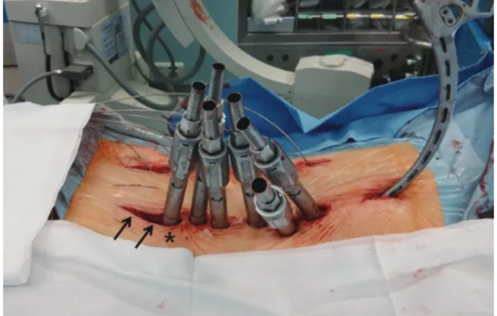
2), and an additional PSO was performed in 5 patients (PSO group). In cases of percutaneous fixation, the most caudal incisions for S1 screw fixation were extended along the posterior superior iliac spine for insertion of the iliac screws and assembly of the rods (Fig. 3).
3. Radiographic evaluation
Whole-spine radiographs were evaluated for the following parameters preoperatively, 2 weeks after the final surgery,
Fig. 1. A 75-year-old woman with lumbar degenerative kyphosis underwent anterior lumbar interbody fusion (ALIF) at L5–
S1 and lateral lumbar interbody fusion (LLIF) at L2–4. Note that lumbar lordosis (LL) increased by 25° after ALIF and LLIF.
During posterior fixation, percutaneous screws were used with iliac screws, and laminectomy was not performed. Com- puted tomography images obtained 2 years postoperatively show that all segments are well fused. PI, pelvic incidence; SS, sacral slope; PT, pelvic tilt; SVA, sagittal vertical axis.
Fig. 2. A 53-year-old woman with lumbar degenerative kyphoscoliosis underwent anterior lumbar interbody fusion at L5–
S1, lateral lumbar interbody fusion at L2–4, and open posterior correction. Note that lumbar lordosis (LL) increased from 30°
to 59°, and the coronal Cobb’s angle (CCA) decreased from 37° to 10° at 1 year postsurgery. PI, pelvic incidence; SS, sacral slope; PT, pelvic tilt; SVA, sagittal vertical axis.
Chong-Suh Lee et al.
1026 Asian Spine J 2016;10(6):1023-1032
and at the last follow-up: PI, sacral slope (SS), pelvic tilt (PT), LL, thoracic kyphosis (TK), sagittal vertical axis (SVA), CCA, and segmental lordosis (SL) at each inter- body fusion level. LL and CCA were also measured on plain radiographs obtained between the 2 surgeries. Spi- nopelvic parameters such as PI, SS, PT, LL, TK, and SVA were measured using current standard methods [27]. A PI-LL within 9° was considered successful correction [20].
For SVA, the perpendicular distance from the C7 plumb line to the superoposterior corner of S1 was measured.
The sagittal balance was considered negative when the C7 plumb line fell behind the reference point. CCA was de- fined as the maximal Cobb’s angle on the anteroposterior plain radiograph. SL was defined as the angle between the upper endplate of the caudal vertebra and the lower end- plate of the cranial vertebra at the level of operation.
4. Hospitalization data and clinical outcomes
Hospitalization data were collected for the following pa- rameters: duration of hospital stay, total surgery time, es- timated blood loss, postoperative blood loss, need for red blood cell transfusion, medical or surgery-related compli- cations, and reoperation status. For clinical evaluation, the Oswestry disability index and the visual analog scale score for the back and leg were recorded and compared among the groups.
5. Statistical analysis
Baseline data were compared between the LDK and LDKS groups with the Mann-Whitney U test. Analysis of vari-
ance was used to compare parameters among the percuta- neous, open, and PSO groups. The Wilcoxon signed rank test was used to compare the overall change in parameters preoperatively and postoperatively. Box and whisker plots provided the first-to-third quartiles of LL increase across the 3 groups. Statistical analysis was performed using SPSS ver. 22.0.0 (IBM Corp., Armonk, NY, USA). A p- values less than 0.05 were considered significant.
Results
The study cohort consisted of 6 men and 26 women aged 67.1±7.4 years (range, 53.0–85.0 years). The preopera- tive diagnoses were LDK in 17 patients and LDKS in 15 patients. ALIF was performed on 1.3 levels per patient (range, 1–2) and LLIF on 2.6 levels per patient (range, 1–4). During posterior surgery, central decompression was performed for 1.0±1.1 levels per patient and forami- notomy for 0.8±1.2 levels per patient. Total fusion levels were 5.4±2.1 (range, 4.0–14.0). The mean follow-up dura- tion was 15.9 months (range, 12.0–35.6 months).
The number of ALIF levels was greatest in the PSO group followed by the open group and the percutaneous group (1.6±0.5 vs. 1.4±0.5 vs. 1.0, respectively; p=0.015).
The number of LLIF levels did not differ among groups (p=0.362). The mean postoperative SL was greater at the ALIF levels than at the LLIF levels (17.5±5.5° vs. 8.1±5.3°, respectively; p<0.001). The mean postoperative increase in SL was also greater at the ALIF levels than at the LLIF levels (11.4±7.9° vs. 4.3±5.3°, respectively; p<0.001).
Compared with preoperative values, the sagittal pa- rameters of SS, PT, LL, TK, and SVA were significantly improved after the final surgery (Table 1). The number of patients with PI-LL less than 9° also increased significant- ly from 4 (12.5%) preoperatively to 30 (93.8%) postopera- tively. At the last follow-up, most sagittal parameters were slightly worse than the immediate postoperative param- eters, even when 5 patients who developed proximal junc- tional kyphosis (PJK) were excluded from the evaluation.
However, compared with the preoperative data, all param- eters in the final follow-up showed improvement, even when the cases of PJK were included. The improvement in the sagittal parameters of SS, PT, LL, TK, and SVA at the last follow-up was significantly greatest in the PSO group followed by the open group and then the percutaneous group. Changes in CCA did not differ significantly among the groups (Table 2).
Fig. 3. The incision for the S1 screw (*) was extended caudally for insertion and assembly of the iliac screws (arrows).
1. Subanalysis of LL increase
After the first-stage operation with ALIF and LLIF, LL in- creased from a mean of 20.1° to a mean of 41.9° (p<0.001).
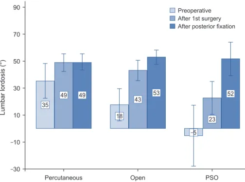
The increase in LL after ALIF and LLIF was 13.6°, 25.5°, and 28.1° for the percutaneous, open, and PSO groups, respectively (Fig. 4). After posterior correction, LL did not increase further in the percutaneous group but increased by 9.8° in the open group and 28.8° in the PSO group compared with angles measured after the first-stage op- eration (Fig. 4). Box and whisker plots demonstrated that the 25%–75% range of LL increase was 4.3°–25.2° for the percutaneous group, 23.2°–41.6° for the open group, and
44.7°–64.9° for the PSO group (Fig. 5).
2. Hospitalization data and clinical outcomes
Table 3 lists the detailed hospitalization data for the study participants. Blood loss and subsequent need for transfu- sion were significantly lowest in the percutaneous group followed by the open group and the PSO group. Com- pared with preoperative measures, all postoperative clini- cal outcome measures such as Oswestry disability index, visual analog scale score, and walking distance were sig- nificantly improved. Final clinical outcomes did not differ among the posterior fixation groups (Table 3).
Table 1. Overall changes in parameters
Parameter Preoperative P1a) After final
surgery P2a) Last follow-up
(excluding PJK) Last follow-up
(including PJK) P3a)
PI (°) 54.7±8.8 - 54.7±8.8 - 54.9±8.2 54.7±8.8 -
SS (°) 23.4±14.7 <0.001 37.9±8.1 0.001 35.1±6.2 33.5±7.8 <0.001
PT (°) 31.3±12.2 <0.001 16.8±5.7 0.001 19.9±5.8 21.2±6.6 <0.001
LL (°) 20.1±24.4 <0.001 51.5±9.3 <0.001 48.3±8.8 46.2±11.8 <0.001
TK (°) 15.1±17.9 <0.001 25.2±11.2 0.029 27.9±11.3 27.5±13.1 <0.001
SVA (mm) 76.5±47.0 <0.001 13.7±25.5 0.061 21.1±27.7 28.0±30.1 <0.001
CCA (°) 14.9±11.3 <0.001 3.0±3.5 0.134 3.5±3.8 3.3±3.6 <0.001
PI-LL (°) 34.6±22.3 <0.001 3.0±6.7 <0.001 6.6±6.5 8.5±10.7 <0.001
No. of patients with
PI-LL ≤9° (%) 4 (12.5) <0.001b) 30 (93.8) 0.070 25 (92.6) 22 (68.8) <0.001 PJK, proximal junctional kyphosis; PI, pelvic incidence; SS, sacral slope; PT, pelvic tilt; LL, lumbar lordosis; TK, thoracic kyphosis; SVA, sagittal verti- cal axis; CCA, coronal Cobb’s angle.
a)P1 was calculated by comparing the parameters preoperatively and after the final surgery, P2 after the final surgery and at the last follow-up, P3 at the last follow-up and preoperatively. All p-values were calculated using the Wilcoxon signed rank test except in cases where PI-LL ≤9°; b)p-values were calculated using the McNemar test.
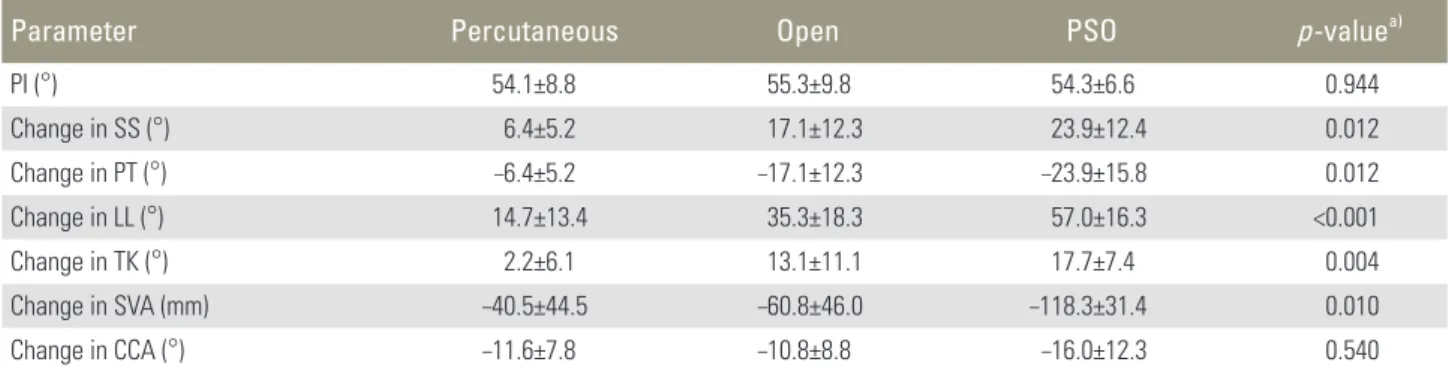
Table 2. Changes in parameters according to correction method
Parameter Percutaneous Open PSO p-valuea)
PI (°) 54.1±8.8 55.3±9.8 54.3±6.6 0.944
Change in SS (°) 6.4±5.2 17.1±12.3 23.9±12.4 0.012
Change in PT (°) –6.4±5.2 –17.1±12.3 –23.9±15.8 0.012
Change in LL (°) 14.7±13.4 35.3±18.3 57.0±16.3 <0.001
Change in TK (°) 2.2±6.1 13.1±11.1 17.7±7.4 0.004
Change in SVA (mm) –40.5±44.5 –60.8±46.0 –118.3±31.4 0.010
Change in CCA (°) –11.6±7.8 –10.8±8.8 –16.0±12.3 0.540
Change of parameters was calculated by values at the last follow-up minus preoperative values.
PSO, pedicle subtraction osteotomy, PI, pelvic incidence; SS, sacral slope; PT, pelvic tilt; LL, lumbar lordosis; TK, thoracic kyphosis; SVA, sagittal ver- tical axis; CCA, coronal Cobb’s angle.
a)p-values were calculated using the analysis of variance test.
Chong-Suh Lee et al.
1028 Asian Spine J 2016;10(6):1023-1032
In terms of complications, 3 revision surgeries were required to repair epidural hematomas with progressive neurologic deficit in 2 patients (1 in the open group and 1 in the PSO group) and for deep infection in 1 patient (open group). Transient weakness developed in 4 patients (2 in the open group, 2 in the percutaneous group) and
persistent weakness in 1 (percutaneous group). During follow-up, PJK developed in 5 patients; 3 of them under- went vertebroplasty, whereas 2 received no treatment. No approach-related complications such as vessel injury or retrograde ejaculation were observed with the exception of 1 case of incisional pseudohernia. No major medical complications occurred.
Discussion
Minimally invasive techniques for corrective surgery for ASD, especially LLIF, have been increasingly used and, compared with the conventional posterior-only approach, have reduced complications [8-10]. Although many au- thors have demonstrated favorable radiographic outcomes after LLIF, the increase in LL is reported to be less than 10° in most studies (Table 4) [9,10,12,14-17]. Acosta et al.
[15] reported a nonsignificant increase in LL from 42.1°
to 46.2°, and the global sagittal alignment also did not change significantly. Similar results have been reported by others. According to a study of pelvic parameters after LLIF by Johnson et al. [16], LLIF does not significantly affect SS, PT, or LL and it increases SL by only 3.3°. Al- though some authors have demonstrated relatively large Fig. 4. Mean and standard deviation of lumbar lordosis at each designated time in the percutaneous,
open, and pedicle subtraction osteotomy (PSO) groups.
Percutaneous Open PSO
90 70 50 30 10 –10
–30
Lumbar lordosis (°) 35
49 49
18
43 53
–5 23
52 Preoperative After 1st surgery After posterior fixation
Percutaneous Open PSO
100 80 60 40 20 0 –20
LL incease (°)
Fig. 5. Box and whisker plot showing the increase in lumbar lordosis (LL). The transverse black line inside the box indicates the median value, and the upper and lower margins outlining the box represent the 75% and 25% quartiles, respectively. PSO, pedicle subtraction osteotomy.
29
increases in LL of 17°–26°, they used hyperlordotic cages (20° and 30°) or performed an anterior release procedure that is not yet commonly available [11,28]. We postulated that these suboptimal sagittal plane results might have oc- curred because the L5–S1 level was not operated or was operated using the posterior fusion techniques.
Because most ASD patients show multilevel degenera- tions, including those in L5–S1, and subsequent loss of SL at this level, the L5–S1 level is commonly included in ASD
surgeries. We performed mini-open ALIF at the most caudal segments (L5–S1 or L4–5, or both) in addition to LLIF to achieve sufficient LL. Compared with TLIF/
PLIF or LLIF, ALIF creates greater SL [22,24], with TLIF/
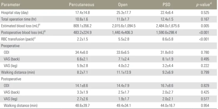
PLIF of the caudal segments being insufficient to restore sagittal balance in long-fusion surgery [1,2]. Our results showed that postoperative SL (17.5°) at the ALIF level was approximately twice that at the LLIF level (8.1°). We as- sumed that the restoration of appropriate SL at the most Table 3. Hospitalization data and clinical outcomes according the correction methods
Parameter Percutaneous Open PSO p-valuea)
Hospital stay (day) 17.4±14.8 25.3±17.7 22.4±6.4 0.525
Total operation time (hr) 10.8±1.6 11.0±1.7 12.4±1.5 0.167
Estimated blood loss (mL)b) 809.1±356.2 2,015.6±1,094.5 2,484.0±1,675.6 0.005
Postoperative blood loss (mL)b) 483.2±224.9 1,440.4±406.3 1,590.6±298.4 <0.001
RBC transfusion (pack)c) 2.2±1.5 5.5±2.8 8.6±5.8 <0.001
Preoperative
ODI 34.4±6.0 33.6±6.5 31.8±9.0 0.780
VAS (back) 6.6±2.1 7.1±2.4 8.1±1.9 0.495
VAS (leg) 5.9±2.8 4.0±3.2 3.2±4.4 0.222
Walking distance (min) 8.2±7.1 11.1±13.9 9.2±6.9 0.799
Postoperative
ODI 14.1±8.6 14.4±7.9 16.7±8.6 0.829
VAS (back) 3.3±1.9 2.5±1.7 2.0±2.7 0.425
VAS (leg) 2.7±2.6 1.9±1.7 2.0±2.1 0.577
Walking distance (min) 48.6±39.7 49.4±34.1 44.0±16.7 0.954
PSO, pedicle subtraction osteotomy; RBC, red blood cell; ODI, Oswestry disability index; VAS, visual analogue scale.
a)p-values were calculated by analysis of variance test; b)Blood loss included both anterior and posterior side; c)The number of transfused pack in- cluded intraoperative and postoperative transfusion.
Table 4. Summary of studies about lateral lumbar interbody fusion for adult spinal deformity
Study No. of
patients
Average lateral lumbar interbody
fusion levels
Preoperative
LL (°) Postoperative
LL (°) Change in LL (°)
Acosta et al. [15], 2011 36 1.8 42.1 46.2 4.1
Phillips et al. [9], 2013 107 3.0 26.7 47.6 19.9
Johnson et al. [16], 2013 30 1.4 42.8 44.4 1.6
Castro et al. [14], 2014 35 3.1 32.0 41.0 9.0
Malham et al. [13], 2016 19 1.6 51.1 45.8 –5.3
Manwaring et al. [11], 2014 9 1.7 43.7 45.9 2.2
Baghdadi et al. [12], 2014 33 5.5 38.0 44.0 6.0
Total (average) 38.4 2.6 39.5 45.0 5.5
LL, lumbar lordosis.
Chong-Suh Lee et al.
1030 Asian Spine J 2016;10(6):1023-1032
caudal segments, L5–S1 or L4–S1, is critical for regional sagittal balance because the lordosis at L4–S1 accounts for approximately 70% of the total LL in normal sagittal curvature. ALIF at the caudal segments also has a biome- chanical advantage in the correction of global kyphotic curvature because the lever arm of correction becomes longer when the lordosis is produced at a more caudal level. Compared to that at the cranial lumbar segments, the ALIF procedure at the L5–S1 level can be performed more easily because it approaches through the window of the great vessel bifurcation, thereby reducing the need for great vessel manipulation. Additionally, we can expect higher union rate at the L5–S1 level using ALIF compared with TLIF/PLIF, particularly in cases of long-level fusion surgery, because ALIF allows for relatively larger cage sizes.
Despite these well-known advantages of ALIF for ASD correction, the procedure inevitably has potential draw- backs. Because ALIF is performed through a separate incision at a different position, it requires 2 or 3 separate approaches (anterior/posterior or anterior/lateral/poste- rior), which can prolong the operation time or necessitate 2-stage operations. Two-stage operations might be con- troversial and burdensome for some surgeons; however, compared with those of posterior-only or 1-stage anteri- or-posterior surgery, 2-stage anterior-posterior surgery showed better radiological results and lower complication rates in a recent study [29].
ALIF can also be accompanied by approach-related morbidity, including vascular injury, ureteral damage, retrograde ejaculation in men, and abdominal muscle de- nervation. However, recent studies have reported that the risk of these complications is low [23,24,30]. In the cur- rent study, we observed no approach-related morbidities with the exception of pseudohernia related to the LLIF approach, although our sample size was small. Compared with the traditional ALIF technique involving a long inci- sion along the abdomen, mini-open ALIF for L5–S1 can be performed through the linea alba via a midline inci- sion, which might reduce damage to the abdominal mus- culature and decrease the subsequent risk of abdominal pseudohernia (focal atony of the abdominal musculature).
Our study showed that LL increased from 20° to 42°
through the first ALIF and LLIF surgeries. We assumed that the majority of this increase was achieved by ALIF given our results showing greater SL at the ALIF level than at the LLIF level. The mean increases in LL after the first
surgery were 13.6°, 25.5°, and 28.1° for the percutaneous, open, and PSO groups, respectively. We attribute these results to the fact that ALIF was performed for the largest number of levels in the PSO group followed by the open group and the percutaneous group (1.6 vs. 1.4 vs. 1.0 lev- els, respectively), and the results support our assumption that the majority of LL restoration was achieved through ALIF. Percutaneous pedicle screw fixation did not alter LL, whereas open fixation increased LL further. This out- come agrees with the results of a previous study [11].
In assessments of the total increase in LL after posterior fixation, box and whisker plots demonstrated that the range of LL increase was 4.3°–25.2° for the percutaneous group, 23.2°–41.6° for the open group, and 44.7°–64.9° for the PSO group (Fig. 5). From these results, we suggest that if the required amount of LL is less than 25°, the percuta- neous method may be adequate to restore sagittal balance.
If the required amount is 25°–45°, open posterior correc- tion with facetectomy is sufficient without PSO. However, if the required amount is greater than 45°, corrective techniques such as PSO should be considered. Our study showed that LL increased by a mean of 35° in the open group (Table 2, Fig. 4). PSO would be necessary to obtain this degree of correction with a posterior-only approach.
Thus, we believe that our technique can reduce the need for PSO if the required amount of LL is less than 45°.
The main limitations of our study are the small number of patients and the short follow-up duration. We acknowl- edge that although we chose the posterior fixation meth- ods based on the required amount of LL, we did not use a definite criterion but instead relied on the surgeons’ ex- perience because, before this study, there were no data to guide the choice of fixation method based on required LL.
Despite these limitations, we successfully demonstrated the utility of ALIF in ASD correction, and the results of this study provide useful suggestions for choosing correc- tion methods that minimize morbidity. Long-term out- comes such as PJK and fusion rate should be investigated further.
Conclusions
Mini-open ALIF combined with LLIF followed by poste- rior fixation may be a feasible technique for achieving op- timal sagittal balance and reducing the necessity of more extensive surgery.
Conflict of Interest
No potential conflict of interest relevant to this article was reported.
References
1. Hasegawa K, Homma T. One-stage three-dimen- sional correction and fusion: a multilevel posterior lumbar interbody fusion procedure for degenerative lumbar kyphoscoliosis: technical note. J Neurosurg 2003;99:125-31.
2. Cho KJ, Suk SI, Park SR, et al. Short fusion versus long fusion for degenerative lumbar scoliosis. Eur Spine J 2008;17:650-6.
3. Cho KJ, Suk SI, Park SR, et al. Complications in pos- terior fusion and instrumentation for degenerative lumbar scoliosis. Spine (Phila Pa 1976) 2007;32:2232- 7.
4. Lenke LG, Sides BA, Koester LA, Hensley M, Blanke KM. Vertebral column resection for the treatment of severe spinal deformity. Clin Orthop Relat Res 2010;
468:687-99.
5. Kim KT, Lee SH, Suk KS, Lee JH, Jeong BO. Out- come of pedicle subtraction osteotomies for fixed sagittal imbalance of multiple etiologies: a retrospec- tive review of 140 patients. Spine (Phila Pa 1976) 2012;37:1667-75.
6. Lee CS, Chung SS, Choi SW, Yu JW, Sohn MS. Criti- cal length of fusion requiring additional fixation to prevent nonunion of the lumbosacral junction. Spine (Phila Pa 1976) 2010;35:E206-11.
7. Daubs MD, Lenke LG, Cheh G, Stobbs G, Bridwell KH. Adult spinal deformity surgery: complications and outcomes in patients over age 60. Spine (Phila Pa 1976) 2007;32:2238-44.
8. Uribe JS, Deukmedjian AR, Mummaneni PV, et al.
Complications in adult spinal deformity surgery: an analysis of minimally invasive, hybrid, and open sur- gical techniques. Neurosurg Focus 2014;36:E15.
9. Phillips FM, Isaacs RE, Rodgers WB, et al. Adult de- generative scoliosis treated with XLIF: clinical and radiographical results of a prospective multicenter study with 24-month follow-up. Spine (Phila Pa 1976) 2013;38:1853-61.
10. Isaacs RE, Hyde J, Goodrich JA, Rodgers WB, Phil-
lips FM. A prospective, nonrandomized, multicenter evaluation of extreme lateral interbody fusion for the treatment of adult degenerative scoliosis: periopera- tive outcomes and complications. Spine (Phila Pa 1976) 2010;35(26 Suppl):S322-30.
11. Manwaring JC, Bach K, Ahmadian AA, Deukmed- jian AR, Smith DA, Uribe JS. Management of sagittal balance in adult spinal deformity with minimally invasive anterolateral lumbar interbody fusion: a preliminary radiographic study. J Neurosurg Spine 2014;20:515-22.
12. Baghdadi YM, Larson AN, Dekutoski MB, et al.
Sagittal balance and spinopelvic parameters after lateral lumbar interbody fusion for degenerative scoliosis: a case-control study. Spine (Phila Pa 1976) 2014;39:E166-73.
13. Malham GM, Ellis NJ, Parker RM, et al. Maintenance of segmental lordosis and disc height in standalone and instrumented extreme lateral interbody fusion (XLIF). Clin Spine Surg 2016 May 26 [Epub]. https://
doi.org/10.1097/BSD.0b013e3182aa4c94
14. Castro C, Oliveira L, Amaral R, Marchi L, Pimenta L. Is the lateral transpsoas approach feasible for the treatment of adult degenerative scoliosis? Clin Or- thop Relat Res 2014;472:1776-83.
15. Acosta FL, Liu J, Slimack N, Moller D, Fessler R, Kos- ki T. Changes in coronal and sagittal plane alignment following minimally invasive direct lateral interbody fusion for the treatment of degenerative lumbar disease in adults: a radiographic study. J Neurosurg Spine 2011;15:92-6.
16. Johnson RD, Valore A, Villaminar A, Comisso M, Balsano M. Pelvic parameters of sagittal balance in extreme lateral interbody fusion for degenerative lumbar disc disease. J Clin Neurosci 2013;20:576-81.
17. Costanzo G, Zoccali C, Maykowski P, Walter CM, Skoch J, Baaj AA. The role of minimally invasive lateral lumbar interbody fusion in sagittal balance correction and spinal deformity. Eur Spine J 2014;23 Suppl 6:699-704.
18. Tempel ZJ, Gandhoke GS, Bonfield CM, Okonkwo DO, Kanter AS. Radiographic and clinical outcomes following combined lateral lumbar interbody fusion and posterior segmental stabilization in patients with adult degenerative scoliosis. Neurosurg Focus 2014;
36:E11.
19. Bridwell KH, Glassman S, Horton W, et al. Does
Chong-Suh Lee et al.
1032 Asian Spine J 2016;10(6):1023-1032
treatment (nonoperative and operative) improve the two-year quality of life in patients with adult symp- tomatic lumbar scoliosis: a prospective multicenter evidence-based medicine study. Spine (Phila Pa 1976) 2009;34:2171-8.
20. Schwab F, Patel A, Ungar B, Farcy JP, Lafage V. Adult spinal deformity-postoperative standing imbalance:
how much can you tolerate? An overview of key pa- rameters in assessing alignment and planning correc- tive surgery. Spine (Phila Pa 1976) 2010;35:2224-31.
21. Glassman SD, Berven S, Bridwell K, Horton W, Di- mar JR. Correlation of radiographic parameters and clinical symptoms in adult scoliosis. Spine (Phila Pa 1976) 2005;30:682-8.
22. Watkins RG 4t, Hanna R, Chang D, Watkins RG 3rd.
Sagittal alignment after lumbar interbody fusion:
comparing anterior, lateral, and transforaminal ap- proaches. J Spinal Disord Tech 2014;27:253-6.
23. Dorward IG, Lenke LG, Bridwell KH, et al. Transfo- raminal versus anterior lumbar interbody fusion in long deformity constructs: a matched cohort analysis.
Spine (Phila Pa 1976) 2013;38:E755-62.
24. Hsieh PC, Koski TR, O’Shaughnessy BA, et al. An- terior lumbar interbody fusion in comparison with transforaminal lumbar interbody fusion: implications
for the restoration of foraminal height, local disc angle, lumbar lordosis, and sagittal balance. J Neuro- surg Spine 2007;7:379-86.
25. Lee CS, Chung SS, Pae YR, Park SJ. Mini-open ap- proach for direct lateral lumbar interbody fusion.
Asian Spine J 2014;8:491-7.
26. Lee CS, Chung SS, Park SJ, Kim DM, Shin SK. Simple prediction method of lumbar lordosis for planning of lumbar corrective surgery: radiological analysis in a Korean population. Eur Spine J 2014;23:192-7.
27. Vaz G, Roussouly P, Berthonnaud E, Dimnet J. Sagit- tal morphology and equilibrium of pelvis and spine.
Eur Spine J 2002;11:80-7.
28. Marchi L, Oliveira L, Amaral R, et al. Anterior elon- gation as a minimally invasive alternative for sagittal imbalance-a case series. HSS J 2012;8:122-7.
29. Kim KT, Lee SH, Lee JH, Kang KJ, Lee JS, Son ES.
Three different methods in deformity correction of degenerative flat back: a single surgeon’s experience with 64 consecutive cases. Asian Spine J 2015;9:361- 9.
30. Sasso RC, Best NM, Mummaneni PV, Reilly TM, Hussain SM. Analysis of operative complications in a series of 471 anterior lumbar interbody fusion proce- dures. Spine (Phila Pa 1976) 2005;30:670-4.