pISSN: 0378-6471 eISSN: 2092-9374 http://dx.doi.org/10.3341/jkos.2012.53.11.1549
= 증례보고 =
한국인에서의 눈물소관과 눈물주머니의 연결 형태
임은혜⋅장선영⋅장재우 건양대학교 김안과병원, 명곡안연구소
목적: 디지털 감산 눈물주머니조영술을 이용하여 한국인에서의 눈물소관과 눈물주머니의 해부학적 연결 형태를 알아보고자 한다.
대상과 방법: 2010년 1월부터 12월까지 눈물흘림을 주소로 내원하여 디지털 감산 눈물주머니조영술을 시행받은 248명(496안)을 대상 으로 눈물소관의 연결형태를 조사하였다. 눈물소관과 눈물주머니의 연결은 type I: 공통눈물소관을 갖는 경우, type II: 눈물소관들이 공통눈물소관을 이루지 않고 하나의 입구를 통해 눈물주머니로 들어가는 경우, type III: 위, 아래 눈물소관들이 독립적으로 두 개의 입구를 갖는 경우로 분류하였다.
결과: 총 496안 중 type I은 90.5% (449/496안)이었으며, type II는 8.3% (41/496안), type III는 1.2% (6/496안)였다. 양안이 서로 다른 형태의 눈물소관의 연결을 보이는 경우는 7.3% (18/248명)였다.
결론: 비록 소수의 경우이지만 공통눈물소관이 존재하지 않을 수 있으며, 눈물소관과 눈물주머니의 연결 형태가 양안이 다른 형태일 수 있음을 주지하는 것은 눈물길 질환 환자를 치료, 접근하는 데 도움이 될 것이다.
<대한안과학회지 2012;53(11):1549-1553>
■ 접 수 일: 2012년 5월 4일 ■ 심사통과일: 2012년 5월 22일
■ 게재허가일: 2012년 9월 24일
■ 책 임 저 자: 장 재 우
서울특별시 영등포구 영신로 136 건양대학교 김안과병원
Tel: 02-2639-7777, Fax: 02-2633-3976 E-mail: jwjang63@gmail.com
눈물은 눈물점을 지나 눈물소관을 통하여 눈물주머니로 들어가는 구조를 갖는다. 약 8 mm 길이의 위, 아래 눈물소 관은 하나로 합쳐져 3-5 mm 길이의 공통눈물소관을 이루 게 되고 눈물주머니의 가쪽벽을 따라 개구하게 된다. 외국 의 보고에 따르면 약 90%에서 공통눈물소관을 갖고, 나머 지 10%에서 위, 아래 눈물소관이 공통눈물소관을 이루지 않고 하나의 입구를 가지거나 각각 독립적으로 눈물주머니 에 연결된다고 알려져 있다.1-5
현재까지 한국인에서의 공통눈물소관의 빈도와 형태에 관한 보고는 없었기에 본 연구에서는 디지털감산 눈물길조 영술을 이용하여 한국인에서의 공통눈물소관의 빈도를 알 아보고, 위, 아래 눈물소관과 눈물주머니와의 연결형태, 양 안의 대칭성에 대하여 알아보고자 하였다.
대상과 방법
2010년 1월부터 2010년 12월까지 눈물흘림을 주소로 본원에 내원하여 디지털 감산 눈물주머니조영술을 시행한 총 346명의 환자 중에서 해부학적으로 눈물소관의 형태를
구분할 수 있는 248명 496안의 눈물주머니조영술 영상을 후향적으로 조사하여 눈물소관의 형태를 분석하였다. 이전 에 실리콘관 삽입술 또는 피부경유, 코경유 눈물주머니코안 연결술을 시행받거나, 기타 코눈물길 수술을 받은 기왕력이 있는 환자의 영상은 본 연구 대상에서 제외되었다.
디지털감산 눈물길조영술(digital subtraction dacryo- cystography, Multistar; Siemens, Erlangen, Germany)은 동일한 검사자에 의해 동일한 술식으로 시행되었다. 환자가 누운 자세에서 손가락 눈물주머니 마사지를 통해 눈물 주 머니를 비운 후, 아래눈물구멍을 통해 수용성 조영제인 VisipaqueTM (Iodixanol, GE Healthcare Inc., Princeton, USA)를 주사하면서 실시간으로 촬영하였다. 위, 아래 눈물 소관 및 눈물주머니와 그 사이의 연결형태가 선명하게 관 찰되는 영상을 선택하여 공통눈물소관의 존재 유무 및 위, 아래 눈물소관으로부터 눈물주머니로의 연결 형태를 관찰 하였다
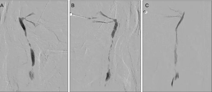
눈물소관과 눈물주머니의 연결은 공통눈물소관을 갖는 경우를 type I, 눈물소관들이 공통눈물소관을 이루지 않고 하나의 입구를 통해 눈물주머니로 들어가는 경우를 type II, 위, 아래 눈물소관들이 독립적으로 두 개의 입구를 갖는 경우를 type III로 나누어(Fig. 1) 눈물소관과 눈물주머니 의 연결 형태를 분류하고,공통눈물소관의 빈도 및 양안의 대칭성 여부를 조사하였다.
A B C
Figure 1. The upper and lower canaliculi can connect to the lacrimal sac by 3 different ways: (A) Type I is a common canaliculus.
(B) Type II is a common opening. (C) Type III is where the upper and lower canaliculi enter the lacrimal sac separately.
Table 1. Types of canalicular connection and symmetry on bilateral dacryocystogram
Canalicular types* Patients no (%) Total patients no (%)
Symmetric Type I & I 217 (87.5)
230 (92.7)
Type II & II 13 (5.2)
Type III & III 0
Asymmetric Type I & II 15 (6.0)
18 (7.3)
Type II & III 3 (1.2)
Type I & III 0
Values are presented as number (%).
*Canalicular types indicates the combination of canalicular types bilaterally indicated in each group of patients.
결 과
총 346명의 환자 중 248명(71.7%)에서 눈물소관의 형 태가 명확하게 관찰되었다.
대상 환자군의 평균 연령은 68.4 ±14.7세였고, 남자가 106명(42.7%), 여자가 142명(57.3%)이었다. 대상환자의 평균 눈물흘림 기간은 33.9 ±10.5개월이었다.
총 496안 중 공통눈물소관이 있는 type I은 90.5%
(449/496안)였으며, type II는 8.3% (41/496안), type III 는 1.2% (6안/496안)으로 조사되었다.
248명의 환자 중 양안이 대칭적으로 같은 눈물소관–눈물 주머니 연결의 형태를 보인 경우는 230명(92.7%)이었고, 양안이 서로 다른 형태의 눈물소관-눈물주머니의 연결 형 태를 보이는 경우는 18명(7.3%)로 관찰되었다. 대칭성을 보이는 경우에는 type I-type I이 87.5% (217/248명)로 가장 많았고, type II-type II는 5.2% (13/248명)였으며 type III-type III는 없었다. 비대칭성으로 나타나는 경우 는 type I-type II가 6.0% (15/248명), type II-type III가 1.2% (3/248명)이었으며 type I-type III는 없었다(Table 1).
고 찰
눈물소관이 눈물주머니로 들어가는 형태의 해부학적 구 조는 예전부터 많은 관심이 있어왔다. 이는 눈물소관–눈물 주머니 연결형태의 다양성을 파악하는 것이 코눈물길 막힘 의 원인과, 코눈물길 수술의 성공률에 영향을 줄 수 있는 인자를 밝혀내어 임상적으로 도움이 될 수 있을 것이라는 기대 때문이었다. 이전 발표되었던 공통눈물소관에 대한 보 고를 살펴보면, 1961년 Jones2가 90%에서 공통 눈물소관 을 보이며, 나머지 10%에서 위, 아래 눈물소관이 합쳐지지 않고 눈물주머니와 연결된다고 하였으나, 그 형태의 분석이 어떻게 이루어졌는지 구체적으로 언급되지 않았다. 이후 1991년 Kurihashi et al6은 사체를 이용하여 현미경적 해부 분석을 하였고 5구의 사체 중 6안에서 공통눈물소관이 존 재했다고 발표하였다.2,6,7이와 반대로 Caldemeyer et al8은 컴퓨터 단층 눈물주머니조영술을 이용한 연구에서 대상이 되었던 모든 환자에서 양안 대칭적으로 공통눈물소관이 존 재하였다고 하였다. 본 연구는 비록 적은 수의 환자(11명) 가 대상이었기 때문에 한계가 있으나 새로운 영상장비를
이용하여 눈물길의 해부학적 형태를 분석하였다는 데 의의 가 있다 하겠다.
오늘날에는 영상기법의 발달로 해부학적 구조를 비교적 정확하게 관찰하는 것이 가능하다. 눈물길조영술은 1909년 Ewing9에 의해 처음 소개된 이후 눈물흘림을 호소하는 환 자에 있어 코눈물길 막힘의 진단에 널리 사용되고 있으며 지속적으로 발전해왔다.10,11 최근에는 무골 영상(bone- free visualization)이 가능한 디지털감산 눈물길조영술 (digital subtraction dacryocystography)이 개발되었다. 이 는 이전에 사용된 방법보다 진단적 해상도나 진단적 가치 가 높고 코눈물길을 통해 조영제가 흘러가는 과정을 실시 간으로 촬영할 수 있다는 장점이 있으며, 특히 조영제 주입 후 눈물주머니에 조영제가 차오르기 전, 초기에 정확한 영 상을 얻을 수 있다. 그러나 영상을 분석함에 있어서 주위를 기울여야 하는데, 저자들의 경험에 의하면 type I 또는 type II 형태의 눈물소관을 갖는 경우, 조영제가 위 눈물소 관을 먼저 나타내 주고, 이후 눈물주머니를 채우게 되는 반 면, type III의 경우 조영제는 아래 눈물소관을 통과한 직후 눈물주머니에 차오르고, 그 이후 위 눈물소관을 보여주게 되므로 촬영 시점에 따라 해석의 오류를 가져올 수 있었다.9
Yazici and Yazici12는 디지털 감산 눈물길조영술을 이용 하여 환자의 코눈물길을 분석한 연구 결과를 보고하였는데, 341안 중 94.1%에서 공통눈물소관을 관찰할 수 있었고 (type I), 3.8%가 공통눈물소관을 형성하지 않고 하나의 입구를 통해 눈물주머니로 들어가는 형태였으며(type II), 아래 눈물소관들이 독립적으로 두 개의 입구를 갖는 경우 (type III)는 2%에 불과하다고 하였다. 본 연구에서는 대상 안의 90.5%에서 공통눈물소관을 갖는 것으로 나타나 Yazici의 연구와 비교하여 다소 낮은 빈도를 보였고, 공통 눈물소관을 갖지 않는 경우는 9.5% (type II and III)로 비 교적 높은 빈도를 보였다.
Type II와 III처럼 공통눈물소관이 없는 경우는 흔하지 않지만 임상적으로 눈물길질환을 접근하는데 고려해야 하 는 경우가 있다. 예를 들어 눈물소관 열상을 복구하는 데 사용하는 pigtail probe는 공통눈물소관이 없는 경우 사용 이 권장되지 않는다.13-15또한 공통눈물소관이 없는 경우는 probing 시행할 때 false passage를 만들기 쉽기 때문에12 silicone intubation이나 probing 시에는 반드시 공통눈물소 관의 해부학적 변이에 대한 고려가 필요하다.16
눈물길의 막힌 위치가 코경유 눈물주머니코안연결술 수 술 결과에 영향을 주는 중요한 인자라는 보고가 있다.17,18 Choi et al18의 보고에 따르면 막힌 위치를 공통눈물소관, 눈물주머니, 눈물관-주머니 연결지점, 코눈물관으로 나누 어 수술성공률을 비교하였고 흥미롭게도 눈물주머니 부위
의 폐쇄(saccal obstruction)가 공통눈물소관의 폐쇄보다 통계학적으로 유의하게 성공률이 낮음을 보고하였다. 공통 눈물소관의 부재가 눈물주머니 부위의 폐쇄에 어떠한 영향 을 미치는지 연구된 바는 아직 없지만, 공통눈물소관에 존재 하는 Rosemuller 판막이가 눈물주머니 고임(retention)12,19 에 병인으로서 중요한 역할을 하는 것을 미루어볼 때 눈물 주머니 막힘과 공통눈물소관의 부재는 관계가 있을 것으로 생각된다. 공통눈물소관이 없는 경우, 특히 위, 아래 눈물소 관이 나뉘어져 눈물주머니와 연결되는 경우(type III)는 본 연구에서도 가장 드물게 관찰되었는데, 과거 Hurwitz는 위, 아래 눈물소관이 나뉘어져 눈물주머니와 연결되는 경우 (type III)는 없었다 하였고,20 이러한 형태의 눈물소관–눈 물주머니 연결은 정상적인 해부학적 변형(variant)이 아닌 아마도 수술 전 또는 수술 중의 탐침자 사용 시 잘못 형성 된 눈물소관 때문일 것이라고 여겨졌다.16,21 Type II 혹은 III의 해부학적 형태와 선천적 또는 후천적 코눈물길 막힘 의 연관성은 불행히도 아직 명확하게 밝혀진 바는 없기 때 문에 공통눈물소관에서 눈물주머니로 연결되는 부위의 해 부학적 변이와 폐쇄부위와의 관계를 밝히기 위한 진행된 추가적인 연구가 필요하다.
본 연구에서는 디지털 감산 눈물길조영술을 이용하여 비 교적 많은 수의 환자를 대상으로 눈물소관-눈물주머니의 연결형태의 빈도를 알아보았다. 한국인을 대상으로 한 이와 같은 보고는 처음이며, 향후 눈물소관-눈물주머니의 연결 형태와 코눈물길 막힘과의 연관성 및 코눈물길 막힘 환자 에게서 눈물주머니코안연결술의 성공률에 형태적 다양성이 미치는 영향을 알아 볼 수 있는 또 다른 연구의 배경으로서 의 의미가 있다.
참고문헌
1) Linberg JV. Surgical anatomy of the lacrimal system. In: Linberg JV, ed. Lacrimal Surgery. New York: Churchill Livingstone, 1988;1-18. Contemporary Issues in Ophthalmology; vol 5.
2) Jones LT. An anatomical approach to problems of the eyelids and lacrimal apparatus. Arch Ophthalmol 1961;66:111-24.
3) Kikkawa DO, Lemke BN. Orbital and eyelid anatomy. In:
Dortzbach RK, ed. Ophthalmic Plastic Surgery: Prevention and Management of Complications. New York: Raven Press, 1994;
1-29.
4) Nowinski TS. Anatomy and physiology of the lacrimal system. In:
Bosniak S, ed. Principles and Practice of Ophthalmic Plastic and Reconstructive Surgery. Philadelphia: WB Saunders, 1996;731- 47.
5) Dale DL. Embryology, anatomy, and physiology of the lacrimal drainage system. In: Stephenson CM, ed. Ophthalmic Plastic, Reconstructive, and Orbital Surgery. Boston: Butterworth-Heinemann, 1997;19-30.
6) Kurihashi K, Imada M, Yamashita A. Anatomical analysis of the human lacrimal drainage pathway under an operating microscope.
Int Ophthalmol 1991;15:411-6.
7) Tucker NA, Tucker SM, Linberg JV. The anatomy of the common canaliculus. Arch Ophthalmol 1996;114:1231-4.
8) Caldemeyer KS, Stockberger SM Jr, Broderick LS. Topical con- trast-enhanced CT and MR dacryocystography: imaging the lac- rimal drainage apparatus of healthy volunteers. AJR Am J Roentgenol 1998;171:1501-4.
9) Ewing AE. Roentgen ray demonstration of the lacrimal abscess cavity. Am J Ophthalmol 1909;26:1-4.
10) Montecalvo RM, Zegel HG, Barnett FJ, et al. Evaluation of the lac- rimal apparatus with digital subtraction macrodacryocystography.
Radiographics 1990;10:483-90.
11) King SJ, Haigh SF. Technical report: digital subtraction dacryocystography. Clin Radiol 1990;42:351-3.
12) Yazici B, Yazici Z. Frequency of the common canaliculus: a radio- logical study. Arch Ophthalmol. 2000;118:1381-5.
13) Walter WL. The use of the pigtail probe for silicone intubation of the injured canaliculus. Ophthalmic Surg 1982;13:488-92.
14) Reifler DM. Management of canalicular laceration. Surv Ophthalmol 1991;36:113-32.
15) Jordan DR, Nerad JA, Tse DT. The pigtail probe, revisited.
Ophthalmology 1990;97:512-9.
16) Hecht S. Dacryocystorhinostomy. In: Guibor P, Smith B, eds.
Contemporary Oculoplastic Surgery. New York: Stratton Intercontinental Medical Book Corp, 1974;41-54.
17) Yung MW, Hardman-Lea S. Analysis of the results of surgical en-
doscopic dacryocystorhinostomy: effect of the level of obstruction.
Br J Ophthalmol 2002;86:792-4.
18) Choi JC, Jin HR, Moon YE, et al. The surgical outcome of endo- scopic dacryocystorhinostomy according to the obstruction levels of lacrimal drainage system. Clin Exp Otorhinolaryngol 2009;2:141-4.
19) Harris GJ, DiClementi D. Congenital dacryocystocele. Arch Ophthalmol 1982;100:1763-5.
20) Hurwitz JJ. Punctum and canaliculus. In: Hornblass A, ed.
Oculoplastic, Orbital and Reconstructive Surgery. Baltimore:
Williams & Wilkins, 1990;1381-93.
21) Cowen D, Hurwitz JJ. Anatomy of the lacrimal drainage system.
In: Hurwitz JJ, ed. The Lacrimal System. Philadelphia:
Lippincott-Raven Publishers, 1996;15-21.
22) Jones LT, Wobig JL. Tear sac foreign bodies and tumors. In: Jones LT, Wobig JL, eds. Surgery of the Eyelids and Lacrimal System.
Birmingham, Ala: Aesculapius Pub Co, 1976;185-93.
23) Veirs ER. Anatomy and embryology of the lacrimal system. In:
Veirs ER, ed. Lacrimal Disorders: Diagnosis and Treatment. St Louis: Mosby, 1976;1-12.
24) Wollf E. Ocularappendages. In: Roger Warwick, ed. Anatomy of the Eye and Orbit, 7th ed. Philadelphia: Saunders, 1976;181-237.
25) Jones LT, Wobig JL. The lacrimal system. In: Jones LT, Wobig JL, eds. Surgery of the Eyelids and Lacrimal System. Birmingham, Ala: Aesculapius Pub Co, 1976;57-70.
26) Hurwitz JJ. The Lacrimal System. Philadelphia: Lippincott-Raven, 1996;261-96.
=ABSTRACT=
Frequency and Characteristic Findings of the Common Canaliculus in Koreans
Eun Hae Lim, MD, Sun Young Jang, MD, Jae Woo Jang, MD, PhD
Myung-Gok Eye Research Institute, Konyang University Kim’s Eye Hospital, Seoul, Korea
Purpose: To investigate the different types of anatomical connection between the lacrimal sac and the canaliculi using digi- tal subtraction dacryocystography (DCG) in Koreans.
Methods: The authors of the present study performed digital subtraction DCG in Korean patients who presented with epi- phora from January 2010 until December 2010. The 248 patients (496 eyes) who achieved a satisfactory DCG image were classified as follows: 1) type I: visible common canaliculus (CC), 2) type II: no visible CC and the canaliculi entering the sac at the point where they meet on the sac wall (common opening), 3) type III: no visible CC and common opening, and each canaliculus entering the sac from different points.
Results: Out of a total of 496 eyes, CC was observed in 449 eyes (type I: 90.5%). In 41 eyes (8.3%), the CC was absent (type II), but the upper and lower canaliculi joined at the wall of the lacrimal sac. In 6 eyes (1.2%), the upper and lower ca- naliculi entered the sac separately (type III). Eighteen patients (7.3%) showed different types of lacrimal drainage system between the 2 eyes.
Conclusions: The CC may not exist in all patients, and the type of anatomical connection between the lacrimal sac and the canaliculi may be different between the eyes in the same individuals. Although such patients comprise a minority, anatomi- cal knowledge of the lacrimal drainage system could be helpful to assess and manage patients with lacrimal drainage disorder.
J Korean Ophthalmol Soc 2012;53(11):1549-1553
Key Words: Common canaliculus, Digital subtraction dacryocystography, Lacrimal drainage system, Lacrimal sac
Address reprint requests to Jae Woo Jang, MD, PhD
Myung-Gok Eye Research Institute, Konyang University Kim’s Eye Hospital
#136 Yeongsin-ro, Yeongdeungpo-gu, Seoul 150-034, Korea
Tel: 82-2-2639-7777, Fax: 82-2-2633-3976, E-mail: jwjang63@gmail.com