pISSN: 0378-6471 eISSN: 2092-9374 http://dx.doi.org/10.3341/jkos.2012.53.6.781
= 증례보고 =
급성 중심장액맥락망막병증 환자에서 유리체내베바시주맙 주입술의 유용성
고경민⋅김주연⋅김종우⋅최문정 건양대학교 김안과병원 안과학교실, 명곡안연구소
목적: 급성 중심장액맥락망막병증 환자에서 유리체내베바시주맙 주입술의 유용성에 대해 알아보고자 하였다.
대상과 방법: 급성 중심장액맥락망막병증으로 진단받은 80명 87안을 후향적으로 분석하였다. 모든 대상 환자의 초진 시와 추적 관찰 시 최대교정시력과 중심황반두께의 변화를 확인하였다.
결과: 경과관찰 기간 동안 모든 환자들이 시력의 호전과 망막하액의 소실을 보였으나 logMAR로 측정한 최대교정시력은 베바시주맙 주입군이 0.11, 보존적 치료군이 0.14로 두 군 사이에는 유의한 차이는 없었다(p=0.13). 중심황반두께도 베바시주맙 주입군이 185.33 μm, 보존적 치료군이 193.41 μm로 역시 통계적으로 유의한 차이가 없었다(p=0.908). 다만 재발률은 베바시주맙 주입군(47%)에서 보 존적 치료군(70%)에 비해 유의하게 낮게 나타났다(p=0.03).
결론: 베바시주맙 주입군과 보존적 치료군에서 최대교정시력과 중심황반두께는 유의한 차이를 보이지 않았다. 하지만 주입군에서 낮 은 재발률(p=0.03)을 보여 베바시주맙 주입술이 급성 중심장액맥락망막병증의 재발률을 낮추는 데 기여할 것으로 생각한다.
<대한안과학회지 2012;53(6):781-785>
■ 접 수 일: 2011년 8월 29일 ■ 심사통과일: 2011년 11월 3일
■ 게재허가일: 2012년 4월 29일
■ 책 임 저 자: 최 문 정
서울특별시 영등포구 영신로 136 김안과병원
Tel: 02-2639-7811, Fax: 02-2633-3976 E-mail: [email protected]
중심장액맥락망막병증(central serous chorioretinop- athy, CSC)은 후극부에 발생하는 장액성 감각신경망막박 리를 특징으로 하는 질환이다.1과거에는 망막색소상피세포 의 결손이나 기능 이상으로 인해 감각망막박리가 생길 것 이라는 가설이 제기되었으나2,3 최근에는 맥락막 혈관의 과 투과성이 병인이라는 주장이 폭넓게 받아들여지고 있다.4,5 인도시아닌그린혈관조영검사의 발전으로 CSC 환자에서 맥 락막의 소엽성 허혈, 맥락정맥충혈, 다발성 맥락막 혈관의 과투과성이 보인다는 것이 알려졌다.6,7맥락막 허혈은 혈관 내피성장인자(vascular endothelial growth factor, VEGF) 의 농도를 높이는 역할을 하게 되고 이는 맥락막혈관의 투 과성을 증가시키며 결국 신경감각박리와 망막색소상피박리 를 야기하는 것으로 여겨진다. 이러한 병인을 근거로 이론 적으로 VEGF가 맥락막 투과성을 감소시키는 데 유용할 것 이라고 여겨지고 있다.8대부분의 경우 급성 CSC은 치료 없 이 자연적으로 호전되는 경과를 보인다.9,10그러나 일부 군 에서는 장액성감각망막박리가 지속되고 낭포황반병성, 색 소망막상피의 기능소실로 인한 지속적 시력감소를 보이기
도 한다.11 시력의 빠른 회복과 질환의 빠른 소실을 원하는 급성 CSC 환자들의 치료에서는 치료효과뿐만 아니라 치료 의 부작용을 감소시키는 것이 중요한 요소라고 할 수 있다.
급성 CSC 환자들을 대상으로 한 무작위위약통제실험에서 절반용량의 verteporfin (3 mg/m2)와 광역동치료를 했을 때 다초점막망전위검사와 빛초점단층촬영검사상 시력과 기 능적, 해부학적 향상이 유의하게 나타났다.12 또 다른 연구 에 의하면 급성 CSC을 광역동치료를 사용하여 치료할 때 30% verteporfin을 사용하면 부작용이 적으면서도 치료효 과를 얻을 수 있다는 것이 밝혀졌다.13 또한 유리체내베바 시주맙 주입은 기존의 레이저광응고술이나 광역학치료 등 에 비해 심각한 부작용이 적기 때문에 급성 CSC의 효과적 인 치료 중 하나로 여겨지고 있다. 여러 연구들을 통해 VEGF가 맥락막 혈관의 과투과성과 관계되며, anti-VEGF agent인 베바시주맙의 유리체강내 주입이 환자의 증상뿐 아니라 감각망막박리의 회복에도 효과가 있음이 보고되고
있다.14-18 하지만 효과가 없다는 상반되는 연구결과8 또한
보고되고 있어 유용성에 대한 연구가 더 필요한 실정이다.
이에 본 연구에서는 3개월 미만의 급성 CSC 환자에서 유리 체내베바시주맙 주입의 유용성을 알아보고자 하였다.
대상과 방법
2009년 1월부터 2010년 1월까지 본원에서 특발성 CSC
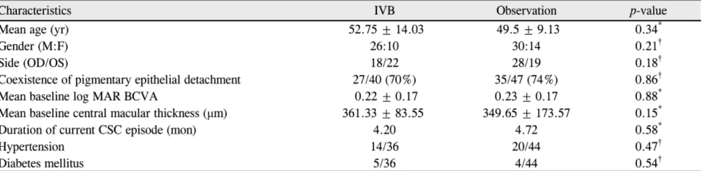
Table 1. Baseline characteristics of patients with acute central serous chorioretinopathy
Characteristics IVB Observation p-value
Mean age (yr) 52.75 ± 14.03 49.5 ± 9.13 0.34*
Gender (M:F) 26:10 30:14 0.21†
Side (OD/OS) 18/22 28/19 0.18†
Coexistence of pigmentary epithelial detachment 27/40 (70%) 35/47 (74%) 0.86†
Mean baseline log MAR BCVA 0.22 ± 0.17 0.23 ± 0.17 0.88*
Mean baseline central macular thickness (μm) 361.33 ± 83.55 349.65 ± 173.57 0.15*
Duration of current CSC episode (mon) 4.20 4.72 0.58*
Hypertension 14/36 20/44 0.47†
Diabetes mellitus 5/36 4/44 0.54†
Values are presented as mean ± SD or number unless otherwise indicated.
IVB = intravitreal bevacizumab injection; log MAR BCVA= logarithm of minimal angle of resolution best-corrected visual acuity; CSC = central serous chorioretinopathy; SD = standard deviation.
*Mann-Whitney U test; †Fisher exact test.
를 진단받은 80명 87안의 의무기록을 후향적으로 분석하 였다. 주입술 치료에 동의했던 40안에 대해서는 유리체내 베바시주맙 주입술을 시행하였으며 주입술 치료를 거부한 47안에 대해서는 보존적 치료를 시행하였다. CSC의 진단 은 고배율 렌즈를 사용한 세극등 현미경검사에서 황반부의 장액성 융기를 보이고, 형광안저촬영에서 병변 부위에 일치 하는 형광고임이 있으며, 빛간섭단층촬영(Stratus OCT, Carl Zeiss Meditec, Dublin, CA; OCT SLO, OTI-OPKO Health Inc, Miami, FL)에서 감각신경망막박리가 확인되는 경우로 하였다. 질환의 호전 여부는 시력, 안저검사와 빛간 섭단층촬영을 통한 황반중심두께 감소로 판단하였다. 경과 관찰기간 동안 망막하액이 소실된 후에 다시 증가되면 재 발한 것으로 정의하였다. 당뇨망막병증, 망막혈관폐쇄, 포 도막염, 연령관련황반변성이 있는 경우, 백내장수술을 포함 한 모든 종류의 안내수술 및 망막수술 병력을 가진 경우는 제외하였으며, 이전에 CSC에 대한 치료 기왕력이 있거나 재발한 경우, 반대쪽 눈에 이환된 적이 있는 경우도 제외하 였다.
베바시주맙(AvastinⓇ, 1.25 mg/0.05 ml)은 각막 변연부 로부터 3.5 mm 떨어진 부위를 통해 유리체강내로 주입하 였으며 주입술 시행 후 Cravit® (levofloxacin 0.5%, Santen, Osaka, Japan) 점안액을 하루 4번 점안하였으며 3 일 뒤에 염증 여부를 확인하였다. 베바시주맙 주입군과 보 존적 치료를 시행한 군에서 초진 시와 1, 3, 6, 9, 12개월 후에 시력측정, 세극등현미경검사, 안저검사, 빛간섭단층촬 영을 시행하였다. 본 연구에서는 측정된 결과는 SPSS for window (version 10.0)를 통해 paired t-test, Fischer’s exact test 및 Mann-Whitney U test를 이용하였다.
p-value가 0.05 미만인 경우를 통계학적 의의가 있는 것으 로 정의하였다.
결 과
대상 환자는 남자 56명, 여자 24명으로 80명이었으며 평 균 연령은 50.3세이었다(Table 1). 성별, 나이, 초진 시 시 력(logMAR)과 초진 시 중심망막두께 등과 같은 환자 군의 통계학적 특성은 두 군 간에 유의한 차이를 보이지 않았다.
증상발현기간은 주입군에서 처음 증상이 생긴 시기부터 유 리체내베바시주맙 주입술을 받기 전까지로 정의하였으며 대조군에서는 증상발현부터 CSC 진단받기 전까지로 정의 하였고 각 군에서 평균 4.20개월, 4.72개월로 유의한 차이 를 보이지 않았다(Table 1). 모든 환자에서 첫 치료 후 1개 월, 그 이후부터는 3개월 간격으로 최소 12개월 동안 정기 적으로 경과관찰을 시행하였다. 재발률은 유리체내베바시 주맙 주입군에서 유의하게 낮은 것으로 나타났다(47%:70%, p=0.031). 초진 시 측정한 초진 환자군 특성에 대해서는 Table 1에 나타나 있다.
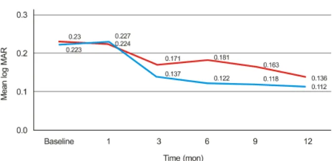
12개월 후 추적관찰 시 모든 환자들에게서 시력의 호전 과 망막하액의 소실이 나타났다. 12개월째 최대교정시력 (logMAR)은 유리체내베바시주맙 주입군에서 0.22 ±0.17 에서 0.11 ± 0.11로 호전되었으며 보존적 치료군에서는 0.23 ±0.17에서 0.136 ±0.21로 호전되었다. 마지막 추 적관찰 시 두 군 간의 평균시력(logMAR)은 유의한 차이를 보이지 않았으며(p=0.132), 중심망막두께는 유리체내베바 시주맙 주입군에서 185.33 ±31.23 μm, 보존적 치료군에 서는 193.41 ±74.33 μm로 나타나 역시 유의한 차이를 보 이지 않았다(Fig. 1, 2). 통계적으로 유의하지 않았지만 베 바시주맙 주입군에서는 망막하액이 소실되는 데 평균 1개 월, 보존적 치료군에서는 3개월이 소요되었다. 다만 재발률 은 베바시주맙 주입술 주입군(47%)에서 보존적 치료군 (70%)에 비해 유의하게 낮게 나타났다(p=0.032). 유리체 내베바시주맙 주입술 후 총 추적기간 동안 안구에 국한되
Figure 1. Graph showing serial changes in the mean logarithm
of the minimum angle of resolution (log MAR) for patients in the intravitreal bevacizumab injection (IVBI) group (blue line) and in the observation group (red line).Figure 2. Graph showing the serial changes in mean central
macular thicknesses (μm) as measured using optical coherence tomography in the IVBI group (blue line) and in the ob- servation group (red line).거나 전신적인 합병증은 관찰되지 않았다.
고 찰
CSC은 망막색소상피 연관 질환으로, 망막색소상피의 특 발적인 누출 또는 맥락막 혈류의 변화로 인해, 장액성망막 박리 및 망막색소상피박리가 유발되는 것으로 알려져 있으 나 아직 명확한 기전은 확립되지 않았다.19 CSC은 대부분 자연적으로 호전되어 대부분의 경우 시력이 20/25 또는 그 이상으로 좋아진다.1,20 그러므로 일반적으로 처음 발생한 급성 CSC은 첫 3개월까지는 특별한 치료 없이 경과 관찰하 게 된다. 하지만 질환이 자연적으로 호전되기까지의 기간 동안은 환자들이 시야흐림, 색각이상, 중심암점의 출현, 대 조민감성의 감소, 원시의 증가 등의 증상으로 고통받게 된 다. 그러므로 환자들은 자주 질환의 더 빠른 호전을 기대한 다. 환자들 대부분이 젊은 층에 속하므로 질환에 의한 직업 적 손실 또한 상당하다.21또한 이 질환의 5%에서는 심각하
고 영구적인 시력소실을 경험하기도 한다.22 단안환자의 경 우나 직업적으로 특별히 빠른 치료를 요하는 경우, 정신과 적 질환의 과거력이 있는 경우와 같이 장액성 황반 박리가 재발할 위험성이 큰 경우 등에서 급성 CSC의 조기 치료가 필요할 수 있다.23
현재까지 유리체내베바시주맙 주입술이 급성과 만성 CSC에 미치는 영향에 대한 연구가 활발이 진행되고 있다.
CSC의 치료로 유리체내베바시주맙 주입술은 안전하고 효 과적이며 기능적, 해부학적으로 호전을 보이게 한다는 연구 결과들이 보고되고 있다.15,24최근 한 연구에 따르면 증상기 간이 3개월 이상인 40안 중 33안(82.5%)에서 첫 유리체내 베바시주맙 주입술 후 3개월 내에 망막하액의 완전한 소실 을 보인 바 있다. 여기서 망막하액의 소실을 보인 안은 형 광안저혈관조영검사와 인도시아닌그린혈관조영검사에서도 호전된 양상을 보였다.8급성 CSC 치료에 대한 연구들을 살 펴보면 Torres-Soriano et al17은 5안에 유리체내베바시주 맙 주입술을 사용하여 부작용 없이 시력 호전 보였다고 보 고한 바 있으며 Seong et al16은 10안에 유리체내베바시주 맙 주입술 후 1개월 안에 신경감각박리가 호전되는 것을 보 고하였다. 그러나 위 연구결과들은 대조군이 없는 소수의 증례보고였다는 한계가 있다. 대조군을 이용한 최근 연구에 서는 유리체내베바시주맙 주입군과 대조군 간에 시력, 중심 망막두께, 관해기간에 있어서 유의한 차이가 없다는 결과가 나타났다(p>0.05). 이 연구는 유리체내베바시주맙 주입술 이 급성 CSC 치료에 미치는 긍정적인 영향은 없었으며 부 작용 또한 없었다고 보고하였다.8유리체내베바시주맙 주입 술과 고전적인 광역동치료를 비교한 한 연구에서는 유리체 내베바시주맙 주입군에서 망막하액의 소실이 더 느리고 불 완전한 양상을 보였지만 안전성 입장에서 보았을 때 유리 체내베바시주맙 주입군에서 광역동치료군보다 중심오목얇 아짐 현상이 더 적게 나타난다고 보고하면서(25%:53%) 저자들은 특정 환자들에게는 유리체내베바시주맙 주입술이 광역동치료에 비하여 더 안전하면서 효과적인 치료방법이 될 수 있다고 주장하고 있다.25 이번 연구에서는 급성 CSC 치료에서 유리체내베바시주맙 주입군이 대조군에 비하여 치료 후 최대교정시력, 중심황반두께에서 유의한 차이를 보 이지 않았지만 유리체내베바시주맙 주입군에서 상대적으로 적은 재발률을 보이는 것으로 나타났다. 그러나 기존 연구 들에서 CSC의 재발률이 첫 발병 1년 내에 30-50% 정도9,22 로 나타난 것에 비해 본 연구에서는 대조군의 재발률이 70%로 높게 나타났고 추적관찰 기간이 길지 않다는 한계 점을 고려했을 때 베바시주맙 주입군에서의 낮은 재발률 (47%)이 유의한지에 대한 논란의 여지가 있다. 본 연구는 후향적인 조사로서의 한계가 있으며 주입술 치료군과 보존
적 치료군을 선택하는 데 있어서 무작위법을 사용하지 않 고 주입술 동의 여부로 나누었기 때문에 양군 간 통계학적 특성이 유의한 차이를 보이지 않았음에도 불구하고 선택바 이어스가 발생할 수 밖에 없었다. 하지만 본 연구는 베바시 주맙 주입술이 급성 중심장액맥락망막병증의 재발률을 낮 출 수 있는 치료로서의 가능성을 보여주었다고 생각되며 앞으로 여기에 대한 대규모의 장기간 추적관찰 연구가 필 요할 것으로 여겨진다.
참고문헌
1) Klais CM, Ober MD, Ciardella AP, et al. Cenral serous chorioretinopathy. In: Ryan SJ, Schachat AP, eds. Retina, 4th ed.
Vol. 2. Philadelphia: Mosby, 2006;1135-62.
2) Marmor MF. New hypotheses on the pathogenesis and treatment of serous retinal detachment. Graefes Arch Clin Exp Ophthalmol 1988;226:548-52.
3) Spitznas M. Pathogenesis of central serous retinopathy: a new working hypothesis. Graefes Arch Clin Exp Ophthalmol 1986;
224:321-4.
4) Costa RA, Farah ME, Freymüller E, et al. Choriocapillaris photo- dynamic therapy using indocyanine green. Am J Ophthalmol 2001;132:557-65.
5) Costa RA, Scapucin L, Moraes NS, et al. Indocyanine green-medi- ated photothrombosis as a new technique of treatment for persis- tent central serous chorioretinopathy. Curr Eye Res 2002;25:
287-97.
6) Stanga PE, Lim JI, Hamilton P. Indocyanine green angiography in chorioretinal diseases: indications and interpretation: an evi- dence-based update. Ophthalmology 2003;110:15-21.
7) Piccolino FC, Borgia L. Central serous chorioretinopathy and in- docyanine green angiography. Retina 1994;14:231-42.
8) Lim JW, Kim MU. The efficacy of intravitreal bevacizumab for idi- opathic central serous chorioretinopathy. Graefes Arch Clin Ophthalmol 2011;249:969-74.
9) Ficker L, Vafidis G, While A, Leaver P. Long-term follow-up of a prospective trial of argon laser photocoagulation in the treatment of central serous retinopathy. Br J Ophthalmol 1988;72:829-34.
10) Yap EY, Robertson DM. The long-term outcome of central serous chorioretinopathy. Arch Ophthalmol 1996;114:689-92.
11) Loo RH, Scott IU, Flynn HW Jr, et al. Factors associated with re-
duced visual acuity during long-term follow-up patients with idio- pathic central serous chorioretinopathy. Retina 2002;22:19-24.
12) Zhao MW, Zhou P, Xiao HX, et al. Photodynamic therapy for acute central serous chorioretinopathy: the safe effective lowest dose of verteporfin. Retina 2009;29:1155-61.
13) Caccavale A, Romanazzi F, Imparato M, et al. Low-dose aspirin as treatment for central serous chorioretinopathy. Clin Ophthalmol 2010;4:899-903.
14) Schaal KB, Hoeh AE, Scheuerle A, et al. Intravitreal bevacizumab for treatment of chronic central serous chorioretinopathy. Eur J Ophthalmol 2009;19:613-7.
15) Artunay O, Yuzbasioglu E, Rasier R, et al. Intravitreal bevacizumab in treatment of idiopathic persistent central serous chorioretinop- athy: a prospective, controlled clinical study. Curr Eye Res 2010;
35:91-8.
16) Seong HK, Bae JH, Kim ES, et al. Intravitreal bevacizumab to treat acute central serous chorioretinopathy: short-term effect.
Ophthalmologica 2009;223:343-7.
17) Torres-Soriano ME, García-Aguirre G, Kon-Jara V, et al. A pilot study of intravitreal bevacizumab for the treatment of central serous chorioretinopathy (case report). Graefes Arch Clin Exp Ophthalmol 2008;246:1235-9.
18) Lim SJ, Roh MI, Kwon OW. Intravitreal bevacizumab injection for central serous chorioretinopathy. Retina 2010;30:100-6.
19) Spaide RF, Campeas L, Haas A. Central serous chorioretinopathy in younger and older adults. Ophthalmology 1996;103:2070-80.
20) Bennett G. Central serous retinopathy. Br J Ophthalmol 1955;39:
605-18.
21) Klatt C, Saeger M, Oppermann T, et al. Selective retina therapy for acute central serous chorioretinopathy. Br J Ophthalmol 2011;95:
83-8.
22) Gass JD. Pathogenesis of disciform detachment of the neuro- epithelium. Am J Ophthalmol 1967;63 Suppl:S1-139.
23) Fok AC, Cahn PP, Lam DS, Lai TY. Risk factors for recurrence of serous macular detachment in untreated patients with central se- rous chorioretinopathy. Ophthalmic Res 2011;46:160-3.
24) Schaal KB, Hoeh AE, Scheuerle A, et al. Intravitreal bevacizumab for treatment of chronic central serous chorioretinopathy. Eur J Ophthalmol 2009;19:613-7.
25) Lee JY, Chae JB, Yang SJ, Yoon YH. Intravitreal bevacizumab ver- sus the conventional protocol of photodynamic therapy for treat- ment of chronic central serous chorioretinopathy. Acta Ophthalmol 2011;89:293-4.
=ABSTRACT=
The Efficacy of Intravitreal Bevacizumab Injection in Patients with Acute Central Serous Chorioretinopathy
Kyung Min Koh, MD, Joo Yeon Kim, MD, Jong Woo Kim, MD, Moon Jung Choi, MD
Myung-Gok Eye Research Institute, Department of Ophthalmology, Konyang University Kim’s Eye Hospital, Seoul, Korea
Purpose: To evaluate the efficacy of intravitreal bevacizumab injection (IVB) in acute central serous chorioretinopathy.
Methods: Retrospective chart review of 87 eyes of 80 patients. They received intravitreal bevacizumab injection (n = 40) and observed without any treatment or any medication (n = 47) for initial treatment of acute CSC. Patient demographics and best-corrected visual acuity (BCVA), central macular thickness (CMT) at baseline were analyzed. Then we had regular follow-ups of BCVA and CMT.
Results: All patients showed improvement in visual acuity and resolution of subretinal fluid following treatment. In the bevacizumab-treated group, the mean BCVA improved from 0.22 ± to 0.11, mean CMT fell from 361.33 to 185.33 (μm) and recurrence rate was 47%. The conservative-treated group showed an improvement in BCVA from 0.23 at baseline to 0.14, mean CMT fell from 349.65 to 193.41 (μm) and recurrence rate was 70% (p = 0.130, p = 0.908 and p = 0.030, respectively).
However, the change of BCVA and CMT of 2 groups showed no differences at any follow-up visit. Recurrence rate was sig- nificantly low in bevacizumab-treated group.
Conclusions: The bevacizumab-treated group had relatively low rates of recurrence in acute CSC patients compared to the observation group. However, intravitreal bevacizumab showed no positive effect in BCVA and CMT of acute CSC pa- tients compared to the observation group. Further investigation will be helpful to understand this therapy in patients with CSC.
J Korean Ophthalmol Soc 2012;53(6):781-785
Key Words: Bevacizumab, Central macular thickness, Central serous chorioretinopathy, Intravitreal injection
Address reprint requests to Moon Jung Choi, MD
Department of Ophthalmology, Konyang University Kim’s Eye Hospital
#136 Yeongsin-ro, Yeongdeungpo-gu, Seoul 150-034, Korea
Tel: 82-2-2639-7811, Fax: 82-2-2633-3976, E-mail: [email protected]