J Korean Soc Radiol 2018;78(5):353-357 https://doi.org/10.3348/jksr.2018.78.5.353
INTRODUCTION
When performing interventional procedure such as transar- terial chemoembolization (TACE), we encounter lots of ana- tomic variations of celiac axis and hepatic arteries. To have bet- ter outcomes and prevent iatrogenic complications, it is essential to be aware of these variations.
Previously published reports have demonstrated that the in- cidence of classical trifurcation pattern of celiac axis was 70.8%
(1); the accessory left gastric artery (LGA) arising from the left hepatic artery (LHA) was 11.1–14.2% (2, 3); the accessory LHA arising from the LGA was 6.25–17.9% (1, 3); and the replaced LHA from LGA in 6.25% (1). The first report was based on visual inspections during surgical operations, angiograms and mag-
netic resonance imaging studies. Other two reports were based on visual inspections during autopsies and on angiograms, re- spectively. However, none of these reports had the analysis using computed tomography (CT), which is considered more effec- tive to precisely display the anatomical detail of blood vessels.
We recently encountered a rare case of the entire LGA arising from the LHA in a 62-year-old man during TACE for recurred hepatocellular carcinoma (HCC), and verified this anatomical variant by 3-phase liver CT with 3-dimensional volume render- ing reconstruction as well as angiography.
Case RepORT
A 62-year-old male, diagnosed with HCC seven years ago and
Entirely Replaced Left Gastric Artery from the Left Hepatic Artery:
A Case Report
좌간동맥에서 기시하는 온전한 좌위동맥의 증례
Suyoung Kim, MD
1, Jung Wook Seo, MD
1, Wonseon Shin, MD
2*
1Department of Radiology, Inje University Ilsan Paik Hospital, Goyang, Korea
2Department of Radiology, Dongsan Medical Center, Keimyung University School of Medicine, Daegu, Korea
Arteries originating from the celiac axis have numerous anatomical variations. When performing interventional and surgical procedures, it is important to be aware of these variations to have better outcomes and to prevent iatrogenic complications.
We report on a case of a 62-year-old man who came to our institution to receive transarterial chemoembolization for hepatocellular carcinoma. The computed to- mography and angiography revealed a rare anatomic variation: the entire left gas- tric artery originated from the left hepatic artery with no other accessory feature of the left gastric artery from celiac axis or aorta was seen. To our knowledge, this is the first report on the entirely replaced left gastric artery from the left hepatic ar- tery that was confirmed by utilizing both computed tomographic and angiographic images.
Index terms Anatomic Variation Angiography
Tomography, X-Ray Computed Celiac Artery
Hepatic Artery
Received August 20, 2017 Revised October 17, 2017 Accepted December 23, 2017
*Corresponding author: Wonseon Shin, MD Department of Radiology, Dongsan Medical Center, Keimyung University School of Medicine, 56 Dalseong-ro, Jung-gu, Daegu 41931, Korea.
Tel. 82-53-250-8374 Fax. 82-53-250-7766 E-mail: [email protected]
This is an Open Access article distributed under the terms of the Creative Commons Attribution Non-Commercial License (http://creativecommons.org/licenses/by-nc/4.0) which permits unrestricted non-commercial use, distri- bution, and reproduction in any medium, provided the original work is properly cited.
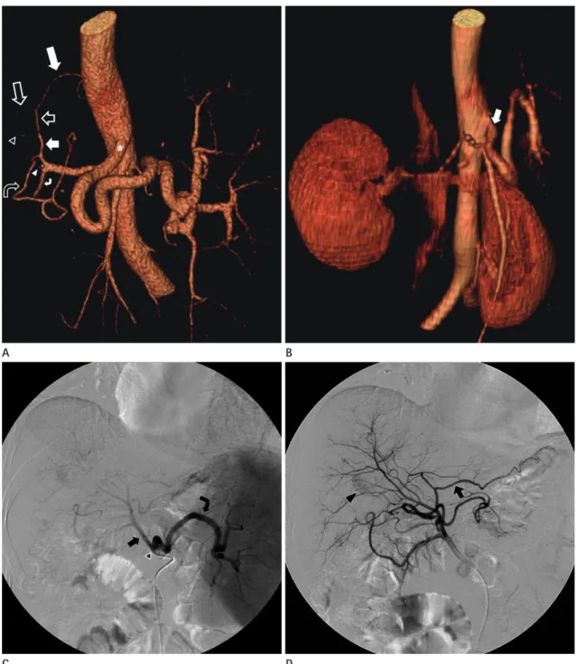
Fig. 1. A 62-year-old male patient with entirely replaced LGA from the LHA, confirmed on liver CT and conventional angiography.
A. In a volume-rendering CT angiographic image, the celiac axis (*) bifurcates into the CHA and the splenic artery, and no other feature of the LGA from celiac axis or aorta is seen. After branching of the GDA (white curved arrow) and the RGA (empty curved arrow), the proper hepatic ar- tery (white short arrow) bifurcates into the RHA (empty arrowhead) and LHA (empty short arrow). The LGA (white long arrow) is directly branch- ing from the LHA, and the LHA is continuing its passage (empty long arrow). Note the unusual communication (white arrowhead) between the RGA and the GDA.
B. In an initial volume-rendering CT image, the celiac axis (arrow) bifurcates into the CHA and the splenic artery, and no other feature of the LGA from the proximal portion of celiac axis or aorta is visible.
C. During the initial TACE, a celiac angiogram shows the CHA (arrow) and splenic artery (curved arrow) are derived from the celiac axis (arrow- head), with no evidence of LGA.
D. A subsequent CHA angiogram shows the LGA (arrow) branching from the LHA. A HCC, in segment 6/7 (arrowhead), is noted.
CHA = common hepatic artery, CT = computed tomography, GDA = gastroduodenal artery, HCC = hepatocellular carcinoma, LGA = left gastric artery, LHA = left hepatic arter, RGA = right gastric artery, RHA = right hepatic artery, TACE = transarterial chemoembolization
C D
A B
a hepatitis B carrier, underwent 3-phase liver CT during his regular surveillance. Previously, he had received TACE two times and radiofrequency ablation five times in a row, and had not experienced surgical operations for HCC. For the following six months, he remained free of recurrence. However, the CT image showed a newly developed mass with typical features of HCC. The mass was measured at 1.4 cm and located in the seg- ment II of the left hepatic lobe. The patient was referred to the interventional radiologist for TACE.
The right common femoral artery was accessed percutane- ously, and celiac angiography using 5-Fr catheter was carried out. The splenic artery and the common hepatic artery (CHA) were arising from the celiac axis without abnormality. However, the LGA did not appear to be in the celiac axis, its most common origin. Subsequent common hepatic arteriogram revealed a ves- sel with ascending course originating from proximal to the um- bilical point of the LHA, and running along the gastrohepatic ligament. At first, we considered it as an accessory LGA from the LHA since the course was about the same as that of an ac- cessory LGA. In addition, there was still the possible presence of the LGA from other mother vessels such as aorta.
After cone-beam CT was taken, superselective arteriograms of the tumor-supplying vessels were obtained using 2-Fr micro- catheter. Chemoembolization using doxorubicin was also per- formed via A2 sub-branch from the LHA passing by the origi- nating point of the LGA.
After the procedure, we reviewed all of the past images ob- tained by 3-phase liver CT with volume rendering reconstruc- tion and angiography. We checked again the fact that the patient had not experienced any kinds of surgical operations at all. The celiac trunk showed an aberrant branching pattern (Fig. 1A); it bifurcated into the CHA and the splenic artery. The course of the CHA was normal. However, the LGA arose from the LHA and was running through the fissure for ligamentum venosum, then along the gastrohepatic ligament. The course was almost the same as that of a usual accessory LGA, which is known to originate commonly from proximal to the umbilical point of LHA (3). The diameter of the LGA was as large as the main LHA, and larger than a usual accessory LGA. Relatively narrower spec- trum of the lesser curvature from the left side of the stomach was supplied by the LGA. The right gastric artery (RGA) was thick- er and longer than a usual RGA and was supplying a broad spec-
trum of the lesser curvature from the right side of the stomach.
Because the patient underwent three times of TACE, we could not exclude the possible degeneration of additional features of the LGA which existed in the past. Therefore, we thoroughly an- alyzed initial CT and angiographic images one by one, and found no any other feature of the LGA from the celiac axis, aorta and other possible mother arteries (Fig. 1B-D). Against this back- drop, we concluded that the LGA was an entirely replaced LGA from the LHA.
DIsCUssION
A case of a completely replaced LGA arising from the LHA has not been documented in the literature regarding anatomic vari- ations of celiac axis and its branching vessels (2, 4, 5). In 1978, Naidich et al. found the case of the replaced LGA arising from the LHA in two of 500 reviewed celiac angiograms (0.4%) in his classic article about the origin of LGA without CT images (6). In comparison with the report of Naidich which covered limited selective angiographic studies of the celiac axis, we confirmed the LGA from the LHA as the solitary main vessel without any accessory features by using CT studies including the volume ren- dering techniques. CT angiography is considered better to pre- cisely display the anatomical detail of blood vessels, and it is re- ported that 70% of accessory LGAs can be diagnosed at the early phase of multidetector CT even with a slice thickness of 5 mm (7).
We are open to the possibility that our imaging findings can be interpreted in two other ways. First, the replaced LGA arises from the proper hepatic artery, then the LHA arises from the LGA. Second, the common trunk of LHA and LGA arises from the proper hepatic artery, then it bifurcates into the LHA and the replaced LGA.
In addition, it is noteworthy that during the review of the im- ages (Fig. 1A) we found an unusual communication between the RGA and the gastroduodenal artery.
Patients who underwent TACE can suffer upper gastrointes- tinal complications such as bleeding, gastritis, and ulceration caused by infused embolic agents into the gastric arteries, espe- cially when anatomic variants exist. It has documented that up- per gastrointestinal bleeding occurred in 3% (range 0–22%) of 2593 patients in 23 trials (8). To reduce the risk of these complica-
tions, it is significant to be aware of anatomical variations and possible communications of hepatic and gastric arteries, prior to performing any procedures.
Furthermore, the catheterization of LGA has a clinical im- portance in controlling of acute gastrointestinal hemorrhage. It is known that the LGA supplies 85% of all angiographically- documented upper gastrointestinal bleeding sites (9). The em- piric embolization of the LGA appears to be helpful when CT or angiography fails to reveal active bleeding foci (10).
The accessory LGA arising from the LHA is one of relatively common anatomical variations, but the case of the replaced LGA arising from hepatic arteries has rarely been documented.
This is the first report that confirmed the completely replaced LGA from LHA by both CT and angiographic images. We be- lieve the awareness of the possible presence of the entirely re- placed LGA without any other additional accessory features can help interventional radiologists and surgeons in planning vari- ous procedures and surgical operations such as TACE, gastric artery embolization, gastrectomy and hepatic transplantation.
RefeReNCes
1. Varotti G, Gondolesi GE, Goldman J, Wayne M, Florman SS, Schwartz ME, et al. Anatomic variations in right liver living donors. J Am Coll Surg 2004;198:577-582
2. Adachi B. Das Arteriensystem der Japaner. 2nd ed. Tokyo:
Kenkyusha Press 1928:11-68
3. Nakamura H, Uchida H, Kuroda C, Yoshioka H, Tokunaga K, Kitatani T, et al. Accessory left gastric artery arising from
left hepatic artery: angiographic study. AJR Am J Roent- genol 1980;134:529-532
4. Gruttadauria S, Foglieni CS, Doria C, Luca A, Lauro A, Mari- no IR. The hepatic artery in liver transplantation and surgery:
vascular anomalies in 701 cases. Clin Transplant 2001;15:
359-363
5. Michels NA. Newer anatomy of the liver and its variant blood supply and collateral circulation. Am J Surg 1966;112:337- 347
6. Naidich JB, Naidich TP, Sprayregen S, Hyman RA, Pudlowski RM, Stein HL. The origin of the left gastric artery. Radiology 1978;126:623-626
7. Ishigami K, Yoshimitsu K, Irie H, Tajima T, Asayama Y, Hiraka- wa M, et al. Accessory left gastric artery from left hepatic artery shown on MDCT and conventional angiography: cor- relation with CT hepatic arteriography. AJR Am J Roentgen- ol 2006;187:1002-1009
8. Marelli L, Stigliano R, Triantos C, Senzolo M, Cholongitas E, Davies N, et al. Transarterial therapy for hepatocellular car- cinoma: which technique is more effective? a systematic review of cohort and randomized studies. Cardiovasc Inter- vent Radiol 2007;30:6-25
9. Kelemouridis V, Athanasoulis CA, Waltman AC. Gastric bleed- ing sites: an angiographic study. Radiology 1983;149:643- 648
10. Lang EV, Picus D, Marx MV, Hicks ME, Friedland GW. Massive upper gastrointestinal hemorrhage with normal findings on arteriography: value of prophylactic embolization of the left gastric artery. AJR Am J Roentgenol 1992;158:547-549
좌간동맥에서 기시하는 온전한 좌위동맥의 증례
김수영
1· 서정욱
1· 신원선
2*
복강 동맥에서 기시하는 혈관은 수많은 해부학적 변이를 보인다. 인터벤션 영상 시술 및 수술 과정 중 치료 결과를 향상시 키고 합병증을 예방하기 위하여 이러한 변이를 숙지하는 것이 중요하다. 62세 남자 환자가 재발한 간암으로 간동맥 화학 색전술을 받기 위하여 내원하였다. 전산화단층촬영술과 혈관조영술에서 좌위동맥은 온전히 좌간동맥에서 기시하는 변이 를 보였으며, 복강 동맥 및 대동맥 등 다른 혈관에서 좌위동맥은 확인되지 않았다. 이는 매우 드문 해부학적 변이로서, 본 연구는 좌간동맥에서 기시하는 온전한 좌위동맥을 전산화단층촬영술 및 혈관조영술 모두를 이용하여 확증한 최초의 보 고이다.
1인제대학교 일산백병원 영상의학과, 2계명대학교 의과대학 동산의료원 영상의학과