J Korean Soc Radiol 2015;73(2):108-112 http://dx.doi.org/10.3348/jksr.2015.73.2.108
INTRODUCTION
Immunoglobulin G4 (IgG4)-related disease is a well-known clinicopathological entity characterized by inflammation that can occur in multiple organ systems with predominance in el- derly men in their 50s or 60s (1). IgG4-related disease has char- acteristic histological features, including lymphoplasmacytic in- flammation, fibrosis, obliterative phlebitis, and an increased number of IgG4-positive plasma cells (2). This disease was first described in patients with autoimmune pancreatitis who also had extra-pancreatic diseases, such as biliary lesions, sialadenitis, retroperitoneal fibrosis, enlarged celiac and hilar lymph nodes, chronic thyroiditis, and interstitial nephritis (3).
IgG4-related disease often affects the dura mater as hypertro- phic pachymeningitis with a pattern of diffuse thickening when the central nervous system (CNS) is involved (2, 4, 5). Nodular
pachymeningial thickening can occasionally present as a mass- like lesion that can be confused with a tumor. The differential diagnosis of dural lesions includes infections and neoplasms in- cluding meningioma, lymphoma, and metastasis. It is impor- tant to discriminate IgG4-related disease from tumors because the former responds well to steroid therapy (4).
Early studies evaluating autoimmune pancreatitis tended to consider that IgG4-positive cell counts of > 10 cells/high pow- ered field (HPF) to > 30 cells/HPF as sufficient for the diagnosis.
However, no criteria have been established for diagnosing extra- pancreatic disease. Some studies on organs, such as the lung, lymph nodes, and salivary glands, advocate using the ratio of IgG4/IgG-positive cells to establish diagnostic cutoff levels. Ra- tios of 30–50% have been applied for diagnosing cases of extra- pancreatic IgG4-related disease. Therefore, a pathological confir- mation is essential and anti-IgG4 antibody immunostaining is
Tumefactive Immunoglobulin G4-Related Disease Involving the Dura Mater: A Case Report and Literature Review
뇌수막종과 유사하게 나타나는 Immunoglobulin G4 연관병증:
증례 보고서와 기존 논문 분석
Jeong Hoon Lee, MD
1, Ji Young Lee, MD
1*, Dong Woo Park, MD
2, Yong Ko, MD
3, Seoung Sam Paik, MD
4, Young-Jun Lee, MD
1Departments of 1Radiology, 3Neurosurgery, 4Pathology, Hanyang University Hospital, Seoul, Korea
2Department of Radiology, Hanyang University Guri Hospital, Guri, Korea
Immunoglobulin G4 (IgG4)-related disease is a well-known disorder characterized by an inflammatory reaction with an increase in the number of IgG4-positive plas- ma cells associated with sclerosis. IgG4-related disease often affects the dura mater with a pattern of diffuse thickening when the central nervous system is involved.
However, some nodular dural thickening requires discrimination from tumors be- cause of obviously different treatment options. We report of a case of IgG4-related disease with tumefactive dural involvement.
Index terms
Immunoglobulin G4-Related Disease Pachymeningitis
Meningioma Dura Mater
Received February 17, 2015 Revised April 2, 2015 Accepted April 21, 2015
*Corresponding author: Ji Young Lee, MD
Department of Radiology, Hanyang University Hospital, 222-1 Wangsimni-ro, Seongdong-gu, Seoul 133-792, Korea.
Tel. 82-2-2290-9373 Fax. 82-2-2293-2111 E-mail: [email protected]
This is an Open Access article distributed under the terms of the Creative Commons Attribution Non-Commercial License (http://creativecommons.org/licenses/by-nc/3.0) which permits unrestricted non-commercial use, distri- bution, and reproduction in any medium, provided the original work is properly cited.
useful for the diagnosis (2).
Here, we describe the radiological features of a case of IgG4- related disease with tumefactive dural involvement and provide a comprehensive literature review of IgG4-related disease involv- ing the dura mater. This report was approved by our Institutional Review Board.
CASE REPORT
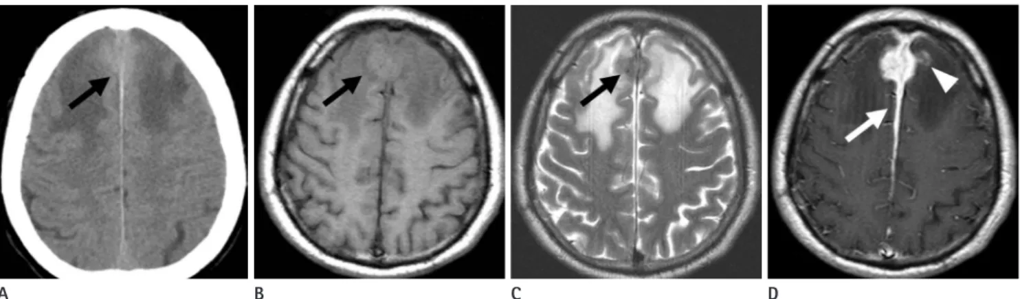
A 57-year-old previously healthy man presented with a first- onset generalized tonic-clonic seizure. The initial non-enhanced brain computed tomography (CT) scan showed a slightly hy- perdense mass along anterior falx cerebri (Fig. 1A). Magnetic resonance imaging (MRI) revealed intermediate signal intensity on T1- and low signal intensity on T2-weighted images with ex- tensive perilesional parenchymal edema (Fig. 1B, C). A post- contrast T1-weighted image showed diffuse thickening and en- hancement of dura mater at the frontal interhemispheric fissure and parasagittal region with involvement of the superior sagittal sinus. The mass showed homogeneous enhancement with a jag- ged border, and adjacent brain parenchymal enhancement was also detected (Fig. 1D). No significant diffusion restriction was observed on a diffusion-weighted image.
With suspicion of a meningioma, the patient underwent a frontal craniotomy, and the mass lesion was removed. Histologic examination revealed a mass with diffuse lymphoplasmacytic in- filtration, severe fibrosis, and phlebitis. An immunohistochemi-
cal study showed up to 26 IgG4-positive cells per HPF, support- ing the diagnosis of IgG4-related disease (Fig. 2). Serum IgG4 level was not available.
The patient was medicated with corticosteroid (250 mg/day methylprednisolone) for the next 6 days after surgery and was discharged on day 10 after surgery without any neurologic defi- cit. The last postoperative follow-up MRI on week 6 after dis- Fig. 1. Computed tomography (CT) and magnetic resonance imaging of immunoglobulin G4-related disease with tumefactive dural involvement in a 57-year-old man. Initial non-enhanced CT (A) demonstrates a slightly hyperdense mass (black arrow) along the anterior falx. The mass (black arrow) shows intermediate signal intensity on T1- (B) and low signal intensity on T2-weighted images (C) with extensive perilesional parenchy- mal edema. A post-contrast T1-weighted image (D) shows, diffuse thickening and enhancement of dura mater (white arrow) at the anterior falx cerebri and parasagittal region. The mass shows homogeneous enhancement with a jagged border and co-involvement of adjacent brain paren- chyma (white arrowhead).
A B C D
Fig. 2. Immunoglobulin G4 (IgG4) immunohistochemical staining (×
400) shows infiltration of abundant IgG4-positive plasma cells (white arrows).
charge revealed significant improvement in the dural lesions.
DISCUSSION
Tumefactive dural involvement of IgG4-related disease was de- tected in this case that could be treated by surgical resection and corticosteroid administration. Intracranial involvement of IgG4- related disease is a relatively rare condition and the tumefactive presentation is even rarer. However, it is important to recognize IgG4-related conditions because they are medically treatable and respond well to corticosteroid therapy.
IgG4-related pachymeningitis was not previously a separate disease entity from idiopathic hypertrophic pachymeningitis.
Linear thickening of the falx and tentorium is the most common finding on an imaging study of idiopathic hypertrophic cranial pachymeningitis, and the next most common finding is focal nodular thickening that simulates a dural mass (6). An increas- ing number of reports suggest that idiopathic hypertrophic pachymeningitis may be part of a disease spectrum, although not all such cases can be categorized as IgG4-related sclerosing disease (7). One report reviewed all pathology specimens diag- nosed as noninfectious hypertrophic pachymeningitis over 25 years at their institution and IgG4-related disease represented 29% of all cases (4). Therefore, dural involvement of IgG4-relat- ed disease is a relatively rare condition (5) and tumefactive du- ral involvement is even rarer, as shown in our literature review in the following paragraphs.
We searched the PubMed database using the keywords: “IgG4- related disease & pachymeningitis” and “IgG4-related disease &
dura mater” and found clinical and radiological information on 43 cases from 27 reports (5, 8). The patients consisted of 27 males (63%) and 16 females (37%) with a mean age of 53.2 ± 11.6 years (range, 32–82 years). The cases of IgG4-related disease most commonly showed more than two sites of dural involvement (18 cases, 41.8%), including the convexity, posterior fossa, skull base, tentorium, or falx. Single lesions were more common at the con- vexity (13 cases, 30.2%) or the skull base (7 cases, 16.3%) (Table 1). MRI revealed relatively low signal intensity on T2-weighted images, representing fibrosis and intermediate signal intensity on T1-weighted image. Post-contrast T1-weighted images showed well-enhanced lesions with diverse features, including diffuse even dural thickening (17 cases, 39.5%) or diffuse dural thicken-
ing with nodularity (13 cases, 30.2%) (Table 2). Co-involvement of brain parenchyma was reported in 9 cases (20.9%) and co-in- volvement of leptomeninges was identified in 3 cases (6.9%).
In our case, the lesion was located in the dura mater encom- passing the anterior falx and parasagittal convexity. A post-con- trast T1-weighted image showed diffuse dural thickening with extreme nodularity that may resemble a tumor. The adjacent su- perior sagittal sinus and brain parenchyma also showed enhance- ment and extensive perilesional parenchymal edema dispropor- tionate to its size on a T2-weighted image. The likelihood of IgG4-related disease rather than a meningioma is suggested by relatively more prominent and thicker hypertrophy of the dura mater than the usual dural tail sign associated with meningioma.
This finding can be helpful to differentiate IgG4-related disease with tumefactive dural involvement from a meningioma.
The differential diagnosis should include conditions, such as Table 1. Locations of Immunoglobulin G4-Related Disease Involving Dura Mater in Literature Review (5, 8)
Involvement Location Patient Number (%)
Single location Convexity 13 (30.2)
Skull base 7 (16.3)
Posterior fossa 4 (9.3)
Tentorium 1 (2.3)
Two or more locations* 18 (41.8)
Total 43 (100)
*Lesions involving two or more locations among convexity, posterior fossa, skull base, tentorium, or falx.
Table 2. Radiologic Features in Immunoglobulin G4-Related Disease Involving Dura Mater on Postcontrast T1-Weighted Image in Litera- ture Review (5, 8)
Radiologic Features Patient Number (%)
Diffuse/even dural thickening 17 (39.5)
Diffuse/nodural dural thickening 13 (30.2) Diffuse/even dural thickening with parenchymal
involvement
4 (9.3)
Diffuse/nodular dural thickening with venous sinus involvement
3 (7.0)
Diffuse/nodular dural thickening with parenchymal involvement
2 (4.7)
Focal/dural thickening with parenchymal involvement 2 (4.7) Diffuse/even dural thickening with leptomeningeal
involvement
1 (2.3)
Diffuse/nodular dural thickening with leptomeningeal involvement
1 (2.3)
Total 43 (100)
lymphoma, metastasis, and infectious meningitis. We considered the possibility of dural lymphoma presenting as a dural mass. Al- though the mass was hyperdense on CT, reflecting a possible highly cellular nature of the lesion, it did not show any diffusion restrictions, so we excluded primary dural lymphoma.
A dural metastasis occurs occasionally and the most frequent- ly reported primary neoplasms are in the prostate, kidney, and breasts. The macroscopic appearance of a dural metastasis is of- ten indistinguishable from a meningioma. Therefore, a metasta- sis should be considered until proven otherwise in a patient with a dural mass and known systemic cancer (9). However, consider- ing that our patient had no history of malignancy and showed no systemic symptoms associated with malignancy, we ruled out a dural metastasis.
Granulomatous diseases, such as fungal diseases, may produce dural masses and pachymeningeal enhancement. However, these granulomatous processes typically affect the basilar menin- ges, rather than the convexities of the cerebral hemispheres. The patient’s clinical presentation provides clues for the differential diagnosis when fever or other signs of infection exist. A lumbar puncture may reveal pleocytosis, and cerebrospinal fluid cul- tures may demonstrate an organism (10). Our patient was im- munocompetent and did not show any signs of infection or labo- ratory findings supporting a fungal infection diagnosis.
In conclusion, IgG4-related disease can demonstrate a dural- based mass in cases of CNS involvement. IgG4-related disease should be kept in the differential diagnosis because it has obvi- ously different treatment options when a dural-based mass ac- companies excessive hypertrophy of adjacent dura mater.
REFERENCES
1. Moss HE, Mejico LJ, de la Roza G, Coyne TM, Galetta SL, Liu GT. IgG4-related inflammatory pseudotumor of the central
nervous system responsive to mycophenolate mofetil. J Neurol Sci 2012;318:31-35
2. Lindstrom KM, Cousar JB, Lopes MB. IgG4-related menin- geal disease: clinico-pathological features and proposal for diagnostic criteria. Acta Neuropathol 2010;120:765-776 3. Lee LK, Sahani DV. Autoimmune pancreatitis in the context
of IgG4-related disease: review of imaging findings. World J Gastroenterol 2014;20:15177-15189
4. Wallace ZS, Carruthers MN, Khosroshahi A, Carruthers R, Shinagare S, Stemmer-Rachamimov A, et al. IgG4-related disease and hypertrophic pachymeningitis. Medicine (Balti- more) 2013;92:206-216
5. Takeuchi S, Osada H, Seno S, Nawashiro H. IgG4-Related In- tracranial Hypertrophic Pachymeningitis: a case report and review of the literature. J Korean Neurosurg Soc 2014;55:
300-302
6. Lee YC, Chueng YC, Hsu SW, Lui CC. Idiopathic hypertro- phic cranial pachymeningitis: case report with 7 years of imaging follow-up. AJNR Am J Neuroradiol 2003;24:119- 123
7. D’Andrea G, Trillò G, Celli P, Roperto R, Crispo F, Ferrante L.
Idiopathic intracranial hypertrophic pachymeningitis: two case reports and review of the literature. Neurosurg Rev 2004;27:199-204
8. Lu LX, Della-Torre E, Stone JH, Clark SW. IgG4-related hy- pertrophic pachymeningitis: clinical features, diagnostic criteria, and treatment. JAMA Neurol 2014;71:785-793 9. Hamid HA, Gee KY, Muhammad R, Abd Rahman ZA, Das S.
Dural metastasis mimicking meningioma: an interesting case. Acta Medica (Hradec Kralove) 2009;52:19-22 10. Smirniotopoulos JG, Murphy FM, Rushing EJ, Rees JH,
Schroeder JW. Patterns of contrast enhancement in the brain and meninges. Radiographics 2007;27:525-551
뇌수막종과 유사하게 나타나는 Immunoglobulin G4 연관병증:
증례 보고서와 기존 논문 분석
이정훈
1· 이지영
1* · 박동우
2· 고 용
3· 백승삼
4· 이영준
1Immunoglobulin G4 (이하 IgG4) 연관병증(IgG4 related disease)은 IgG4-양성 형질세포로 구성된 염증반응과 경화증을 특징으로 하는 질환으로, 중추신경계를 침범하는 경우 경막이 미만성으로 두꺼워질 수 있다. 그러나 경막이 결절성으로 두 꺼워지는 경우에 영상의학적으로 종양과 감별이 어려울 수 있다. 따라서 저자들은 뇌수막종과 유사하게 나타난 종괴양 경 막침범을 보이는 IgG4 연관병증의 증례와 영상학적 소견에 대한 문헌고찰을 보고한다.
한양대학교병원 1영상의학과, 3신경외과, 4병리과, 2한양대학교 구리병원 영상의학과