Introduction
During strength training, squats strengthen the buttocks, thighs, and trunk muscles, which are importantfor running, jumping, and lifting and increase bone density. Squats are basic exercises that strengthen the muscles, ligaments, and tendons of the lower extremities[1], and form important components of training programs during physical therapy and various sports programs[2]. Squat exercises with incorrect posture cause injuries to the lower back and knees[3];
flat feet are associated with lower muscular activities in the medial, lateral, and tibialis anterior muscles than in normal feet[4]. Weakness of the tibialis anterior and
tibialis posterior leads to valgus and weakens the ability to control pronation of the foot[5]. As such, the imbalance of the lower extremity muscles changes the alignment and musculoskeletal system[6] and can cause patellofemoral pain syndrome (PFPS) in the knee joint.PFPS causes pain around the knee joint during excessive flexion and extension of the knee joint, which is caused by pelvic anterior tilt, overactivity of the vastuslateralis, delayed onset of the vastusmedialis, and increased valgus force of the knee joint[7, 8].
The foot affects the biomechanical alignment and dynamic function of the lower extremities and contributes to changes in the musculoskeletal system[6]. The medial longitudinal arch of the foot is an important structure https://doi.org/10.14474/ptrs.2021.10.3.367
eISSN 2287-7584 pISSN 2287-7576
Phys Ther Rehabil Sci 2021, 10(3), 367-373
www.jptrs.org
Effect on Squatting with Short Foot Exercise on Muscle
Activation and Onset of Contraction in the Quadriceps Femoris
Hyunwoo Noh
a, Jihye Jung
b, Seungwon Lee
b,c*a
Department of Physical Therapy, Graduate School of Sahmyook University
b
Institute of SMART Rehabilitation, Sahmyoook University
c
Department of Physical Therapy, College of Health and Welfare, Sahmyook University
Objective: Short foot exercise(SFE) is frequently used to increase the medial longitudinal arch of the foot, as well as the intrinsic foot muscles. This studyinvestigated the effects of SFE onmuscular activity and the onset of contraction of the quadriceps femoris muscle during squats in healthypeople. It also aimed to compare and analyze the results with those of the general squat method and propose a more efficient squat method.
Design: Cross-sectional study.
Methods: This study compared 20 adults (male=10, female=10) who statisfied the inclusion criteria for the muscle activity and onset of the muscle contraction of the quadriceps femoris using surface EMG under two conditions: general squats and SFE squats.
Results: Separate analyses and comparisons of the outcomes of the SFE squat and the general squat, showed a significant increase in the muscle activities of the rectus femoris and vastus medial muscles in both males and females (p<0.05). The onset of muscle contraction was significantly delayed for the vastus lateralis relative to that for the vastus medialis (p<0.05). However, it delayed significantly in females, but not in males.
Conclusions: The SFE squats induced selective muscular activities of the rectus femoris and vastus medialis muscels and affected the onset of contraction of the vastus medialis and lateralis muscles.
Key Words: Quadriceps muscle, Electromyography, Exercise, Foot
Received: Sep 10, 2021 Revised: Sep 28, 2021 Accepted: Sep 28, 2021
Corresponding author: Seungwon Lee(ORCID https://orcid.org/0000-0002-0413-0510) Sahmyook University, 815 Hwarang-ro, Nowon-gu, Seoul, Republic of Korea [01795]
Tel: +82-2-3399-1630, Fax: +82-2-3399-1639, E-mail: [email protected]
This is an Open-Access article distributed under the terms of the Creative Commons Attribution Non-Commercial License (http://creativecommons.org/licenses/
by-nc/4.0) which permits unrestricted non-commercial use, distribution, and reproduction in any medium, provided the original work is properly cited.
Copyright © 2021 Korean Academy of Physical Therapy Rehabilitation Science
responsible for the efficient transmission and distribution of force through the foot and is maintained by the ligament and fascia and the intrinsic and extrinsic muscles of the foot[9].
In the foot, the medial longitudinal arch and intrinsic muscle are the core of the ankle[10], and the arch of the foot is controlled by a local stabilizer and a global mover. Local stabilizers provide primary stability to the arch of the foot, but mechanical deformation can result in pronated and supinated feet with abnormal structures and functions[11]. These changes lead to foot instability, causing closed-chain pathodynamic problems in the lower extremities and compensatory effects, which can negatively affect body balance and gait[12]. The short foot exercise (SFE) typically maintains the stability of the intrinsic muscles and structures of the foot. The intrinsic muscle exercise of the foot activates postural stabilization and reflex responses, improves muscle proprioception and postural sway, and changes the muscle activation pattern[13].
In a previous study[14], it was observed that the intrinsic muscles of the foot and the medial longitudinal archact as stabilizers of the ankle and contribute to the alignment and stabilization of the lower extremities, thereby changing the pattern of muscle activation. To date, the SFE has focused on the activation of the intrinsic muscle of the foot and the improvement of the medial longitudinal arch. Only a few studies have reported that the activation of the medial longitudinal arch and intrinsic foot muscle affects the muscle activities of the lower extremities.
Therefore, this study aimed to propose a more efficient and safe method for performing squats based on assessment of muscle activities of the lower extremities and the onset of muscle contraction associated with SFE during squats. This study hypothesized that SFE squats will have significant effects on the muscle activities of the quadriceps and the onset of muscle contraction.
Method Participants
The participants of this study were 25 healthy young adults aged 20-30 years who had no history of
orthopedic surgery or neurosurgery within the last 6 months and no pain in the knee and ankle joints. The participants were recruited through oral publicity and flyers. Twenty participants (10 males, 10 females) were selected from the applicants through publicity;
five patients with musculoskeletal disorders were excluded based on a preliminary questionnaire. The number of participants was calculated using the G*power program (3.1, Heinrich-Heine-Universität Düsseldorf, Germany) with an effect size of 0.08, a significance level of 0.05, and power of 0.80, with a minimum sample of 15 or more.
Procedures
This study was a cross-sectional study, and participants who voluntarily agreed to participate in the experiment were recruited after fully explaining the purpose, research method, and scope of use as a result. The researcher proceeded with the study after explaining the details and purpose of the study to all the participants.
It was also highlighted that consent to participate could be withdrawn at any time during the study. This study was approved by the Research Ethics Committee of Sahmyook University(2-1040781-AB-N-01-2017099HR).
After fully explaining the procedure of this study to the participants, written informed consent was obtained.
The muscle activity andonset of muscle contraction were compared and analyzed for two conditions: when a general squat was performed and a squat was performed after the SFE. General squat and SFE squat were performed five times each, and the muscle activity and the onset of muscle contraction were assessed five times each.
The electrodeattachment muscle is the quadriceps
femoris muscle[15]. The electrode attachmentswere as
follows: 1) the rectus femoris formed a vertical line
from the anterior superior iliac spine to the patella at
the midpoint; 2) the vastus medialis was located 2cm
on the medial side above the patella; 3) the vastus
lateralis formed the midline of the femur and was
3-5cm lateral to the patella. The measurement site was
the dominant muscle, and for an accurate attachment
site, the electrode was attached parallel to the muscle
where the maximal muscle contraction occurred after
measurement using the manual muscle test. All
attachments were performed by the same assessor, and the stratum corneum was removed using thin sandpaper after shaving the electrode attachment site for accurate electrode attachment. Foreign substances were also removed with an alcohol swab to reduce skin resistance.
Intervention
The participants were 20in total, and they wore shorts for EMG measurement to prevent the electrodes from being covered. Exercise training was conducted for 10 min before the experiment, and the movements were shown. Two exercise conditions, the general and SFE squats, were randomly performed, and each condition was assessed five times. The SFE instructed the participant to shorten the foot in an anterior-posterior direction while actively attempting to bring the head of the first metatarsal toward the heel without curling the toes. First, the physical therapist allowed the patient to assume state of partial weight-bearing; subsequently, the patient actively performed in a state of complete weight-bearing[13].
In this study, the general and SFE squats were performed and compared. The general squat is a half squat in which the knee and hip joints are parallel to the floor[16]. The general squat refers to a half squat position in which the knee and hip joints are parallel to the floor.
Outcome measure
The muscle activity and the onset of muscle contraction of the quadriceps femoris were measured using a surface electromyogram (Datalog, Biometrics, Gwent UK). The measured data were analyzed using the MyoResearch software (MR.10.8, Noraxon, USA).
The measurement unit was μV, the sampling frequency was set to 1000Hz, and the range of the 60 Hz notch filter and bandpass filtering was set to 20-500Hz after filtering and rectification. The root mean square (RMS) was calculatedbased on the obtained data, and the
%MVIC value, which was converted to a percentage by dividing by the voluntary maximum isometric contraction (MVIC) value, was used.
The maximum isometric contraction was measured before performing the squat exercise, and the method
used in the previous study[17] and musculoskeletal assessment[18] were used as reference for the MVIC.
The MVIC was measured five times for each posture, and the average value was used. The average EMG signal volume for 3 s, excluding the first and last seconds after collecting for 5 seconds was used to represent the muscle activity during the maximum isometric contraction.
The onset of muscle contraction was determined as the time at which a signal with a standard deviation of more than three times the standard deviation of the average EMG signal was generated for 200 ms before the isometric contraction was generated for 25 ms or more.To calculate the difference between the onset of muscle contraction for the vastus medialis and vastus lateralis, the value obtained by subtracting the onset of muscle contraction for the vastus lateralis from that for the vastus medialis was used. If the difference was positive, the vastus medialis contracted late or the vastus lateralis contracted first[19].
Statistical analysis
For all statistical analyses, the mean and standard error were calculated using a statistical program (SPSS ver.18.0, IBM, USA)for all tasks and statistics. The Shapiro-Wilk test for normality verification were used, and all the data were normally distributed. For the general characteristics of the participants, descriptive statistics were used, and a paired sample t-test was used to comparethe two conditions. Statistical significance was set at 0.05.
Result
General characteristics of the participants
The general characteristics of the participants are presented in Table 1.
Comparison of the muscle activities of the quadriceps associated with the general and SFE squats
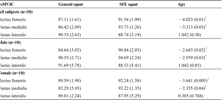
Only the muscle activities of the rectus femoris and vastus medial muscles were higher significantly during the SFE squat than during the general squat (p<0.05).
However, the muscle activity of the vastus lateralis
muscle was lower and the difference was not
statistically significant (Table 2).
Comparison of the onset of muscle contraction of vastus medialis and lateralis during the general and SFE squats
Compared withthe general squat, the SFE squat was associated with a statistically significant delay of the onset of muscle contraction only in the vastus lateralis
(p<0.05). In the male, the onset of muscle contraction was delayed during the SFE squat, but it was not significantly different from that during the general squat. In the female, the onset of muscle contraction was significantly delayed for the SFE squat compared with that for the general squat(p<0.05)(Table 3).
Characteristics Male (n=10) Female (n=10)
Age (years) 26.28 (2.67) 24.00 (3.70)
Height (cm) 173.30 (5.14) 165.00 (5.14)
Weight (kg) 69.30 (9.67) 52.80 (7.06)
The values are presented as mean (SD).
Table 1. General Characteristics of Participants (n=20)
On set time(ms) General squat SFE squat t(p)
All subjects (n=20)
VMO-VL 0.01 (0.12) -0.5 (0.06) -3.654 (0.002)
*Male (n=10)
VMO-VL -0.22 (0.13) -0.39 (0.08) -0.225 (0.82)
Female (n=10)
VMO-VL 0.21 (0.16) -0.62 (0.07) -4.355 (0.002)
*The values are presented as mean (SD). SFE: short foot exercise, VMO: vastus medialis, VL: vastus lateralis
*
p<0.05.
Table 3. Onset of muscle contraction during general squat and SFE squat (n=20)
%MVIC General squat SFE squat t(p)
All subjects (n=20)
Rectus femoris 87.11 (1.61) 91.54 (1.09) -4.023 (0.01)
*Vastus medialis 86.42 (2.89) 93.73 (1.26) -3.313 (0.03)
*Vastus lateralis 90.35 (2.63) 88.74 (3.19) 1.042 (0.30)
Male (n=10)
Rectus femoris 84.64 (3.02) 90.84 (2.03) -2.643 (0.02)
*Vastus medialis 90.55 (2.71) 94.69 (2.24) -2.559 (0.03)
*Vastus lateralis 91.69 (5.78) 88.53 (5.41) 1.042 (0.85)
Female (n=10)
Rectus femoris 89.59 (1.98) 92.24 (1.58) -3.641 (0.005)
*Vastus medialis 82.29 (5.95) 93.22 (1.35) -2.355 (0.04)
*Vastus lateralis 89.01 (2.24) 87.95 (5.29) 0.305 (0.768)
The values are presented as mean (SD). SFE: short foot exercise
*