Tyrosinase Inhibitory Activities of Meso-dihydroguaiaretic Acid from Machilus thunbergii
Hyun Sook Kwon1, Kyung Dong Lee2, Su Cheol Kim3 and Soo Jeong Cho4*
1Korea Promotion Institute for Traditional Medicine Industry, Gyeongsan 712-260, Korea
2Department of Oriental Medicine Materials, Dongshin University, Naju 520-714, Korea
3Amicogen Inc., Jinju 660-852, Korea
4Department of Pharmaceutical Engineering, Gyeongnam National University of Science and Technology, Jinju 660-758, Korea Received July 28, 2015 /Revised October 30, 2015 /Accepted October 30, 2015
Machilus thunbergii (Lauraceae) is an evergreen tree cultivated in Korea and Japan. M. thunbergii has long been used as a traditional medicine in Korea, China, and Japan to treat various diseases, includ- ing edema, abdominal pain, and abdominal distension. In this study, dried stem bark of M. thunbergii extracted in methanol and extract was partitioned into n-hexane, CHCl3, and BuOH. The CHCl3-solu- ble extracts chromatographed on silica gel column using a CHCl3/acetone and n-hexane/EtOAc mix- ture to afford Compound 1 and 2. Two dibenzylbutane lignans, macelignan (1) and meso-dihy- droguaiaretic acid (2), were isolated from the CHCl3-soluble extract of M. thunbergii stem bark. The structures of 1 and 2 were determined by 1H- and 13C-NMR spectroscopic data analyses and a com- parison with literature data. The tyrosinase inhibitory activity of the isolated compounds was evaluated. Among these compounds, Compound 2 strongly inhibited the monophenolase (IC50=10.2 μM) activity of tyrosinase. A kinetic analysis showed that Compound 2 was a competitive inhibitor. The apparent inhibition constant (Ki) for Compound 2 binding to free enzyme was 4.8 μM. Based on these results, it can be concluded that meso-dihydroguaiaretic acid (2) is a potential candidate for the treat- ment of melanin biosynthesis-related skin diseases.
Key words : Dibenzylbutane lignans, macelignan, Machilus thunbergii, meso-dihydroguaiaretic acid
*Corresponding author
*Tel : +82-55-751-3397, Fax : +82-55-751-3399
*E-mail : [email protected]
This is an Open-Access article distributed under the terms of the Creative Commons Attribution Non-Commercial License (http://creativecommons.org/licenses/by-nc/3.0) which permits unrestricted non-commercial use, distribution, and reproduction in any medium, provided the original work is properly cited.
Journal of Life Science 2015 Vol. 25. No. 11. 1298~1303 DOI : http://dx.doi.org/10.5352/JLS.2015.25.11.1298
Introduction
Machilus thunbergii (Lauraceae) is the evergreen tree grown in areas of Korea and Japan. The bark of this plant has been used as a folk medicine for the treatment of leg oederma, abdominal pain and abdominal distension in Korea [15]. Lignans, alkaloids [20], flavonoids [9], butanolids [10] and essential oils [11] have been reported as components from M. thunbergii, some of which have hepatoprotective ac- tivity as antioxidants [27], antibacterial activity [7], and in- hibitory activity on nitric oxide synthesis in activated macro- phages [9]. Macelignan and meso-dihydroguaiaretic acid (MDGA), an active lignan compounds were isolated from the stem bark of M. thunbergii [20]. Macelignan has been re- ported to possess antioxidant [14], anti-inflammatory [5], an-
ticarinogenic, and hepatoprotective effects [24] and cause al- teration in hepatic enzyme activities [21]. MDGA has been reported to have antioxidant [27], antifungal [1], anti- microbial [8], antiallergic [18], neuroprotective [16], and hep- atoprotective activities [19].
Tyrosinase (EC 1.14.18.1), also known as polyphenol oxi- dase, is a copper-containing mixed-function oxidase widely distributed in microorganism, animals, and plants. This oxi- dase catalyzes two distinct reactions of melanin synthesis, the hydroxylation of a monophenol and the conversion of an O-diphenol to the corresponding O-quinone. Tyrosinase is responsible for browning in plants and is considered to be deleterious to the color quality of plant-derived foods and beverages [4]. This unfavorable darkening from enzymatic oxidation generally results in a loss of nutritional and eco- nomic values and has been of great concern. However, Tyrosinase is not only the key enzyme in the browning of fruits and vegetables, but also the key enzyme of the darken- ing of skin, hair and eyes in animals. Hence, the discovery of new and safe tyrosinase inhibitors should have broad applications. In recent years, tyrosinase inhibitors have at- tracted concern owing to the hyperpigmentation [3], result- - Note -
ing from the increased use of tyrosinase enzyme in medici- nal and cosmetic products [17], and their identification and isolation from natural sources have been also increased [25].
Natural tyrosinase inhibitors are generally considered to be free of harmful side effects and can be produced at reason- able low costs. Therefore, the development and utilization of more effective tyrosinase inhibitors of natural origin are desired.
In our continuous search for new tyrosinase inhibitors from M. thunbergii, the MeOH extracts were subsequently partitioned and isolated. As a result, two dibenzylbutane li- gnans, macelignan (1) and MDGA (2) were isolated from M. thunbergii. In this study, the isolation and structural deter- minations of these two compounds are described. All the isolated compounds were evaluated for their tyrosinase in- hibitory activities.
Materials and Methods
Plant material
The stem bark of M. thunbergii was purchased from an oriental drug store in Pohang, Gyeongbuk, Korea, in July 2009. A voucher specimen (MT2009-01) has been deposited at the Laboratory of Molecular Neurophysiology, POSTECH, Pohang, Korea.
Instruments
NMR experiments were conducted on a Bruker AM 300 or 500 MHz FT-NMR instrument with tetramethylsilane (TMS) as internal standard. EIMS was collected on Jeol JMS-700 spectrometer. Optical rotations were measured on Perkin-Elmer 343 polarimeter. Silica gel (230-400 mesh, Merck), RP-18 (ODS-A, 12 nm, S-150 m, YMC) and Sephadex LH-20 (Amersham Biosciences, Uppsala, Sweden) were used for column chromatography. Thin-layer chromatographic (TLC) analysis was performed on Kieselgel 60 F254 (silica gel, 0.25 mm layer thickness, Merck) and RP-18 F254S (Merck) plates.
Extraction and isolation
The dried stem bark (500 g) of M. thunbergii were chopped and extracted three times with 100% methanol (1 l ×3) for 7 days at room temperature. The combined methanol extract was concentrated in vacuo to yield a brown gum (11.3 g).
The methanol extract was suspended in H2O (500 ml), then partitioned in turn with n-Hexane, CHCl3 and BuOH (each
3×500 ml) to afford n-Hexane extract (1.8 g), CHCl3 extract (3.5 g), and BuOH extract (1.5 g). The CHCl3-soluble extract was silica gel column chromatography (230-400 mesh) using CHCl3/Acetone (100:1→1:1) mixtures to yield seven sub- fraction (D1-D7). Fraction D1 (900 mg) was applied to silica gel column (230-400 mesh) chromatography with n-Hexane/
EtOAc (49:1→1:1) to yield fourteen subfractions (E1-E14).
Subfractions E6 was subjected to silica gel column (230-400 mesh) chromatography with n-Hexane/EtOAc (49:1→9:1) to afford compounds 1 (21 mg) and 2 (20 mg).
Compound 1: colorless prisms; [α] +3.0o (c 1.8 in CHCl3);
EIMS m/z 328; IR (KBr) νmax 3466, 1514, 1489, 931 cm-1, UV λmax nm 213, 230, 285 (MeOH); 1H-NMR (500 MHz, CDCl3) δ 0.84 (6H, t, J=6.0 Hz, 9-CH3 and 9'-CH3), 1.75 (2H, m, H-8 and H-8'), 2.31 (2H, m, H-7b and H-7'b), 2.74 (2H, dd, J=4.5, 13.6 Hz, H-7a and H-7'a), 3.86 (3H, s, OCH3-3'), 5.45 (1H, s, OH-4'), 5.91 (2H, s, OCH2O), 6.60 (1H, s, H-6'), 6.62 (1H, d, J=1.9 Hz, H-2'), 6.63 (1H, s, H-6), 6.65 (1H, s, H-2), 6.73 (1H, d, J=7.8 Hz, H-5), 6.83 (1H, d, J=7.9 Hz, H-5'); 13C-NMR (125 MHz, CDCl3) δ 16.1 (C-9), 16.2 (C-9'), 38.9 (C-7'), 39.1 (C-7), 39.3 (C-8), 39.4 (C-8'), 55.9 (OCH3-3'), 100.7 (OCH2O), 107.9 (C-5), 109.4 (C-2), 111.5 (C-2'), 114.0 (C-5'), 121.7 (C-6), 121.8 (C-6'), 133.8 (C-1'), 135.7 (C-1), 143.6 (C-4'), 145.5 (C-4), 146.3 (C-3'), 147.5 (C-3).
Compound 2: colorless crystals, [α] +0o (c 0.1 in CHCl3);
EIMS m/z 330; IR (KBr) νmax 3559, 3371, 1648, 1592 cm-1;
1H-NMR (500 MHz, CDCl3) δ 0.85 (6H, d, J=6.6 Hz, 9-CH3
and 9'-CH3), 1.75 (2H, m, H-8 and H-8'), 2.30 (2H, dd, J=9.2, 13.5 Hz, H-7b and H-7'b), 2.74 (2H, dd, J=5.0, 13.5 Hz, H-7a and H-7'a), 3.85 (6H, s, OCH3-3 and OCH3-3'), 5.40 (2H, s, OH-4 and OH-4'), 6.61 (2H, s, H-2 and H-2'), 6.66 (2H, d, J=8.0 Hz, H-6 and H-6'), 6.83 (2H, d, J=8.0 Hz, H-5 and H-5');
13C-NMR (125 MHz, CDCl3) δ 16.2 (C-9, 9'), 38.9 (C-7, 7'), 39.2 (C-8, C-8'), 55.9 (OCH3 ×2), 111.5 (C-2, 2'), 114.0 (C-5, 5'), 121.7 (C-6, 6'), 133.8 (C-1, 1'), 143.6 (C-4, 4'), 146.3 (C-3, 3').
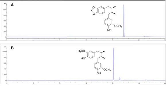
High-performance liquid chromatography (HPLC) analysis of compounds
Samples were filtered through a 0.45 μm syringe filter (Millipore, Billerica, MA, USA) and analyzed by HPLC (Agilent 1260, Agilent Technologies, Waldbronn, Germany).
The analytical column was a Kinetex C18 (4.6 × 150 mm, Phenomenex, CA, USA). The detection was set at ELSD (Evaporative Light Scattering Detector) and the solvent flow rate was held constant at 0.5 ml/min. The mobile phase used
A
B
Fig. 1. HPLC chromatograms of compounds 1(A) and 2(B) from M. thunbergii.
for the separation consisted of solvent A (0.1% trifluoroacetic acid in water) and solvent B (0.1% trifluoroacetic acid in ace- tonitrile). A gradient elution procedure was used as 0 min 98% A, 3 min 95% A, 20 min 100% B, 23 min 98% A, 30 min 98% A. The injection volume was 3 μl for analysis. All samples were analyzed in triplicate.
Tyrosinase inhibitory activity
Mushroom tyrosinase (EC 1.14.18.1) was assayed as de- scribed previously with slight modifications [4], using L-ty- rosinase or L-3,4-dihydroxyphenylalanine (L-DOPA) as sub- strate. In a spectrophotometric experiment, the enzyme ac- tivity was monitored by dopachrome formation at 475 nm with a Spectra MAX plus spectrophotometer (Molecular de- vice, Sunnyvale, CA) at 30℃. All test samples were dis- solved in dimethyl sulfoxide (DMSO) and used for the ex- periment with dilution. The final concentration of DMSO in the test solution was 1.5%. First, 200 μl of a 4.5 mM L-ty- rosine or 12 mM L-DOPA aqueous solution was mixed with 2785 μl of 0.25 M phosphate buffer (pH 6.8) and 10 μl of the test sample, incubated at 30℃ for 10 min. Then, 5 μl of tyrosinase solution (130 units) was added to the phos- phate buffer and incubated for additional 20 min. DMSO without test samples was used as the control, and kojic acid was used as a positive control. The assay was conducted in triplicate of separate experiments. The data analysis was performed by using Sigma Plot 2000 (SPSS Inc., Chicago, IL). The inhibitory concentration leading to 50% activity loss (IC50) was obtained by fitting experimental data to the logis- tic curve by the equation as follows:
Activity (%) = 100[1/(1+([I]/IC50))]
Inhibition mode was analyzed by Enzyme Kinetics Module 1.0 (SPSS Inc.) equipped with Sigma Plot 2000.
Results and Discussion
The MeOH extract of the M. thunbergii was partitioned successively with n-Hexane, CHCl3, and BuOH soluble frac- tions, respectively. Then, we were evaluated for the in- hibitory activity of the partitioned fraction on tyrosinase.
Among the partitioned fractions, the CHCl3 soluble fraction showed significant tyrosinase inhibitory activity (IC50=97.9±
5.2 μg/ml), whereas n-Hexane and BuOH fractions showed weaker activity. So, CHCl3 soluble fraction was subject to chromatographed on a normal-phase column and a re- verse-phase column, to give two dibenzylbutane lignans, macelignan (1) and meso-dihydroguaiaretic acid (2). Subse- quently, HPLC was performed as it is more sensitive, se- lective, and speedy. The use of ELSD detector is convenient method to identify. The chromatogram of the result of purity analysis by HPLC is shown in Fig. 1. The purity of com- pounds 1 and 2 were more than 95%.
Compound 1 was obtained as colorless prisms. The EIMS of 1 had a molecular ion peak at m/z 328 [M+], consistent with the molecular formula of C20H24O4. The 1H-NMR spec- trum of 1 showed one hydroxyl group δH 5.45 (1H, s), one methoxy group δH 3.86 (3H, s), two methyl group δH 0.84 (6H, t, J=6.0 Hz, H-9, 9'), two benzylic methylene protons δH 2.31 (2H, m, H-7b, 7'b) and 2.74 (2H, dd, J=4.5, 13.6 Hz,
Table 1. Tyrosinase inhibitory activity of compounds
Compounds UV475
L-Tyrosine IC50(μM) L -DOPA IC50(μM) 1
2 Kojic acid
82.81 10.20 11.36
>200 145.32
14.05
A B C
Fig. 2. Inhibitory effect of MDGA (2) on tyrosinase. (A) Effect of MDGA (2) on the tyrosinase catalyzed oxidation of L-tyrosine.
Inset: Plot of 1/ʋ vs. concentration of MDGA (2). (B) Dixon plots for the inhibition of the monophenolase activities of tyrosinase by MDGA (2). Concentrations of substrates for curves were 300 (●) and 450 (○) μM, respectively. (C) Time dependent inhibition of tyrosinase in the presence of MDGA (2). Concentrations of MDGA (2) from top to bottom were 0 (0), 3 (1), 10 (2), 25 (3) μM.
H-7a, 7'a)], one methylenedioxyl group δH 5.91 (2H, s), two methine group δH 1.75 (2H, m, H-8, 8') and six aromatic pro- tons δH 6.60 (1H, s, H-6'), 6.62 (1H, d, J=1.9 Hz, H-2'), 6.63 (1H, s, H-6), 6.65 (1H, s, H-2), 6.73 (1H, d, J=7.8 Hz, H-5) and 6.83 (1H, d, J=7.9 Hz, H-5'). The 13C-NMR spectrum showed one methoxy carbon δC 55.9 (OCH3), two methyl carbons δC 16.1 (C-9) and 16.2 (C-9'), two methylenes δC 38.9 (C-7'), 39.1 (C-7), one methylenedioxy carbon δC 100.7 (OCH2O), eight methins δC 39.39 (C-8), 39.4 (C-8'), 107.9 (C-5), 109.4 (C-2), 111.5 (C-2'), 114.0 (C-5'), 121.7 (C-6) and 121.8 (C-6'), and six quaternary carbons δC 133.8 (C-1'), 135.7 (C-1), 143.6 (C-4'), 145.5 (C-4), 146.3 (C-3') and 147.5 (C-3).
These spectral data suggested 1 to be a dibenzylbutane lignan. Based on of the spectral data, compound 1 was iden- tified as (8R,8'S)-7-(3,4-methylene-dioxyphenyl)-7'-(4-hydroxy- 3-methoxyphenyl)-8,8'-dimethylbutane (macelignan) by com- paring its spectroscopic data with literature data [20, 26].
Compound 2 was obtained as colorless crystals. The EIMS of 2 had a molecular ion peak at m/z 330 [M]+, consistent with the molecular formula C20H26O4. The 1H-NMR spec- trum showed two hydroxyl group δH 5.40 (2H, s), two me- thoxy groups δH 3.85 (6H, s), two methyl groups δH 0.85 (6H, d, J=6.6 Hz, H-9, 9'), four benzylic methylene groups δH 2.30 (2H, dd, J=9.2, 13.5 Hz, H-7b, 7'b) and 2.74 (2H, dd, J=5.0, 13.5 Hz, H-7a, 7'a)], two methine groups [δH 1.75 (2H, m, H-8, 8')] and six aromatic protons δH 6.61 (2H, s, H-2, 2'), 6.66 (2H, d, J=8.0 Hz, H-6, 6') and 6.83 (2H, d, J=8.0 Hz, H-5, 5')]. The 13C-NMR spectrum showed two methoxy groups δC 55.9 (OCH3), two methyl groups δC 16.2 (C-9, 9'), two methylene groups δC 38.9 (C-7, 7'), eight methin groups δC 39.2 (C-8, C-8'), 111.5 (C-2, 2'), 114.0 (C-5, 5') and 121.7
(C-6, 6'), and six quaternary carbons δC 133.8 (C-1, C-1'), 143.6 (C-4, 4') and 146.3 (C-3, 3'). These spectral data sug- gested that 2 was a dibenzylbutane lignan. As a result, com- pound 2 was identified as (8R,8'S)-7,7′-di-(4-hydroxy-3-me- thoxyphenyl)-8,8′-dimethylbutane (meso-dihydroguaiaretic acid, MDGA) by comparing its spectroscopic data with the previously reported data [2, 20].
The isolated compounds were evaluated for their in- hibitory activities on tyrosinase. As shown in Table 1 and Fig. 2A, MDGA (2) exhibited a dose-dependent inhibitory effect on the monophenolase activity of mushroom ty- rosinase (IC50=10.2 μM). The result obtained indicates that MDGA (2) exhibited significantly higher inhibitory activity than positive control kojic acid. Also, MDGA (2) inhibited mushroom tyrosinase diphenolase activity with an IC50 of 145.32 μM (Table 1). In the previous reports, phenyl- propanoids, stilbenes, and other derivatives, including 3,4-dihydroxycinnamic acid [13], oxyresveratrol were identi- fied as tyrosinase inhibitors. Among them, phenylpropa- noids containing a hydroxyl group possess potent tyrosinase inhibitory activity. In the case of oxyresveratrol the presence of a hydroxyl group in the ring seems to be related to their inhibitory potency [22]. In this study as well, we suggest that the inhibitory effect of 2 was determined by the guaiacyl
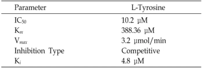
Table 2. Kinetics and inhibition constants of mushroom ty- rosinase by MDGA
Parameter L-Tyrosine
IC50
Km
Vmax
Inhibition Type Ki
10.2 μM 388.36 μM 3.2 μmol/min Competitive 4.8 μM
group.
The kinetic behavior of mushroom tyrosinase during the oxidation of L-tyrosine was studied first. Under the con- dition used in the present investigation, the oxidation of L-tyrosine by mushroom tyrosinase followed the Michaelis- Menten kinetics. The kinetic parameters for mushroom ty- rosinase were obtained from the Dixon plot. The results il- lustrated in Fig. 2B show that MDGA (2) is a competitive inhibitor because increasing the MDGA (2) concentration re- sulted in a family of lines with a common intercept on the 1/ʋ axis but with different slopes. The equilibrium constant for inhibitor binding, Ki of L-tyrosine obtained from the Dixon plot were 4.8 μM (Table 2). The time dependence of 2 on the tyrosinase catalyzed oxidation of L-tyrosine was studied. MDGA (2) showed time dependent inhibition (Fig.
2C). As shown in Fig. 2C, the lag time is known for the oxidation of monophenol substrates such as L-tyrosine and this lag can be shortened. This lag time can be extended by monophenolase inhibitors such as tropolone [6] and gal- angin [12]. MDGA (2) did extend this lag phase, indicating that 2 inhibit the hydroxylation of L-tyrosine.
In summary, two of dibenzylbutane lignans were isolated from CHCl3-soluble fractions of M. thunbergii. The structures were identified as macelignan (1) and MDGA (2) by the physicochemical and spectroscopic data. The isolated com- pounds were evaluated for their tyrosinase inhibitory activities. Among them, MDGA (2) inhibited the oxidation of L-tyrosine catalyzed by mushroom tyrosinase. The in- hibition mechanism obtained from the Dixon plot show that MDGA (2) is a competitive inhibitor. MDGA (2) only binds the free enzyme to form an EI complex rather than bind the ES complex.
Use of tyrosinase inhibitors is becoming increasingly im- portant in the cosmetic industry due to their skin whitening and preventive effects. Besides being used in the treatment of some dermatological disorders associated with melanin hyperpigmentation, tyrosinase inhibitors have found an im- portant role in the cosmetic and pharmaceutical industries
for their skin-whitening effect and depigmentation after sun- burn [23]. In this study, it can be concluded that MDGA (2) can be potential candidate for the treatment of melanin biosynthesis related skin diseases, likely hyper-pigmentation of human as well as animals.
Acknowledgment
This work was supported by natural products bank of KOTMIN (Korea Promotion Institute for Traditional Medicine Industry) and “Cooperative Research for Agriculture Science & Technology Development (Project No. PJ010129)”
Rural Development Administration, Republic of Korea.
References
1. Cho, J. Y., Choi, G. J., Son, S. W., Jang, K. S., Lim, H. K., Lee, S. O., Sung, N. D., Cho, K. Y. and Kim, J. C. 2007.
Isolation and antifungal activity of lignans from Myristica fragrans against various plant pathogenic fungi. Pest Manag.
Sci. 63, 935-940.
2. Forrest, J. E., Heacock, R. A. and Forrest, T. P. 1974. Diaryl- propanoids from nutmeg and mace (Myristica fragrans Houtt.). J. Chem. Soc. Perkin 1 2, 205-209.
3. Friedman, M. 1996. Food browning and it’s prevent; an overview. J. Agric. Food Chem. 44, 631-653.
4. Ha, T. J., Tamura, S. and Kubo, I. 2005. Effects of mushroom tyrosinase on anisaldehyde. J. Agric. Food Chem. 53, 7024- 7028.
5. Jin , D. Q., Lim, C. S., Hwang, J. K., Ha, I. and Han, J. S.
2005. Biochem. Biophys. Res. Commun. 331, 1264-1269.
6. Kahn, V. and Andrawis, A. 1985. Inhibition of mushroom tyrosinase by tropolone. Phytochemistry 24, 905-908.
7. Karikome, H., Mimaki, Y. and Sashida, Y. 1991. A butano- lide and phenolics from Machilus thunbergii. Phytochemistry 30, 315-319.
8. Kawaguchi, Y., Yamauchi, S., Masuda, K., Nishiwaki, H., Akiyama, K., Maruyama, M., Sugahara, T., Kishida, T. and Koba, Y. 2009. Antimicrobial activity of stereoisomers of bu- tane-type lignans. Biosci. Biotechnol. Biochem. 73, 1806-1810.
9. Kim, N. Y. and Ryu, J. H. 2003. Butanoilds from Machilus thunbergii and their inhibitory activity on nitric oxide syn- thesis in activated macrophages. Phytother. Res. 17, 372-375.
10. Kim, W., Lyu, H. N., Kwon, H. S., Kim, Y. S., Lee, K. H., Kim, D. Y., Chakraborty, G., Choi, K. Y., Yoon, H. S. and Kim, K. T. 2013. Obtusilactone B from Machilus Thunbergii targets barrier-to-autointegration factor to treat cancer. Mol.
Pharmacol. 83, 367-376.
11. Komae, H. and Hayashi, N. 1972. Terpenes from Actino- daphne, Machilus and Neolitsea species. Phytochemistry 11, 1181-1182.
12. Kubo, I. and Kinst-Hori, I. 1999. Flavonols from saffron flower. J. Agric. Food Chem. 47, 4121-4125.
초록:후박나무에서 분리한
meso
-dihydroguaiaretic acid의 tyrosinase 저해활성권현숙1․이경동2․김수철3․조수정4*
(1한국한방산업진흥원, 2동신대학교 한약재산업학과, 3아미코젠, 4경남과학기술대학교 제약공학과)
후박나무(녹나무과)는 한국과 일본 등지에 서식하는 상록 교목으로 한국, 중국, 일본에서 부종, 복통, 복부 팽만 등의 질병 치료를 위해 오랫동안 사용되어오고 있다. 본 연구에서는 후박나무 껍질을 메탄올에 추출하고 메탄올 추출물을 헥산, 클로로포름, 부탄올에 순차적으로 분획하였다. 클로로포름 분획물로부터 2종의 화합물을 분리하 였으며 분리된 화합물1과 2의 구조는 1H-, 13C-NMR과 참고 문헌 데이터에 의해 dibenzylbutane lignin 화합물인 macelignan (1)과 meso-dihydroguaiaretic acid (2)로 동정되었다. 분리된 화합물들의 tyrosinase 저해 활성을 측정 한 결과, 화합물 2는 tyrosinase 저해 활성 중 monophenolase (IC50 = 10.2 μM)에 대해 높은 저해활성을 나타내는 경쟁적 저해제였으며 효소에 결합하는 화합물 2의 저해 상수(Ki 값)는 4.8 μM였다. 따라서 meso-dihydroguaiaretic acid (2)는 멜라닌 생합성과 관련된 피부 질환 치료를 위한 잠재적 후보가 될 수 있을 것으로 판단된다.
13. Lee, H. S. 2002. Tyrosinase inhibitors of Pulsatilla cernua root-derived materials. J. Agric. Food Chem. 50, 1400-1403.
14. Lee, J. Y., Han, Y. B., Woo, W. S. and Shin, K. H. 1990.
Antioxidant activity of Diarylbutanes. Kor. J. Pharmacogn. 21, 270-273.
15. Li, G., Lee, C. S., Woo, M. H., Lee, S. H., Chang, H. W.
and Son, J. K. 2004. Lignans from the bark of Machilus thun- bergii and their DNA topoisomerases I and II inhibition and cytotoxicity. Biol. Pharm. Bull. 27, 1147-1150.
16. Ma, C. J., Sung, S. H. and Kim, Y. C. 2004. Neuroprotective lignans from the bark of Machilus thunbergii. Planta Med. 70, 79-80.
17. Maeda, K. and Fukuda, M. 1991. In vitro effectiveness of several whitening cosmetic components in human melano- cytes. J. Soc. Cosmet. Chem. 42, 361-368.
18. Moon, T. C., Seo, C. S., Ha, K., Kim, J. C., Hwang, N. K., Hong, T. G., Kim, J. H., Kim, D. H., Son, J. K. and Chang, H. W. 2008. meso-Dihydroguaiaretic acid isolated from Saururus chinensis inhibits cyclooxygenase-2 and 5-lip- oxygenase in mouse bone marrow-derived mast cells. Arch.
Pharm. Res. 31, 606-610.
19. Park, E. Y., Shin, S. M., Ma, C. J., Kim, Y. C. and Kim, S.
G. 2005. meso-Dihydroguaiaretic acid from Machilus thunber- gii down-regulates TGF-β1 gene expression in activated hepatic stellate cells via inhibition of AP-1 activity. Planta Med. 71, 393-398.
20. Park, B. Y., Min, B. S., Kwon, O. K., Oh, S. R., Aha, K. S.,
Kim, J. T., Kim, D. Y., Bae, K. and Lee, H. K. 2004. Increase of caspase-3 activity by lignans from Machilus thunbergii in HL-60 cells. Biol. Pharm. Bull. 27, 1305-1307.
21. Paul, S., Hwang, J. K., Kim, H. Y., Jeon, W. K., Chung, C.
and Han, J. S. 2013. Multiple biological properties of maceli- gnan and its pharmacological implications. Arch. Pharm. Res.
36, 264-272.
22. Seo, S. Y., Sharma, V. K. and Sharma, N. 2003. Mushroom tyrosinase: recent prospects. J. Agric. Food Chem. 51, 2837- 2853.
23. Parvez, S., Kang, M. K., Chung, H. S. and Bae, H. S. 2007.
Naturally occurring tyrosinase inhibitors: mechanism and applications in skin health, cosmetics and agriculture industries. Phytother. Res. 21, 805-816.
24. Shin, K. H. and Woo, W. S. 1986. Hepatic Drug metabolism modifier from arils of Myristica fragrans. Kor. J. Pharmacogn.
17, 91-99.
25. Son, S. M., Moon, K. D. and Lee, C. Y. 2000. Rhubarb juice as a natural antibrowing agent. J. Food Sci. 65, 1288-1289.
26. Woo, W. S., Shin, K. H., Wagner, H. and Lotter, H. 1987.
The structure of macelignan from Myristica fragrans.
Phytochemistry 26, 1542-1543.
27. Yu, Y. U., Kang, S. Y., Park, H. Y., Sung, S. H., Lee, E. J., Kim, S. Y. and Kim, Y. C. 2000. Antioxidant lignans from Machilus thunbergii protect CCl4-injured primary cultures of rat hepatocytes. J. Pharm. Pharmacol. 52, 1163-1169.