내안난• 근싱악외 시 지lb긴 체 Z오 ZOON Jo니ma I of Korean Arthroscopy Soc.
Volume,Numbed, December, 2CO2
생분해성 간섭나사를 이용한 전방 십자 인대 재건술 후 발생한 결절종 - 증례보고 -
전남대학교 의 과대학 정 형외과학교실
송은규- 심상돈• 김명선
Pretibial Ganglion afterAnterior Cruciate Ligament Reconstruction with Bioabsorbable Interference
Screw fixation (Bioscrew®) -ACase Report-
Eun Kyoo Song, M.D., SangDon 아lim, M.D., Myung SunKim, M.D.
Department of Orthopedic Surgery, Chonnam National University, Gwangju, Korea
ABSTRACT: The complication caused by a bioabsorbable interference screw composed of Poly-L-Lactic- Acid is rare after anterior cruciate ligament (ACL) reconstruction. We reported a case of a pretibial ganglion at the orifice of the tibial tunnel where the graft tendon had been fixed with a bioabsorbable interference screw (Bioscrew1-) fbr ACL reconstruction using autogenous hamstring tendon. The patient was underwent ganglion excision and interference screw removal.
KEY WORDS: Anterior cruciate ligament, Bioabsorbable interference screw, Ganglion
서 론
전방 십자 인대 손상의 치료로 자가 슬괵건을 이용한 전 방 십자 인대 재건술이 많이 사용되고 있으며% 이 때 이식 건과 골터널 사이의 고정을 위해 간섭나사를 많이 사용하고 있다. 금속성 간섭나사의 경우 골-슬개건-골을 고정하기 위 해 많이 사용되었으며 슬괵건의 고정에도사용되었으나 가 섭나사로 인한 이식건의 손상, 간섭나사 제거를 위한 재수 술이 필요하다는 단점이 있어 최근에는 간섭나사의 형태를
* Address reprint requests to Eun Kyoo Song, M.D.
Department of Orthopaedic Surgury Chonnam University Hospital 8 Hak dong, Gwangju 501-757, Korea Tel: 82-62-220-6336, Fax: 82-62-225-7794 E-mail: [email protected]
개량하여 이식건의 손상을 줄이고 그 구성 성분 또한 생분 해성 재료를 이용한 간섭나사를 많이 사용하고 있다. 생분 해성 간섭나사의 구성 성분에는 Poly-Glycolic-Acid Poly-D-Lactic-Acid, Poly-L-Lactic-Acid 등이 있으며 이로인해 슬관절부위에 부작용이 발생했다는 보고는 매우 드물다〜⑵ 저자들은 자가 슬괵건을 이용한 전방 십자인대 재건술시 생분해성 간섭나사로 경골터널에 이식건을 고정한 후 경골 터널 입구에 발생한 전경골부 결절종 1예를 경험 하여 자기공명영상 및 조직소견과 함께 보고하는바이다.
증 례
35세 남자 환자로 약 1 닌 전부터 발생한 우측슬관절 동 통 및 우측 전경골부 무통성 종괴를 주소로 내원하였으며.
과거력상 1년 3개월 전 낙상사고로 발생한 우측 슬관절의 전방 십자 인대 손상, 내측 및 외측 반월상 연골판 손상에 대하여 수상 후 3개월째 본원에서 자가 슬괵건을 이용한
—188 —
생분해성 간섭나사를 이용한 전방 십자' 인대 재건술 후 발생한 결절종• 송은규 외
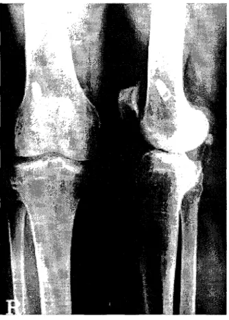
Fig. 1. Anteroposterior and lateral radiographs of the right knee showing no tibial tunnel widening at 1 year follow up after ACL reconstruction with autogenous hamstring ten
don and tibial fixation with bioabsorbable interference
Fig. 3. Microscopic finding of the cyst wall shows synovial lin
ing and diagnosed to ganglion.
screw.
Fig. 2. Magnetic resonance images that were checked 1 year after ACL reconstruction show the 1cm sized oval-shapd lesion in the tibial tunnel site in which the graft tendon had been fixed with a bioabsorbable interference screw, which represented low sig
nal intensity in T1-weighted saggital image (A), high signal intensity in T2-weighted saggital image (B) and non-enhanced lesion by contrast (C).
一189 —
대 한관절 경학회지 제 6 권 제 2 호 2002년
관절경하 전방 십자 인대 재건술을 시행하였으며, 동시에 meniscal arrow-' 를 이용한 내측 반월상 연골판봉합 및 외측 반월상 연골판 부분 절제술을 시행하였다. 수술 당시
전신 마취하에서 슬관절의 불안정성에 대한 이학적 검사 결 과 Lachmac 검사상 3+ 및 Pivot-Shi闭사상 1+의 소 견을 보였다. 대퇴 터널에는 Ligament Anchor (LA) sere渺로 고정하고 경골부에는 이식건을 슬관절 신전상태 에서 8X25 mm Poly-L-Lactic-Acid interference screw(Bioscre'W Linvatec, USA)로 이식건을 고정하 였다. 전방 십자 인대 재건술 후 1년째 우측 전경골부에 무 통성 종괴가 촉지 되었다. 단순방사선 사진상 터널 확장소 견은 보이지 않았으며(Fig. 1), 자기 공명 영상촬영 결과 T1 강조 영상에서 지신호 강도를, T2 강조 영상에서 고신 호 강도를, 조영증강 영상에서 조영 증강되지 않는 1cm 크기의 낭종성 종괴가 관찰되어 (Fig. 2-A, B, C) 종괴제 거술 및 간섭나사 제거를 시행하였다. 수술 소견상 BioscreW 전방에 逐 1x0.5 cm 크기의 피막에 잘 둘러 싸인 종괴가 관찰되었고 젤라틴과 같은 물질을 함유하고 있 었다. 제거된 생분해성 간섭나사는 거의 분해되지 않은 상 태였고 경골 터 널과슬관절과는서로교통하고 있지는 않았 다. 우측 슬관절에 대한 관절경소견상 감염이나 염증의 소 견은 없었으며, 슬관절내 재건된 이식건의 상태는 비교적 양호하였다. 낭종에 대한 조직학적 검사상 결절종에 합당한 소견이 관찰되었다(Fig. 3).
고 찰
전방 십자 인대 재건술시 이식건의 고정시 생분해성 간섭 나사가 금속나사의 단점을 극복할 수 있으며 금속나사를 사 용했을 때보다 술후 향상된 영상을 얻을 수 있다고 생각되 어 최근 생분해성 간섭 나사를 이용한 이식건의 고정 방법 이 사용되고 있다그러나 생분해성 간섭나사의 경우 생 체내 분해 및 흡수과정에서 염증반응 및 간섭나사 파단이 생길 수 있는 단점들이 지적되고 있다. Poly-glycolic acid (PGA) 합성물의 경우 약 8%에서 심각한 염증이 발 생하였다는 보고가 있으나 Poly-D, L-Lactic-Acid 합성 삽입물의 경 우 염증과 관련한 합병증을 약 1.5%例서 보고 하고 있다이는 두 합성물의 분해 및 흡수과정에서의 반 감기 및 분해과정의 차이에 기인하는 것으로, Poly- Lactic-Acid(PLA) 합성물의 경우 가수분해 과정을 통해 분해되며 인체에서의 반감기 또한7~1 題인PGA 합성물 에 비해 약 6개월로 긴 편이다. Poly-D-Lactic Acid 간 섭나사못의 흡수 기간은 몇 달인 반면". Poly-L-Lacr.ic- Acid가 분해되는데 걸리는 기긴은 몇 년이상이 소요되는 것으로 알려져 있으나 Poly-L-Lactic-Ac와!분해되는 긴 기간동안 생리 적 분해기전에 대한 연구는 드물다%
Martine快은 골-슬개골 동종 이식건을 이용한 전방십자
인 대 재건술 후 2.5년동안 완전한 형태로 보존된 Poly-L- Lactic-Acid 간섭나사에 대한 증례를 보고했으며, 슬관절 에서 Poly-L-Lactic-Ac와! 생분해가 육안상으로나 현미 경상으로도 어떤 염증반응이나 이물질 반응을 일으키지 않 았다고보고하고 있다. 현재 PLA 합성물이 인체 내에서 가 장 합병증이 적은 것으로 보고되어 널리 사용되고 있으며, PLA 중합체로 구성된 생분해성 간섭나사는 만족할만한 임 상적 결과를 보이고 있다”,저자 등이 사용한 흡수성 간 섭 나사의 성분은 Poly-L-Lactic-Acid 였으며 수술 당시 거의 분해되지 않았다.
지금까지 전방 십자인대 재건술 후 경골 터널 말단에 증 상을야기하여 고정기구 또는돌출된 이식건의 제거를 요했 던 고정기구나 이식건의 돌출에 대해 수편의 보고가 있다1,.
Sga잉ione 등은 반 건양건을 이용한 보강술로 전방 십자 인대 재건술을 시행한 72 명의 환자들 중 경골 터널 입구 에서 1예의 낭종 발생을 보고하였으며 , Simonian 등'°,은 자가 슬개건 또는 자가슬괵건을 이용한 전방십자인대 재 건술후 경골 터널과 통하는 전경부 피하에 발생한 결절종 4예를 보고 하였다. Victoroff 등⑴은 동종 이식 건을 이 용 하여 경골부 고정은 Staple 과 추가적으로 screw 및 washed- 사용하여 전방 십자 인대 재건술을 실시한 후 경골 터널 말단에서 돌출되는 피하 전경골부 결절종에 대한 4예를 보고한 바 있다. 본 증례의 경우에는 술 후 1년째 우측 전경부에 결절종이 발생하여 제거술을 시행하였으며 수술소견상 Bioscre浦 상부에 lx lx。。cm 크기의 잘 피막에 둘러싸인 젤라틴과 같은 물질을 함유한 결절종 관찰 도]었다.
전방 십자 인대 재건술과 관련한 전경부의 낭종 발생의 원인으로 동종 이식조직의 골터널에서의 불완전한 결합 혹 은 이식건 섬유들의 압력에 의한 괴사 때문이라는 보고가 있으며小”气 Martinek 등”은 이완된 나사못의 기계적인 자극이 이물질 반응을 자극하여 발생한 것으로 보인다고도 하였다. Imhoff 등。은 전방 십자 인대 재건술 후 강력한 재활치료를 시행했던 2명의 여성 운동선수들에서 대퇴골 고정부위에서 생분해성 Poly-L-Lactic-Acid 나사못 주위 에서 골용해성 변화를 보고 하였다.
저자들의 경우 경골 터널입구의 피하 결절종이 경골과 고 정시 사용된 생분해성 간섭나사못의 성분사이의 이상 반응 이나 슬관절 내부의 관절액이 경골 터널과 이식건 사이의 미세한 틈으로 유출되어 발생했을 것으로 생각하며 , 생흡수 성 간섭나사를 이용하여 경골부를 고정한 다른 예에 대해서 도 세심한 추시가 필요하리라 생각된다.
REFERENCES
1) Bach BR: Potential pitfalls of Kurosaka screw interfer
ence fixation for ACL surgery. Am J Knee Surg. 2, 76-
— 190 —
생분해성 간섭나사를 이용한 전방 십자 인대 재건술 후 발생한 결절종• 송은규 외
78,1985.
2) Bergsma JE, de Bruijn WC, Rozema FR, Bos RR and Boering G: Late degradation tissue responce to poly(L- lactide) bone plates and screws. Biomaterials. 16, 25-31,
1995.
3) Bostman OM: Current concepts review: Absorbable implants for the fixation of the fracture. J Bone Joint Surg.
73-A,148, 1991
4) Imhoff AB, Martinek V, Schwamborn T and Me리T:
Bioabsorbable interference screws in ACL reconstruction:
A prospective clinical and MRI study. Oral presentation, I. European Society of Sports Traumatology, Knee Surgery, and Arthroscopy Nice, France, April-May. 1998.
5) Kurosaka M, Yoshiya S and Andrich JT: A biomechan
ical comparison of different surgical techniques of graft fixation in anterior cruciate ligament reconstruction. Am J Sports Med. 15, 225-229,1987.
6) Kurzweil PR, Frogameni AD and Jackson DW: Tibial interference screw removal following anterior cruciate lig
ament reconstruction. Arthroscopy. 11, 289-291,1995 7) Martinek V and Friederich NF: Tibial and pretibi지 cyst
formation after anterior cmciate ligament reconstruction with bioabsorbable interference screw fixation. Arthroscopy.
15,317-320, 1999.
8) Martinek V, Seil R, Latterman C, Watkins SC and Fu FH: The fate of the Poly-L-Lactic Acid interference screw after anterior cruciate ligament reconstruction.
Arthroscopy. 17, 73-76, 2001.
9) Sgaglione NA, Warren RF, Wickiewicz TL, Gold DA and Panariello RA: Primary repair with s이nitendinos니s tendon augmentation of acute anterior cruciate ligament injuries. Am J sports Med. 18, 64-73, 1990.
10) Simonian PT, Wickiewicz TL, O' B리en SJ, Diens JS, Schatz JA and Warrren RF: Pretibial cyst formation after anterior cruciate ligament surgery with soft tissue autografts. Arthroscopy. 14, 215-220, 1998.
i I) Stahelin AC, Weiler A, Rufenacht H, Hoffmann R, Gcissmann A and Feinstein R: Clinical degradation and biocompatibility of different bioabsorbable Interference screw: A report of six cases. Arthroscopy. 13, 238-244, 1997.
12) Takizawa T, Akizuki S, Hiriu사讨H and Yasukawa Y:
Foreign body gonitis caused by a broken poly-L-lactide acid screw. Arthroscopy. 14, 329-330, 1998.
13) Victoroff BN, Paulos LE, Beck C and Goodfellow DB:
Subcutaneous cyst formation associated with anterior cru
ciate ligament allograft: A report of four cases and litera
ture review. Arthroscopy. 11,486-494, 1995.
자가슬괵건을 이용한 전방 십자인대의 재건술시 사용되는 P이y-L-Lactic-Acid 성분의 생분해성 간섭나사로 인 한 합병증은 많지 않다, 저자들은 전방십자 인대 재건술시 생분해성 간섭나사로 이식건을 고정 후 경골 터널 전방 에 발생한 결절종에 대해 결절종 절제술고卜동시에 간섭나사를 제거한 1예에 대해 보고하는 바이다.
—191 —