http://dx.doi.org/10.5763/kjsm.2014.32.2.92
Received: April 11, 2014 Revised: November 10, 2014 Accepted: November 13, 2014 Correspondence: Myung-Ku Kim
Department of Orthopedic Surgery, Inha University School of Medicine, 366 Seohae-daero, Jung-gu, Incheon 400-103, Korea Tel: +82-32-890-3043, Fax: +82-32-890-3047, E-mail: [email protected]
Copyright ©2014 The Korean Society of Sports Medicine
CC
This is an Open Access article distributed under the terms of the Creative Commons Attribution Non-Commercial License (http://creativecommons.org/
licenses/by-nc/3.0) which permits unrestricted non-commercial use, distribution, and reproduction in any medium, provided the original work is properly cited.
전방십자인대의 경골 고정 시 생흡수성 봉합나사 및 금속나사 고정의 기능적 결과
인하대학교 의과대학 정형외과학교실
김명구ㆍ박주용ㆍ안치훈
Functional Outcome of Bioabsorbable Suture Anchor and Metal Screw Fixation on Tibial Side for Anterior Cruciate Ligament Reconstruction
Myung-Ku Kim, Ju-Yong Park, Chi-Hoon Ahn
Department of Orthopedic Surgery, Inha University School of Medicine, Incheon, Korea
The aim of this study was to evaluate the postoperative outcomes of anterior cruciate ligament (ACL) reconstruc- tionuction using 2 additional fixation technique on tibial side. Between October 2008 and February 2012, sixty consecutive patients who underwent ACL reconstruction with allograft for ACL injuries were retrospectively enrolled.
All patients were reconstructed with fresh frozen achilles tendon or posterior tibialis tendon allograft. Fixation on tibial side with bioabsorbable suture anchor (BSA) was in 30 patients (group A) and metal screw fixation was in 30 patients (group B). The data was collected at preoperatively and at least 1 years postoperatively, which included KT-2000 arthrometer objectively, and Tegner score, Lysholm score, and International Knee Documentation Committee (IKDC) scores subjectively. At the final follow up, the KT-2000 arthrometer improved significantly with an average of 3.28 mm anterior translation in the group A, 3.56 mm in group B. The preoperative mean Lysholm, Tegner and IKDC score was 46.14, 4.86, 63.17 in the group A, and 45.30, 4.40, 54.07 in the group B. The postoperative mean Lysholm, Tegner and IKDC score was 83.80, 8.14, 75.57 in the group A, and 88.75, 7.62, 65.10 in the group B. All functional outcomes were improved significantly (p=0.004) in both groups, but no differences were noted between the 2 groups (p>0.05).
Both additional fixation techniques using BSA or metal screw fixation on tibial side in ACL reconstruction improved functional outcomes significantly. BSA technique seems to provide adequate strength suitable for early rehabilitation after ACL reconstruction.
Keywords: Anterior cruciate ligament reconstruction, Suture anchor, Screw
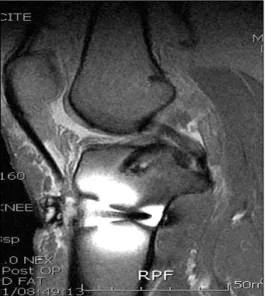
Fig. 1. Metallic artifact on tibial side in group A patient that using metallic screw.
서 론
전방 십자 인대 재건술은 급성 및 만성 전방 십자 인대 파열로 기능을 상실한 불안정한 슬관절의 일차 치료법으로 인정되고 있다 1,2) . 전방 십자 인대 재건 시 조기 재활 치료를 위해서는 이식건의 초기 고정력 , 특이 경골부의 약한 고정력이 초기실패의 중요한 원인으로 알려져 있다 . 생체 흡수성 간섭 나사를 이용한 전방 십자 인대 이식편 고정은 간섭나사로 인한 이식건의 손상이 적은 것을 비롯하여 여러 장점들이 있으나 , 인장 강도가 약한 단점이 있다 3) . 따라서 수술 후 초기에 강한 고정력을 얻기 위하여 경골 고정시 혼합 고정이 사용되고 있는데, 통상적으로 생체 흡수성 간섭 나사 및 다양한 금속 나사들을 이용하여 고정한다 4-7) . 하지만 금속 나사를 이용하여 고정하는 경우 하퇴 전내측부의 연부조직이 적어서 발생하는 금속 돌출이나 피부 및 연부조직의 자극으로 인해 금속물 제거가 필요한 경우가 많으며 , 자기공명영상 추시 검사를 하는 경우 금속물에 의한 간섭 현상이 나타나는 단점이 있다(Fig.
1) 8,9) .
저자들은 과거 돼지 대퇴골 및 슬개건을 이용한 실험에서 생체 흡수성 봉합 나사를 이용한 혼합 고정 방법에 대한 생역학 적 결과를 보고하였는데 , 생체 흡수성 봉합 나사를 이용한 혼합 고정 방법은 간섭 나사 단독으로 사용한 경우보다 약 3배의 최대 파손 강도 및 강성도를 보였으며 이는 지주 금속 나사를 사용했을 때와 유사한 결과를 보였다 10) . 이에 저자들은 생역학
적 결과에서 금속 나사 고정과 유사한 강도를 보인 생체 흡수성 봉합 나사의 임상적 결과에 대해 보고하고자 한다.
연구 방법
1. 연구 대상
2008년 10월부터 2012년 2월까지 본원에서 전방 십자 인대 재건술을 시행 받은 60명의 환자를 대상으로 조사하였다. 동측 슬관절의 안정성에 영향을 줄 수 있는 후방 십자 인대 및 내측, 외측 측부 인대의 손상이 동반되어 있는 환자들은 제외 하였다. 경골 터널부위에 생체 흡수성 간섭 나사로 이식건을 고정한 이후 부가적인 혼합 고정 시 생체 흡수성 봉합 나사를 사용한 30예를 그룹 A로, 지주 금속 나사 및 spike washer를 이용한 30예를 그룹 B로 나누어 구분하였다. 60예 모두 신선 동결 아킬레스건 또는 후경골근 동종건을 이용하여 전방 십자 인대 재건을 시행하였으며 그룹 A는 27예에서 후경골근 동종 건을, 3예에서 아킬레스 동종건을 이용하였으며, 그룹 B는 30예 모두 아킬레스 동종건을 이용하였다. 두 그룹의 평균 추시 관찰 기간은 15개월(범위: 12–47개월)이었고, 그룹 A의 수술 당시 평균 나이는 32세(범위: 17–71세)이며, 성별은 남자 24예, 여자 6예였다. 그룹 B의 평균 나이는 36세(범위: 19–61세) 이며, 남자 23예, 여자 7예였으며, 두 그룹 간의 인구학적 자료 에서 통계적으로 유의한 차이는 없었다.
2. 평가 방법
슬관절 안정성에 대한 평가로는 수술 전 및 최종 추시 시 KT-2000 관절 계측계를 이용하여 건측 과의 최대 도수 전방 전위 차를 정량적으로 측정하였으며 , 임상적 결과에 대하여는 슬관절 동통, 부종, 계단 쪼그려 앉기, 파행, 잠김, 보조기 사용 여부, 불안정성 등 여덟 가지 점수를 합산하는 Lysholm score와 환자의 활동 정도를 판정하는 Tegner score 및 International Knee Documentation Committee (IKDC)의 평가 방법을 이용하 여 최종 추시 시의 측정 결과와 비교하였다. 통계학적 검증은 그룹 간 비교를 위해서 SPSS 통계 프로그램(SPSS ver. 12.0, SPSS Inc., Chicago, IL, USA)을 이용한 Mann-Whitney test를 사용하였으며, p<0.05인 경우 통계학적으로 유의한 것으로 판단하였다.
3. 이식건의 고정 방법
신선 동결 아킬레스건을 이용한 환자의 경우 대퇴 터널로
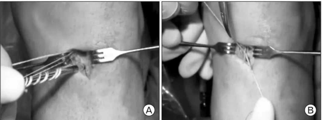
Fig. 3. (A) Bioabsorbable suture anchor was inserted on tibial drill- ing site. (B) FiberWire on both sides was tied.
Fig. 2. (A) Bioabsorbable suture anchor (Duet Suture Anchor, Linvatec Corp., Largo, FL, USA). (B) Separated suture anchor tip and FiberWire (Arthrex, Naples, FL, USA) connecting the tendon. (C) Reassembled bioabsorbable suture anchor.
이식골편을 진입시키고 전내측 삽입구를 통해 생체 흡수성 간섭나사(BioScrew, Linvatec Co., Largo, FL, USA)로 대퇴부 골편을 고정하였으며 , 후경골근 동종건을 이용한 환자의 경우 이식건 근위부 고정은 Endobutton CL Ultra (Smith & Nephew Inc, Andover, MA, USA)을 사용하여 대퇴골부 고정을 시행하 였다. 그룹 A의 경골부 이식건의 고정은 생체 흡수성 간섭 나사를 이용한 고정을 먼저 시행한 뒤 생체 흡수성 봉합 나사 (Duet Suture Anchor, Linvatec Co.)에 FiberWire (Arthrex, Naples, FL, USA)를 끼워 넣은 후(Fig. 2) 고정할 위치에 drilling을 시행하였다 . 이후 생체 흡수성 봉합 나사를 삽입 후(Fig. 3A) 양측의 FiberWire를 봉합하였다(Fig. 3B). 그룹 B의 경골부 이식건의 고정은 동일하게 생체 흡수성 간섭나사를 이용한 고정을 먼저 시행한 뒤 지주 금속 나사 및 spike washer를 이용한 혼합 고정 방법을 사용하였다.
4. 수술 후 재활
수술 직후 슬관절을 30 o 가량 굴곡시킨 상태에서 장하지 부목 고정을 시행하였으며 수술 후 3일 째 장하지 보조기를 착용한 후 수동적 슬관절 굴곡 운동을 시작하였다 . 1주째 30 o , 2주째 45 o , 3주째 60 o , 4주째 90 o 까지의 슬관절 굴곡을 허용하였 으며, 체중 부하의 경우 수술 후 1 주째부터 부분 체중 부하를 시행하며 점차 체중 부하 정도를 늘려 8주째 완전 체중 부하를 시행하였다.
결 과
1. 슬관절 안정성
KT-2000 최대 도수 전방 전위 차이는 생체 흡수성 봉합나사 를 이용한 그룹 A에서 건측에 비해 수술 전 평균 5.1 mm (범위: 2.0–10.0 mm)에서 최종 추시 시 3.28 mm (범위: 1.0–8.0 mm)로 안정되었으며, 15예에서 3 mm 미만의 차이를, 11예에 서 3–5 mm의 차이를, 2예에서 각각 7 mm, 8 mm의 차이를 보였다. 금속 고정을 이용한 그룹 B에서는 건측에 비해 수술 전 평균 5.72 mm (범위: 1.0–12.0 mm)에서 최종 추시 시 3.56 mm (범위: 0.0–10.0 mm)로 안정되었으며, 15예에서 3 mm 미만의 차이를, 12예에서 3–5 mm의 차이를, 2예에서 7 mm, 1예에서 10 mm의 차이를 보였다. 하지만 두 그룹간의 통계학 적으로 유의한 차이는 보이지 않았다(p=0.84).
2. 임상적 결과
Lysholm score는 그룹 A에서 수술 전 평균 46.14점(범위:
33–52점)에서 최종 추시 시 평균 83.8점(범위: 74–89점)으로
향상되었으며 Tegner score는 수술 전 평균 4.86점(범위: 2–7점)
에서 최종 추시 시 평균 8.14점(범위: 7–9점)으로 향상되었다.
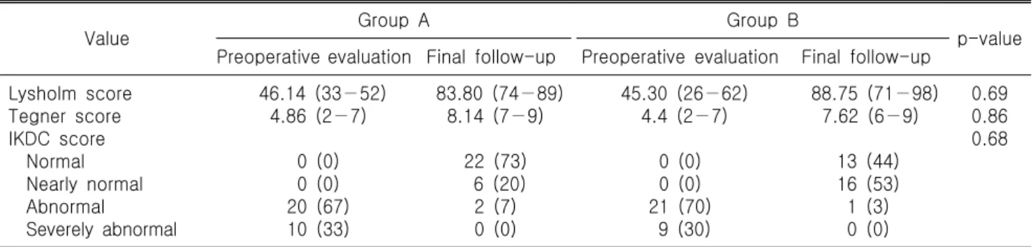
Table 1. Comparison of clinical outcome of both group
Value Group A Group B
p-value Preoperative evaluation Final follow-up Preoperative evaluation Final follow-up
Lysholm score Tegner score IKDC score
Normal Nearly normal Abnormal
Severely abnormal
46.14 (33−52) 4.86 (2−7)
0 (0) 0 (0) 20 (67) 10 (33)
83.80 (74−89) 8.14 (7−9)
22 (73) 6 (20) 2 (7) 0 (0)
45.30 (26−62) 4.4 (2−7)
0 (0) 0 (0) 21 (70) 9 (30)
88.75 (71−98) 7.62 (6−9)
13 (44) 16 (53) 1 (3) 0 (0)
0.69 0.86 0.68
Values are presented as average (range) or number (%).
IKDC: International Knee Documentation Committee.
그룹 B의 Lysholm score는 수술 전 평균 45.03점(범위: 26–62점) 에서 최종 추시 시 평균 88.75점(범위: 71–98점)으로, Tegner score는 수술 전 평균 4.40점(범위: 2–7점)에서 최종 추시 시 평균 7.62점(범위: 6–9점)으로 향상되었으나 두 그룹 간의 통계학적으로 유의한 차이는 없었다(p=0.69, 0.86).
두 그룹의 IKDC subjective score는 그룹 A에서 수술 전 비정상 20예, 중증 비정상 10예였지만, 최종 추시 시 22예에서 정상 , 6예에서 유사 정상, 2예에서 비정상의 결과를 보였으며, 그룹 B에서는 수술 전 비정상 21예, 중증 비정상 9예에서, 최종 추시 시 13예에서 정상, 16예에서 유사 정상, 1예에서 비정상의 결과를 보여 양 그룹 모두 90% 이상에서 유사 정상 이상의 결과를 보였다 . 하지만 이 또한 두 그룹 간의 통계학적 인 차이는 없었다(p=0.68) (Table 1).
고 찰