Differential Sensitivities of Human Multidrug-resistant Cancer Cells to BIIB021 and Modulation of Hsp90 Inhibitors by NSAIDs and Niclosamide
Hyun-Jung Moon, Su-Hoon Lee, Sun-Hee Kim* and Chi-Dug Kang*
Department of Biochemistry, Pusan National University School of Medicine, Yangsan 626-870, Korea Received June 29, 2018 /Revised August 16, 2018 /Accepted August 17, 2018
The critical role of heat shock protein 90 (Hsp90) in tumorigenesis led to the development of several first- and second-generation Hsp90 inhibitors, which have demonstrated promising responses in cancers. In this study, we found second-generation Hsp90 inhibitor BIIB021-resistant multidrug-resistant (MDR) human cancer cells, although BIIB021 was shown to be active in first-generation Hsp90 inhibitor 17- allylamino-17-demethoxygeldanamycin (17-AAG)-resistant MDR cells. MCF7-MDR and HeyA8- MDR cells were more resistant to BIIB021 than their parental counterparts, indicating that BIIB021 cannot be applicable to all cancer cells expressing MDR proteins. We revealed that dimethyl-celecoxib (DMC), one of the non-steroidal anti-inflammatory drugs (NSAIDs), potentiated cytotoxicity of BIIB021 against both BIIB021-resistant and BIIB021-sensitive MDR cells. The effectiveness of NSAIDs involving cele- coxib and DMC in combination with BIIB021 led to the autophagic degradation/down-regulation of mutant p53 (mutp53) that overexpressed MDR cells and the suppression of Hsp70 induction. This re- sulted in sensitization of MDR cells to BIIB021. Moreover, autophagy induction by sulindac sulfide, another type of NSAID, and niclosamide, an FDA-approved anthelmintic drug, potentiated 17-AAG- mediated autophagic degradation/down-regulation of mutp53 and c-Myc, client proteins of Hsp90.
Therefore, our results suggest that NSAIDs and niclosamide positively enhance the anticancer activity of Hsp90 inhibitors through an autophagic pathway. They may also be new candidates for sensitizing MDR cells to Hsp90 inhibitors.
Key words : Autophagy, BIIB021, Hsp90 inhibitor, NSAIDs, Niclosamide
*Corresponding authors
*Tel : +82-51-510-8081, Fax : +82-51-510-8086
*E-mail : [email protected] (Sun-Hee Kim)
*Tel : +82-51-510-8082, Fax : +82-51-510-8086
*E-mail : [email protected] (Chi-Dug Kang)
This is an Open-Access article distributed under the terms of the Creative Commons Attribution Non-Commercial License (http://creativecommons.org/licenses/by-nc/3.0) which permits unrestricted non-commercial use, distribution, and reproduction in any medium, provided the original work is properly cited.
Journal of Life Science 2018 Vol. 28. No. 10. 1212~1219 DOI : https://doi.org/10.5352/JLS.2018.28.10.1212
Introduction
Heat shock proteins (Hsps) are molecular chaperones that are consistently increased to help cells survive under con- ditions of stress. As a member of the Hsps, Hsp90 is a molec- ular chaperone that plays an important role in the mod- ification and stabilization of a variety of proteins implicated in tumor cell proliferation and survival. Particularly, Hsp90 is unexpectedly abundant to maintain levels of proteins in cancer cells. Hsp90 inhibitors can target the ATP domain of Hsp90 and prohibit its exchange of ADP for ATP, leading to the degradation of client proteins and disruption of sig- naling cascades. Many of the known clients are protein kin-
ases or transcription factors involved in multiple signal transduction pathways including tyrosine kinases; Bcr-Abl, epidermal growth factor receptor family members, c-Met, in- sulin-like growth factor-1 receptor and pp60c-src, serine/
threonine kinases; Akt, Cdk4, Raf-1 steroid hormone re- ceptors, p53, Stat3, Mdm2 and telomerase. Concomitantly, Hsp90 inhibitors induce tumor cell apoptosis, promote cell cycle arrest and abrogate microenvironmentderived cytopro- tection [18].
Geldanamycin and its derivative, 17-allylamino-17-deme-
thoxy-geldanamycin (17AAG), were first developed as Hsp
90 inhibitors (first-generation Hsp90 inhibitors), and they ex-
hibited effective anticancer potency but severe side effects
and low solubility restricted their application at the clinical
level. Moreover, the antitumor activity of 17-AAG and other
ansamycins are significantly curtailed by the expression of
multidrug resistance (MDR) proteins, a major contributor to
drug resistance commonly observed in heavily pretreated
cancer patients. Many types of cancer express relatively high
levels of P-glycoprotein (P-gp), a major type of MDR protein,
which include colon, kidney, adrenocortical and hepatocel-
lular cancers whereas breast, lung, neuroblastoma and other
tumors express P-gp at intermediate levels [1, 5]. In many cases, untreated tumors, which do not express MDR pro- teins, could be induced to express P-gp and other MDR pro- teins to a high level upon treatment with drugs that are P-gp substrates, including chemotherapeutic drugs and molecu- larly targeted compounds [4, 15]. Previous studies have shown that the 17-AAG and ansamycin derivatives of Hsp90 inhibitors are inactive in P-gp/MDR1- and/or MRP1-ex- pressing cell lines [16, 19]. 17-AAG might be transported by P-gp in a distinct manner or that the magnitude of the influ- ence of P-gp on ansamycin activity may be related to the slow kinetics of action of these Hsp90 inhibitors in cells.
Second-generation Hsp90 inhibitor BIIB021 is an orally available, fully synthetic novel small-molecule Hsp90 in- hibitor that has shown strong antitumor activities in a large number of preclinical models and is now under clinical in- vestigation [3, 14]. It has been reported that over-expression of P-gp is associated with resistance to 17-AAG but not to BIIB021, and BIIB021 is active in P-gp and/or MRP-1 ex- pressing drug-resistant cancer cells [19]. Moreover, BIIB021 potentiate the effects of other therapeutics beyond degrading oncogenic protein [7]. Since the cytotoxicity of BIIB021 in MDR cells showed no correlation with P-gp level in only two tested MDR variants [19], it is necessary to test various MDR cells derived from different types of tumors. In this study, we determined whether BIIB021 could be active against various MDR cells derived from different types of tumors, and also developed new sensitizers that enhance cy- totoxicity of Hsp90 inhibitors and thus reverse Hsp90 in- hibitor resistance of MDR cells.
Materials and Methods
Cell culture and reagents
Human MDR variants such as CEM/VLB
100cells isolated from CEM human lymphoblastic leukemia cells, MCF7-MDR cells isolated from human breast cancer MCF-7 cells and HeyA8-MDR cells isolated from HeyA8 human ovarian can- cer (moderately differentiated papillary cystadenocarcinoma of the ovary) cells were used, which were kindly provided by Dr. Fiedler (MD Anderson, TX, USA). These cells were maintained in DMEM supplemented with 10% fetal bovine serum and were incubated at 37℃, 5% CO
2and 95% humid- ity. 17-allylamino-17-demethoxy-geldanamycin (17-AAG) (Enzo Life Sciences Inc., Farmingdale, New York, USA), celecoxib (CCB), 2,5-dimethyl-celecoxib (DMC) (Sigma-Aldrich, St.
Louis, MO, USA). BIIB021 (Selleckchem, Houston, TX, USA), niclosamide and sulindac sulfide (Sigma-Aldrich, St. Louis, MO, USA) were used in this study.
Cell proliferation assay
Cell proliferation was measured by counting viable cells by using the 3-(4,5-dimethylthiazol-2-yl)-2,5-diphenyltetra- zolium bromide (MTT) colorimetric dye-reduction method.
Exponentially growing cells (1- or 2×10
4cells/well) were plated in a 96-well plate and incubated in growth medium treated with the indicated concentration of BIIB021 and DMC (or CCB) at 37℃. After 96 hr, the medium was re- moved using centrifugation, and MTT-formazan crystals solubilized in 100 μl DMSO. The optical density of each sam- ple at 570 nm was measured using ELISA reader. The optical density of the medium was proportional to the number of viable cells. Inhibition of proliferation was evaluated as a percentage of control growth (no drug in the medium). All experiments were repeated in at least two experiments in triplicate.
Western blot analysis
Cell lysates from control and indicated drug-treated cells (1×10
6cells) were prepared using M-PER Reagent (Thermo Scientific Inc., USA). The lysates were clarified by centrifuga- tion. Equal amounts of protein were resolved by SDS-PAGE and immunoblotted with indicated antibodies. Western blot analysis was performed with specific primary antibodies against LC3 and p62 (Novus Biologicals, Littleton, CO, USA), b-actin (Sigma-Aldrich, St. Louis, MO, USA), Hsp70 (Enzo Life Sciences, Inc., NY, USA), c-Myc (Cell Signaling Technol- ogy Inc., Beverly, Massachusetts, USA), p53 (Santa Cruz Biotechnology, CA, USA), The p53 antibody (DO-1) is a mouse monoclonal antibody raised against amino acids 11- 25 of p53 of human origin (Santa Cruz Biotechnology), which was recommended for detection of wild and mutant p53 of human origin.
Apoptosis assessment by Annexin V staining
The effects of BIIB021 and/or CCB (or DMC) on apoptosis
were measured by Annexin V staining. MCF7-MDR cells
(2×10
5cells/ml) were treated with BIIB021 in the presence
or absence of CCB and/or DMC for 24 hr. Then cells were
centrifuged and resuspended in 100 μl of the staining sol-
ution containing Annexin V-fluorescein (FITC Apoptosis de-
tection kit; BD ParMingen San Diego, CA, USA) and propi-
A
B
C
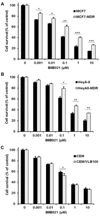
Fig. 1. Differential responses of human MDR variants to BIIB021. MCF-7, HeyA-8 and CEM cells and their MDR variants MCF7-MDR (A), HeyA8-MDR cells (B) and CEM/VLB100 (C) were treated with serial concentrations of BIIB021. Percentage of cell survival was determined after 96 hr of incubation using MTT assay. Results are the means ± SEs of three experiments. *p<0.05, **p<0.01 and ***p<0.001.
dium iodide (Sigma-Aldrich, St. Louis, MO, USA) in a Hepes buffer. After incubation at room temperature for 20 min, the percentage of early (annexin V positive/PI negative) and late apoptotic cells (annexin V positive/PI positive) was quanti- fied by FACS for Annexin-V and PI staining.
Statistical analysis
A Student’s t-test was used to calculate the statistical sig- nificance of the experimental data and the level of sig- nificance was set as *p<0.05, **p<0.01 and ***p<0.001.
Results
Differential responses of three MDR cells to Hsp90 inhibitor BIIB021 and enhancement of BIIB021 sensi- tivity by NSAIDs
Since previous study showed that BIIB021 was active in two MDR variants such as NCI/ADR-RES and MES-SA DX5 cells [19], we therefore examined the changed cytotoxicity of BIIB021 in three MDR variants isolated from tumors of various types such as MCF7-MDR cells, HeyA8-MDR cells and CEM/VLB
100cells when compared to BIIB021 cytotoxicy of their parental counterparts. Our data showed that two variants of MDR such as MCF7-MDR and HeyA8-MDR cells were more resistant to BIIB021 than their parental counter- parts (Fig. 1A, Fig. 1B), whereas BIIB021 sensitivity of both CEM/VLB
100and its parental counterpart was not different, and BIIB021 was active against both cells (Fig. 1C), indicating it is necessary to establish methods for sensitization of BIIB021-resistant MDR cells.
Previous we reported that sensitivity of Hsp90 inhibitor 17-AAG was enhanced by celecoxib (CCB) and its analog 2,5-dimethyl-celecoxib (DMC), non-steroidal anti-inflamma- tory drugs (NSAIDs) class, via autophagic inducing ability, which resulted in autophagic degradation of mutant p53 (mutp53) that over-expressed MDR cells. [12]. Therefore, we examined whether BIIB021 cytotoxicity in MDR cells could be modulated by DMC. Our data showed that DMC po- tentiated the cytotoxicity of BIIB021 in BIIB021-sensitive CEM/VLB
100cells (Fig. 2A). Importantly, BIIB021-resitant MCF7-MDR cells showed that BIIB021 cytotoxicity was sig- nificantly enhanced by DMC or CCB treatment (Fig. 2B, Fig.
2C). Moreover, we estimated the percentage of early and late apoptotic cells in BIIB021-treated MCF7-MDR cells in the presence of absence of CCB (or DMC) cells using conven- tional flow cytometry (Fig. 3). In this experiment, the per-
centage of cells in both the lower and upper right quadrants
of the Annexin/PI analysis was significantly increased by
co-treatment with BIIB021 and CCB (or DMC) versus
BIIB021 alone, indicating sensitization of MDR cells to
BIIB021-induced apoptosis by NSAIDs. These results suggest
A
B
C
Fig. 2. Enhancement of BIIB021 cytotoxicity in MDR variants by DMC. CEM/VLB100 cells (A) and MCF7-MDR (B and C) were treated with serial concentrations of BIIB021 in the presence or absence of dimethyl celecoxib (DMC;
5- and 10 μM). Percentage of cell survival was de- termined after 96 hr of incubation using MTT assay.
Results are the means ± SEs of three experiments.
*p<0.05, **p<0.01 and ***p<0.001.
Fig. 3. Enhancement of BIIB021-induced apoptosis in MDR cells by NSAIDs. MCF7-MDR cells were treated with 0.5 μM BIIB021 and/or in the presence or absence of 5 μM CCB (or DMC) for 24 hr, and early and late apoptotic cells were analyzed by flow cytometry. Apoptosis index is de- fined as a percentage of early and late apoptotic cells.
The lower right quadrant shows annexin positive cells (early apoptotic) and the upper right quadrant shows annexin and PI positive cells (late apoptosis cells).
the possibility that NSAIDs such as DMC and CCB could be candidate sensitizers for BIIB021-resistant MDR cells.
Induction of autophagy and suppression of Hsp70 expression in BIIB021-treated MDR cells by NASIDs We determined whether CCB and DMC could induce au- tophagic activity when CEM/VLB
100cells were co-treated with BIIB021 and CCB (or DMC). To measure autophagic flux in CEM/VLB
100cells co-treated with BIIB021 and CCB (or DMC), LC3 is the most widely used autophagosome marker because the amount of LC3-II reflects the number of autophagosomes and also degradation of p62 is another widely used marker to monitor autophagic activity because p62 directly binds to LC3 and is selectively degraded by au- tophagy and autophagy-related structures. Therefore, we evaluated the autophagic degradation of mutp53 in CEM/
VLB
100cells by assessing changes in the levels of LC3-II and p62 (Fig. 4). When LC3 conversion and p62 level in CEM/
VLB
100cells were not responded by BIIB021, CCB induced
an increase of LC3 conversion and a decrease of p62 level
in BIIB021-treated cells. Similarly, DMC also enhanced LC3
conversion and reduced p62 level in BIIB021-treated cells.
Fig. 4. Induction of autophagy and acceleration of autophagic mutp53 degradation and suppression of Hsp70 activa- tion in MDR cells treated with BIIB021 by CCB/DMC.
CEM/VLB100 cells were treated with BIIB021 (0.1 or 0.2 μM) in the presence or absence of 25 μM CCB (or 25 μM DMC) for 24 hr. Change levels of LC3BI/II, p62, mutp53 and Hsp70 were determined by Western blot analysis. Actin was used as a loading control.
A
B
C
Fig. 5. Induction of autophagy and acceleration of autophagic mutp53 degradation in MDR cells treated with 17-AAG by sulindac sulfate (SS) and niclosamide (NC). CEM/
VLB100 cells were treated with serial doses of sulindac and niclosamide for 24 hr (A), and were treated with 17-AAG (2- and 10 μM) in the presence or absence of 50 μM SS or 0.5 μM NC for 24 hr (B). MCF7-MDR cells were treated with17-AAG (2- and 10 μM) in the presence or absence of 50 μM SS or 0.5 μM NC for 24 hr (C).
Change levels of LC3BI/II, p62, mutp53, Hsp70 and c-Myc were determined by western blot analysis. Actin was used as a loading control.
In parallel with the autophagy-inducing effect of CCB and DMC, the level of mutp53 in BIIB021-treated cells was de- creased by treatment of CCB or DMC, and importantly BIIB021-mediated induction of Hsp70 was significantly sup- pressed by CCB or DMC. DMC is more effective than CCB against down-regulation of mutp53 and suppression of Hsp70 induction in BIIB021-treated cells. These results in- dicated the possibility that combined effect of BIIB021 and CCB/DMC on the induction of autophagy would reduce BIIB021-mediated mutp53, which triggered apoptosis in CEM/VLB
100cells. These results suggest the possibility that NSAID including CCB and DMC could be a new class of BIIB021 sensitizer through suppression of Hsp70 induction that limits the clinical benefits of Hsp90 inhibitors.
Induction of autophagy and potentiation of 17-AAG- mediated autophagy by sulindac sulfide and niclosa- mide
Next, to further find new modulators of autophagy in- ducer for potentiation of Hsp90 inhibitor, we developed su- lindac sulfide (SS) that belongs to a class of NSAIDs that has anti-tumorigenic and anti-inflammatory activities [9] and niclosamide (NC), an FDA approved oral anti-helminthic drug that has cytotoxicity in a broad spectrum of cancer cells [2]. We evaluated the autophagy inducing effect of SS and NC in MDR cells by assessing changes in the levels of LC3-II and p62. When CEM/VLB
100cells were treated with serial doses of SS, treatment of CEM/VLB
100cells with SS resulted in an increase of LC3 conversion (LC3-II) and a decrease of p62 level in a dose dependent manner, indicating autoph-
agy-inducing ability of SS, which is associated with degrada- tion/down-regulation of mutp53 (Fig. 5A, left). Similar re- sults were observed in CEM/VLB
100cells treated with serial doses of NC, leading to induce autophagy as demonstrated by an increase of LC3 conversion and a decrease of p62 level, which causes degradation/down-regulation of mutp53 (Fig.
5A, right). We further examined whether combination of 17-AAG and SS could modulate 17-AAG-mediated-LC3- II/LC-I and p62 levels in CEM/VLB
100cells (Fig. 5B, right).
SS significantly augmented 17-AAG-mediated level of LC3-II
and accelerated reduction of p62 level, indicating the com-
bined effect of 17-AAG and SS on the induction of autoph-
agy. Moreover, the autophagy-inducing effect of SS accel-
erated 17-AAG-mediated down-regulation/degradation of
mutp53 and c-Myc, a target gene of mutp53. Our results
showed that SS-induced autophagy was associated with deg-
radation/down-regulation of c-Myc as well as mutp53 (Fig.
5B, left). When MCF7-MDR cells were co-treated with NC and 17-AAG, NC treatment significantly enhanced 17-AAG- mediated down-regulation/degradation of mutp53 and c-Myc via induction of autophagy (Fig. 5B, right). Next, we examined the effect of the presence or absence of SS (or NC) on 17-AAG-treted MCF7-MDR cells (Fig. 5C). Similarly, the autophagy-inducing effect of SS and NC accelerated 17- AAG-mediated down-regulation/degradation of mutp53 in the MDR cells. Moreover, SS and NC suppressed 17-AAG-induced Hsp70 induction, indicating possibility that reversal of Hsp90 inhibitor resistance in MCF7-MDR cells by SS and NC. Therefore, our results suggest that SS and NC could potentiate the activity of Hsp90 inhibitors via au- tophagy pathway.
Discussion
Hsp90 inhibitors are a group of promising antitumor agents that lead to the selective degradation of proteins in- volved in multiple oncogenic processes [11], and they hinder the growth of many types of tumors in both in vitro and
in vivo tumor models [13]. BIIB021, a second-generationHsp90 inhibitor, is a purine scaffold-based and a fully syn- thetic Hsp90 inhibitor that binds to the ATP-binding pocket of Hsp90 and interferes with Hsp90 chaperone function, which caused cell death in conjunction with alterations in expression of Hsp90 client proteins, resulting in client pro- tein degradation and tumor growth inhibition. BIIB021 that binds selectively to Hsp90 has been evaluated in a phase I clinical trial involving solid tumors [14] and a phase II clin- ical trial involving gastrointestinal stromal tumors [3].
Previous study showed that BIIB021 was relatively in- dependent of MDR1 expression and was active against P-gp expressing cell lines [19]. In this study, we found that BIIB021 cannot be applicable to all cancer cells expressing MDR proteins since MCF7-MDR and HeyA8-MDR cells were more resistant to BIIB021 than their parental counter- parts but BIIB021 was sensitive to CEM/VLB
100cells, indicat- ing differential responses of MDR cells to BIIB021. Moreover, we showed that CCB and its derivative DMC that belongs to the NSAIDs could be as new sensitizers of BIIB021 as well as 17-AAG in MDR cells. Indeed, NSAID-induced au- tophagy has diverse anticancer effects in different type of cancer cells by regulating Beclin-1, LC3-II, p62, and Atg5-12 and can modulate tumor autophagy through various signal-
ing pathways involving PI3K/Akt/mTOR cascade [17].
Previously, we reported that autophagy inducing ability of NSAIDs involving CCB, DMC and IBU were effective for the sensitization of MDR cells to 17-AAG through inhibition of Akt/mTOR and STAT3 pathways [12]. The combined ef- fect of BIIB021 and CCB/DMC reduce BIIB021-mediated mutp53 via the induction of autophagy, which might trigger cell death in MDR cells. In addition, our data showed that sulindac sulfide (SS) and niclosamide (NC) had autophagic autophagy-inducing ability, which may contribute to the ability of enhancing BIIB021 sensitivity. Present study showed that non-steroidal anti-inflammatory drugs (NSAIDs) such as CCB, DMC and SS induced autophagy irrespective of their COX selectivity since both DMC and SS are non-se- lective COX inhibitors but CCB is a cyclooxygenase (COX)-2 selective inhibitor. Indeed, COX-independent mechanisms may contribute to, or be fully responsible for their anticancer properties.
Autophagy, a self-destructive response of cells to stress, leads to degradation of endogenous cellular protein ag- gregates and damaged organelles by lysosomes. LC3 is con- verted into LC3-I, following proteolytic cleavage at the C-ter- minal region by autophagy-related protein-4. LC3-I is then transformed by the lipid phosphatidyl ethanolamine to LC3-II. LC3-II is deployed and attaches to the membrane of an autophagosome. LC3-II remains attached to the mem- brane, until the autophagosome fuses with a lysosome [6].
CCB, DMC and SS commonly enhanced LC3-II level and conversely reduced p62 level, indicating autophagy inducing ability by NSAIDs. Interestingly, we showed that an FDA- approved drug NC that is given orally to helminthosis pa- tients also increased autophagic activity in MDR cells. It has been reported that NC is able to block the multiple signaling pathways that govern cancer initiation and progression, and also NC has potent in vitro and in vivo anti-tumor growth activities [8]. Our study indicates the possibility that autoph- agy-inducing activity of NSAIDs including CCB, DMC, SS and NC could modulate Hsp90 inhibitors. We found that CCB and DMC induced an increase of LC3 conversion and a decrease of p62 level in BIIB021-treated MDR cells.
Moreover, autophagy-inducing activity of DMC was asso-
ciated with down-regulation of mutp53, and suppression of
Hsp70 induction, which can trigger BIIB021-mediated cell
death by inducing apoptosis in MDR cells. In addition, both
SS and NC also enhanced 17-AAG-mediated level of LC3-
II/LC-I and accelerated reduction of p62 in MDR cells.
Moreover, the autophagy-inducing effect of SS and NC ac- celerated 17-AAG-mediated down-regulation/degradation of mutp53 and c-Myc, a target gene of mutp53, indicating the combined effect of 17-AAG and SS/NC on the induction of autophagy and enhancement of 17-AAG cytotoxicity by SS or NC treatment.
It has been reported that BIIB021 is not susceptible to me- tabolism NQO1/DT-diaphorase enzymes or to efflux by P-glycoprotein, thus avoiding some of the liabilities of 17- AAG [19]. But BIIB021 up-regulated expression of the Hsp70 and Hsp27, cause of resistance to HSP90 inhibitors, in both tumor tissue and spleen [10], indicating that BIIB021 has the same mechanism of action as the natural product geldana- mycin and its derivative 17-AAG as well as other Hsp90 inhibitors. Interestingly, CCB and DMC suppressed BIIB021- mediated Hsp70 induction of BIIB021-resistant MCF7-MDR cells, indicating their ability of reversing BIIB021 resistance of the cells.
In conclusion, we found potentiating effect of NSAIDs on BIIB021 activity in MDR cells, and sensitization of BIIB021- resistant MDR cells to BIIB021 through autophagic degrada- tion of mutp53 and suppression of Hsp70 induction by NSAIDs. In addition, SS and NC possessing autophagy-in- ducing activity could be new sensitizers of Hsp90 inhibitors including BIIB021.
Acknowledgement
This work was supported by a 2-Year Research Grant of Pusan National University.
References
1. Bansal, T., Jaggi, M., Khar, R. K. and Talegaonkar, S. 2009.
Emerging Significance of Flavonoids as P-Glycoprotein Inhibitors in Cancer Chemotherapy. J. Pharm Pharm Sci. 12, 46-78.
2. Cheng, B. X., Morales, L. D., Zhang, Y. H., Mito, S. and Tsin, A. 2017. Niclosamide induces protein ubiquitination and inhibits multiple pro-survival signaling pathways in the human glioblastoma U-87 MG cell line. PLoS One 12, e0184324.
3. Dickson, M. A., Okuno, S. H., Keohan, M. L., Maki, R. G., D'Adamo, D. R., Akhurst, T. J., Antonescu, C. R. and Schwartz, G. K. 2013. Phase II study of the HSP90-inhibitor BIIB021 in gastrointestinal stromal tumors. Ann. Oncol. 24, 252-257.
4. Fischer, V., Einolf, H. J. and Cohen, D. 2005. Efflux trans- porters and their clinical relevance. Mini Rev. Med. Chem.
5, 183-195.
5. Gottesman, M. M., Fojo, T. and Bates, S. E. 2002. Multidrug resistance in cancer: Role of ATP-dependent transporters.
Nat. Rev. Cancer 2, 48-58.
6. Hsieh, Y. C., Athar, M. and Chaudry, I. H. 2009. When apoptosis meets autophagy: deciding cell fate after trauma and sepsis. Trends Mol. Med. 15, 129-138.
7. Kim, S. H., Kang, J. G., Kim, C. S., Ihm, S. H., Choi, M.
G., Yoo, H. J. and Lee, S. J. 2016. Synergistic cytotoxicity of BIIB021 with triptolide through suppression of PI3K/Akt/ mTOR and NF-kappa B signal pathways in thy- roid carcinoma cells. Biomed. Pharmacother. 83, 22-32.
8. Li, Y. H., Li, P. K., Roberts, M. J., Arend, R. C., Samant, R. S. and Buchsbaum, D. J. 2014. Multi-targeted therapy of cancer by niclosamide: A new application for an old drug.
Cancer Lett. 349, 8-14.
9. Liggett, J. L., Zhang, X. B., Eling, T. E. and Baek, S. J. 2014.
Anti-tumor activity of non-steroidal anti-inflammatory drugs:
Cyclooxygenase-independent targets. Cancer Lett. 346, 217- 224.
10. Lundgren, K., Zhang, H., Brekken, J., Huser, N., Powell, R.
E., Timple, N., Busch, D. J., Neely, L., Sensintaffar, J. L., Yang, Y. C., McKenzie, A., Friedman, J., Scannevin, R., Kamal, A., Hong, K., Kasibhatla, S. R., Boehm, M. F. and Burrows, F. J. 2009. BIIB021, an orally available, fully syn- thetic small-molecule inhibitor of the heat shock protein Hsp90. Mol. Cancer Ther. 8, 921-929.
11. Maloney, A. and Workman, P. 2002. HSP90 as a new ther- apeutic target for cancer therapy: the story unfolds. Expert Opin. Biol. Ther. 2, 3-24.
12. Moon, H. J., Kim, H. B., Lee, S. H., Jeun, S. E., Kang, C.
D. and Kim, S. H. 2018. Sensitization of multidrug-resistant cancer cells to Hsp90 inhibitors by NSAIDs-induced apop- totic and autophagic cell death. Oncotarget 9, 11303-11321.
13. Neckers, L. 2002. Hsp90 inhibitors as novel cancer chemo- therapeutic agents. Trends Mol. Med. 8, S55-S61.
14. Saif, M. W., Takimoto, C., Mita, M., Banerji, U., Lamanna, N., Castro, J., O'Brien, S., Stogard, C. and Von Hoff, D. 2014.
A Phase 1, Dose-Escalation, pharmacokinetic and pharma- codynamic study of BIIB021 administered orally in patients with advanced solid tumors. Clin. Cancer Res. 20, 445-455.
15. Shtil, A. A. and Azare, J. 2005. Redundancy of biological regulation as the basis of emergence of multidrug resistance.
Int. Rev. Cytol. 246, 1-29.
16. Taldone, T., Gozman A., Maharaj R. and Chiosis, G. 2008.
Targeting Hsp90: small-molecule inhibitors and their clinical development. Curr. Opin. Pharmacol. 8, 370-374.
17. Yu, C., Li W. B., Liu J. B., Lu J. W. and Feng, J. F. 2018.
Autophagy: novel applications of nonsteroidal anti-inflam- matory drugs for primary cancer. Cancer Med. 7, 471-484.
18. Zhang, H. and Burrows, F. 2004. Targeting multiple signal transduction pathways through inhibition of Hsp90. J. Mol.
Med. (Berl). 82, 488-499.
19. Zhang, H., Neely, L., Lundgren, K., Yang, Y. C., Lough, R., Timple, N. and Burrows, F. 2010. BIIB021, a synthetic Hsp90 inhibitor, has broad application against tumors with ac- quired multidrug resistance. Int. J. Cancer 126, 1226-1234.
초록:항암제 다제내성(MDR) 암세포의 Hsp90 저해제 BIIB021에 대한 감수성의 차이 및 NSAIDs 및 Niclosamide에 의한 Hsp90 저해제의 활성 변화
문현정․이수훈․김선희*․강치덕*
(부산대학교 의학전문대학원 의과학과 생화학교실)