The Carboxyl-terminal Tail of a Heterotrimeric Kinesin 2 Motor Subunit Directly Binds to β2-tubulin
Young Joo Jeong1, Sung Woo Park2, Sang-Jin Kim3, Won Hee Lee4, Mooseong Kim4, Sang-Hwa Urm5 and Dae-Hyun Seog1*
1Departments of Biochemistry, Inje University College of Medicine, Busan 47392, Korea
2Departments of Convergence Biochmedical Science, Inje University College of Medicine, Busan 47392, Korea
3Departments of Neurology, Inje University College of Medicine, Busan 47392, Korea
4Department of Neurosurgery, Inje University College of Medicine, Busan 47392, Korea
5Department of Preventive Medicine, Inje University College of Medicine, Busan 47392, Korea Received October 1, 2018 /Revised October 10, 2018 /Accepted October 17, 2018
Microtubules form through the polymerization of α- and β-tubulin, and tubulin transport plays an im- portant role in defining the rate of microtubule growth inside cellular appendages, such as the cilia and flagella. Heterotrimeric kinesin 2 is a molecular motor member of the kinesin superfamily (KIF) that moves along the microtubules to transport multiple cargoes. It consists of two motor subunits (KIF3A and KIF3B) and a kinesin-associated protein 3 (KAP3), forming a heterotrimeric complex.
Heterotrimeric kinesin 2 interacts with many different binding proteins through the cargo-binding do- mains of the KIF3s, but these binding proteins have not yet been specified. To identify these proteins for KIF3A, we performed yeast two-hybrid (Y2H) screening and found a specific interaction with β2- tubulin (Tubb2), a microtubule component. Tubb2 was found to bind to the cargo-binding domain of KIF3A but did not interact with KIF3B, KIF5B, or kinesin light chain 1 in the Y2H assay. The carbox- yl-terminal region of Tubb2 is essential for interaction with KIF3A. Other Tubb isoforms, including Tubb1, Tubb3, Tubb4, and Tubb5, also interacted with KIF3A in the Y2H screening. However, α1-tubu- lin (Tuba1) did not interact with KIF3A. In addition, an antibody to KIF3A specifically co-immun- oprecipitated the KIF3B and KAP3 associated with Tubb2 from mouse brain extracts. In combination, these results suggest that a heterotrimeric kinesin 2 motor protein is capable of binding to tubulin and may transport it in cells.
Key words : Binding protein, kinesin, microtubule, transport, tubulin
*Corresponding author
*Tel : +82-51-890-6974, Fax : +82-51-894-5801
*E-mail : [email protected]
This is an Open-Access article distributed under the terms of the Creative Commons Attribution Non-Commercial License (http://creativecommons.org/licenses/by-nc/3.0) which permits unrestricted non-commercial use, distribution, and reproduction in any medium, provided the original work is properly cited.
Journal of Life Science 2019 Vol. 29. No. 3. 369~375 DOI : https://doi.org/10.5352/JLS.2019.29.3.369
Introduction
The assembly of microtubule-based cytoskeleton is im- portant to the organelles transport in cells. Microtubules are the major longitudinal cytoskeletal filament and form through the polymerization of α-tubulin and β-tubulin [17]. In the cells, microtubules have two structurally distinct ends, plus- end and minus-end [9]. The minus-end of microtubule is lo- cated at MTOC and the plus-end is oriented cell peripherally [9]. Microtubule grows at the plus-end. Kinesins are plus- end-directed molecular motor proteins that move along mi- crotubule tracks [7]. Microtubule-based motor, kinesins, play
the key roles in moving of various kinds cargoes, including membrane vesicles, organelles, proteins and their complexes, and mRNAs [7].
Kinesin 2 represents two conserved subfamilies of hetero- trimeric and homodimeric motors found in various organ- isms [9, 18]. It is essential for the intracellular transport and intraflagellar transport that is important for left and right determination of embryos [9]. Several diseases are linked to the loss of activity of kinesin 2, such as cystic kidney dis- eases, hydrocephalus, and polydactyly [8, 9, 13]. KIF3, a member of the kinesin 2 family, forms a heterotrimeric com- plex that consists of two different kinesin motor proteins (KIF3A and KIF3B), which moved toward the plus ends of microtubule and a kinesin-associated protein 3 (KAP3), which was associated with the tail region of KIF3A and KIF3B [9, 18, 21]. KIF3A and KIF3B are abundantly ex- pressed in nerve tissue and ubiquitously expressed in other tissues [9, 21]. Microinjection and immunoprecipitation by anti-KIF3B antibody revealed that KIF3s is thought to play
a role in anterograde transport in cells [20].
In the formation of cilia and flagella, various proteins, such as tubulins need to be transport along microtubules tracks [9]. Previously studies suggest that KIF3s plays an important role in the intraflagellar transport (IFT) complex and the cilia and flagella assembly [11, 16, 19, 22]. In Chlamy- domonas, tubulin is transported by a combination of IFT com- plex [4]. Similarly, in the Caenorhabditis elegans, tubulin is transported into the sensory cilia by KIF3s [6, 15]. Also, tubu- lin is shown to bind to the IFT complex through co-operative interaction [12]. KIF3s knock-out mice exhibit randomized establishment of left-right asymmetry [8, 9]. It is implicated in certain chronic human disorders such as polycystic kidney disease [8, 9]. The molecular mechanisms of this left-right asymmetry were approached microscopically by observing the ventral node, which is a ciliated organ transiently ex- posed on the surface of the ventral midline [8]. Thus, KIF3s plays important roles for anterograde IFT into the flagella and cilia formation in many species and in many cell types [8, 9].
Understanding how KIF3s binds to tubulin is important question. In this study, we screened for proteins that bind with the cargo-binding domain of KIF3A, and found protein interacting with β2-tubulin type (Tubb2), which is micro- tubule component [3, 17]. The KIF3A and Tubb2 interaction suggests that tubulins could be transported by heterotrimeric kinesin 2.
Materials and Methods
Plasmid constructs
Full-length mouse α1-tubulin (Tuba1), Tubbs, Calcium- calmodulin-dependent kinase II a (CaMKIIa), kinesin light chain 1 (KLC1), and the carboxyl (C)-terminal region of KIF5B were amplified by polymerase chain reaction (PCR) from Marathon-ReadyTM cDNA library (Clontech, Palo Alto, CA, USA) and cloned into pGEM T-easy vector (Promega Corp, Madison, WI, USA). Mouse KIF3A and cargo-binding domain (aa 585-701) were subcloned from pCAGGS-KIF3A obtained from Prof. Kaibuchi K. Nagoya University, Nagoya, Japan [14] into the EcoRI and XhoI restriction sites of the pLexA.
Screening of KIF3A-binding proteins by yeast two- hybrid assay
The Matchmaker LexA two-hybrid system was used for
screening according to the manufacturer’s manual (Clon- tech). In brief, pLexA-KIF3A was transformed into yeast strain EGY48 carrying the p8op-lacZ gene. Transformed cells were transformed with the mouse brain cDNA library [9]
and grown on synthetic dextrose (SD) plates supplemented with glucose but with no histidine, tryptophan, or uracil (SD/-His/-Trp/-Ura). The selection of positive clones was performed on an SD/-His/-Trp/-Ura/-Leu plate containing galactose, raffinose, X-gal, and BU salts. Plasmids from pos- itive clones were analyzed by EcoRI and XhoI restriction digestion. Unique inserts were sequenced and protein se- quence analysis was performed with the BLAST algorithm at the National Center for Biotechnology Information (NCBI).
Sequence-verified clones were tested again for interactions of with the bait in yeast by the retransformation.
β-Galactosidase activity in liquid cultures of yeast The β-galactosidase activity of yeast was assayed as de- scribed previously [21]. Mid-log phase yeast cells were col- lected and permeabilized with 0.1% sodium dodecyl sul- phate (SDS) and chloroform. An excess amount of o-nitro- phenyl-β-D-galactoside (ONPG) (Sigma-Aldrich, St. Louis, MO, USA) was added to yeast lysate, and the mixture was incubated for each time at 30℃, and then the reaction was stopped by increasing pH to 11 by the addition of 1 M Na2CO3. The formation of the reaction product, o-nitro- phenol, was determined by measuring absorbance at 420 nm on a spectrophotometer and normalizing for the reaction time. The units of enzyme activity were calculated by the following equation: units=1,000× [(OD420– 1.75 × OD550)]/
(reaction time x culture volume × OD600). All experiments were independently performed at least three times [1].
Co-immunoprecipitation and immunoblot analysis Mouse brains were homogenized in ice-cold homoge- nization buffer (0.32 M sucrose, 4 mM HEPES, pH 7.3) sup- plemented with protease inhibitor cocktail (Sigma-Aldrich).
Mouse brain lysate was diluted in the same volume of 2X binding buffer (50 mM HEPES, 240 mM KCl, 2 mg/ml BSA, 0.2% Triton X-100, pH 7.4) and incubated with anti-KIF3A antibody [20] or with control IgG overnight at 4oC, followed by precipitation with protein-A Sepharose (Amersham Pharmacia, Piscataway, NJ, USA). The beads were collected by brief centrifugation and washed three times with TBS-T (20 mM Tris-HCl, pH 7.5, 0.15 M NaCl, 0.1% Tween 20).
The washed beads were resuspended with Laemmli’s load-
A
B
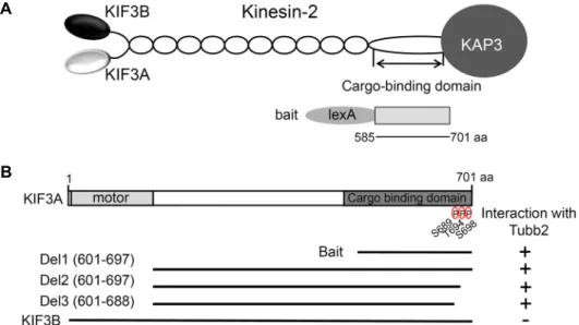
Fig. 1. Identification of the proteins interacted with KIF3A by yeast two-hybrid screening. (A) Schematic diagram of heterotrimeric kinesin 2. The cargo-binding domain of KIF3A used for the yeast two-hybrid screen. (B) Tubb2 binding region in KIF3A.
KIF3A has the motor domain and cargo binding domain, indicated in gray. The truncated forms of KIF3A were assessed in the yeast two-hybrid assay for interaction with Tubb2. +, interaction; -, no interaction; KIF3A, kinesin superfamily protein 3A; KIF3B, kinesin superfamily protein 3A; Tubb2, β2-tubulin type; aa, amino acids.
ing buffer and the proteins were eluted and denatured by boiling for 2 min. The proteins were processed for 10%
SDS-PAGE and immunoblot analysis with antibodies against KIF3B [20], KAP3 [20] and Tubb2 (Invitorgen, Carlsbad, CA, USA). The animal study was approved by the institutional review board (IRB), and the approval number was 05-12 of Inje University animal center.
Results
Identification of KIF3A interacting proteins by yeast two-hybrid assay
The amino (N)-terminal motor domain of heterotrimeric kinesin 2 is responsible for the motor activity and the C-ter- minal tail domain of heterotrimeric kinesin 2 is responsible for interacting to various proteins or cargoes [7, 18]. To iden- tify the specific binding proteins of heterotrimeric kinesin 2, we used the C-terminal cargo-binding domain (aa 585-701) of KIF3A fused to the DNA-binding domain of pLexA as a bait and isolated positive clones from a mouse brain cDNA library (Fig. 1A). From 8×10⁶ colonies screened, we obtained 3 positive clones. Two positive clones (clone 1, and 2) turned out to possess Tubb2 cDNA fragments (Fig. 2A). The pos- itive clones possessed cDNA fragments corresponding to the C-terminal region of Tubb2. Microtubules are assembled from heterodimer of α- and β-tubulin in cells [17]. Tubb2
form heterodimers with Tuba that incorporate into micro- tubules [17]. Tubb2 is three distinct domains: a N-terminal, intermediate, and a C-terminal domain (Fig. 1B) [3, 17]. The N-terminal domain (tubulin/FtsZ) interacts with GTP [3, 17].
The intermediate domain is role for microtubule assembly.
The C-terminal domain interacts with microtubule-asso- ciated proteins (MAPs) such as Tau protein, or microtubule- based motor proteins [3, 17]. To determine the minimal bind- ing domain of Tubb2 that is required for the interaction with KIF3A, we constructed several deletion mutants of Tubb2.
Yeast two-hybrid assays showed that the minimal domain required for binding was dependent on the C-terminal do- main of Tubb2 (Fig. 2A).
Previously study confirms that the C-terminal cargo-bind- ing domain of KIF3A has three phosphorylation sites. S689 was phosphorylated by PKA and T694, S698 were phos- phorylated by CaMKIIa both in vitro and in vivo [10]. The phosphorylation of KIF3A facilitates cargo loading and transport in cells [10]. Next, we examined the effects of the C-terminal phosphorylation on binding with KF3A and Tubb2. We constructed several deletion mutants of KIF3A and tested the interaction with Tubb2 and KIF3A by yeast two-hybrid assay. As shown in Fig. 1B, the deletion mutants of the C-terminal phosphorylation sites interact with Tubb2.
This data suggests that the phosphorylation sites of KIF3A did not affect the interaction of KIF3A with Tubb2. Next,
A
B C
Fig. 2. Minimal Tubb2 binding region in KIF5A. (A) KIF3A binding region in Tubb2. The positive clones isolated from the yeast two-hybrid screening possesses the cDNA for Tubb2. Tubb2 has tubulin/FtZ domain, indicated in gray. The truncated forms of Tubb2 were assessed in the yeast two-hybrid assay for interaction with KIF3A. (B, C) The tail region of KIF5B and KIF3A, KIF3B the full length KLC1, and the full length Tubbs were tested for the interaction in the yeast two-hybrid assay. KIF3A interacted with Tubbs but not with Tuba1. Tubb2 specifically interacted with KIF3A but not with KIF5B, KIF3B or KLC1.
CaMKIIa served as a positive control for interaction with KIF3A. +, interaction; -, no interaction; KIF3A or Tubb2, KIF3A, kinesin superfamily protein 3A; KIF3B, kinesin superfamily protein 3A; KLC, kinesin light chain; Tuba1, α1-tubulin type;
Tubbs, β-tubulin type; CaMKIIa, Calcium-calmodulin-dependent kinase II a; aa, amino acids.
we investigated whether Tubb2 interacts with KIF3B, the other KIF3. KIF3B did not interact with Tubb2 (Fig. 1B). This data suggest that KIF3A specifically interacts with Tubb2.
Microtubules are assembled from mixtures of isotypes: 9 α-tubulin isotypes and 9 β-tubulin isotypes [3, 17]. The N- terminal domain of tubulin isotypes is highly conserved [17].
However, the C-terminal region of tubulin isotypes shows amino acid sequence variation between isotypes [3, 17].
Next, we investigated whether KIF3A interacts with tubulin isotypes. As shown in Fig. 2B, KIF3A interact with Tubbs.
However, KIF3A did not interact with Tuba1. CaMKIIa, known to interact with KIF3A [10], served as a positive control. This data suggest that KIF3A selectively bind to the β-tubulin isotypes. Next we investigated whether kinesin 1 (KIF5B, and KLC1) interact with Tubb2. As shown in Fig.
2C, there was no detectable binding between KIF5B and KLC1 with Tubb2. Together, these results show that Tubb2 specifically interacts with KIF3A.
Tubb2 is associated with heterotrimeric kinesin 2 To quantify the binding affinity of Tubb2 to KIF3A, KIF3B, KIF5B or KAP3 expression plasmids were trans- formed into yeast and the β-galactosidase activity was meas- ured in liquid cultures. The interaction of Tubb2 with KIF3A yielded approximately 450 units of β-galactosidase activity (Fig. 3A). This result shows that Tubb2 directly bound to KIF3A but not to the other KIFs and KAP3.
Heterotrimeric kinesin 2 is composed of a KIF3A/KIF3B heterodimer and KAP3, forming a heterotrimeric complex [9, 18]. To address the question whether the direct inter- action of KIF3A to Tubb2 mediates the interaction of hetero- trimeric kinesin 2, we performed co-immunoprecipitation analyses. Lysates from mouse brain were incubated with an- ti-KIF3A antibody. Protein G-agarose beads precipitated the immuno-complexes, which were then subsequently sepa- rated by SDS-PAGE and immunoblotted with anti-KIF3B, anti-KAP3 and anti-Tubb2 antibodies (Fig. 3B). As shown in Fig. 3B, anti-KIF3A antibody efficiently precipitated with Tubb2 and heterotrimeric the kinesin 2 complex, KIF3A/
A B
Fig. 3. Association of heterotrimeric kinesin 2 with Tubb2 in co-immunoprecipitation. (A) The strength of interaction between Tubb2 and KIF3s, KIF5B or KAP3 was examined quan- titatively using β-galactosidase activity in yeast two-hybrid reporter assay. (B) Mouse brain lysates were immunoprecipitated with an anti-KIF3A antibody or preimmune se- rum, and then the precipitates were immu- noblotted with anti-KIF3B, KAP3 or Tubb2 antibodies. KIF3A, kinesin superfamily pro- tein 3A; KIF3B, kinesin superfamily protein 3A; KAP3, kinesin-associated protein 3;
Tubb2, β2-tubulin type.
KIF3B heterodimer and KAP3. These results suggest that the interaction of Tubb2 to heterotrimeric kinesin 2 is mediated by KIF3A.
Discussion
In this study, we show that KIF3A interacts with Tubb2.
Using the cargo-binding domain of KIF3A as bait, we identi- fied Tubb2 in a yeast two-hybrid assay. The C-terminal re- gion of Tubb2 interacted with the cargo-binding domain of KIF3A. Furthermore, using a co-immunoprecipitation, we showed that heterotrimeric kinesin 2 can be co-precipitated with Tubb2. Taking all of these results together, we hereby suggest that heterotrimeric kinesin 2 transports tubulins through the interaction between KIF3A and Tubbs.
In mammalian, nine different α-tubulin isotypes and nine different β-tubulin isotypes have been identified [3]. Each tubulin isotypes have a high conserved. Some isotypes differ only in a couple of amino acid [3]. The N-terminal domain and intermediate domain of tubulin isotypes interact with GTP and are high similarity [3]. Although tubulin isotypes are highly homologous, the C-terminal domain sequences provide distinct subunit and isotypes [3]. The heterogeneity of the C-terminal tail region directly affects the outer surface microtubule and regulate the interaction of microtubule with their binding proteins [3]. We found that Tubbs interacts on- ly with KIF3A. KIF3A and KIF3B has a high conserved mo- tor domain and stalk domain [9, 18]. However, the C-termi- nal cargo-binding domain that mediate the interaction with various binding proteins or cargoes has low similarity [10].
We found that Tubb2 interacts only with the C-terminal car- go-binding domain of KIF3A. In this study, we found that Tubb2 directly interacts only with KIF3A. Also, we found that KIF3A link heterotrimeric kinesin 2 and Tubb2 by co-immunoprecipitation.
What would be the meaning of the interaction between KIF3A and Tubb2? Primary cilia and flagella are cellular ap- pendages [9]. The assembly of microtubule critical roles the cilia and flagella growth [9]. Various tubulin isotypes are shown to localize in the flagella and at cilium [3, 17]. The cytoplasmic tubulin is found as a stable dimer of α-tubulin and β-tubulin subunits [3]. Tubulin dimer assembles at the microtubule plus-end in peripheral region of cells [3, 9]. The tubulin transport plays an important role of microtubule growth inside the cilia and flagella [9]. In flagella, tubulin dimer is transported by a combination of IFT and free dif- fusion [4]. Also, tubulin dimer is shown to bind to the IFT 74/81 complex [2]. These results indicated that tubulin dim- er could be transported into the cilia and flagella through the anterograde transport.
Kinesins bind to various cargoes through it’s the C-termi- nal tail region and a variety of different adaptor proteins [7]. Previously study has been shown that kinesin 1 binds to tubulin dimer through an adapter protein, CRMP-2, to transport them into the axon [12]. Here, we found that the heterotrimeric kinesin 2 directly bind to Tubb2 through the C-terminal cargo-binding domain of KIF3A. Thus, we sug- gest that heterotrimeric kinesin 2 tail may play crucial role in intracellular transport of tubulin.
Acknowledgment
This research was supported by Basic Science Research Program though the National Research Foundation of Korea (NRF) funded by the Ministry of Education, Science and Technology (NRF-2015R1D1A1A01056820).
References
1. Ausubel, F. M., Brent, R., Kingston, R. E., Moore, D. D., Seidman, J. G., Smith, J. A. and Struhl, K. 1998. Current Protocols in Molecular Biology, pp13.6.1-13.6.5, John Wiley &
Sons, NY, USA.
2. Bhogaraju, S., Cajanek, L., Fort, C., Blisnick, T., Weber, K., Taschner, M., Mizuno, N., Lamla, S., Bastin, P., Nigg, E. A.
and Lorentzen, E. 2013. Molecular basis of tubulin transport within the cilium by IFT74 and IFT81. Science 341, 1009-1012.
3. Chakraborti, S., Natarajan, K., Curiel, J., Janke, C. and Liu, J. 2016. The emerging role of the tubulin code: From the tubulin molecule to neuronal function and disease. Cytoske- leton 73, 521-550.
4. Craft, J. M., Harris, J. A., Hyman, S., Kner, P. and Lechtreck, K. F. 2015. Tubulin transport by IFT is upregulated during ciliary growth by a ciliumautonomous mechanism. J. Cell Biol. 208, 223-237.
5. Davenport, J. R., Watts, A. J., Roper, V. C., Croyle, M. J., van Groen, T., Wyss, J. M., Nagy, T. R., Kesterson, R. A.
and Yoder, B. K. 2007. Disruption of intraflagellar transport in adult mice leads to obesity and slow-onset cystic kidney disease. Curr. Biol. 17, 1586-1594.
6. Hao, L., Thein, M., Brust-Mascher, I., Civelekoglu-Scholey, G., Lu, Y., Acar, S., Prevo, B., Shaham, S. and Scholey, J.
M. 2011. Intraflagellar transport delivers tubulin isotypes to sensory cilium middle and distal segments. Nat. Cell Biol.
13, 790-798.
7. Hirokawa, N., Noda, Y., Tanaka, Y. and Niwa, S. 2009. Kinesin superfamily motor proteins and intracellular transport. Nat.
Rev. Mol. Cell Biol. 10, 682-696.
8. Hirokawa, N. and Tanaka, Y. 2015. Kinesin superfamily pro- teins (KIFs): Various functions and their relevance for im- portant phenomena in life and diseases. Exp. Cell Res. 334, 16-25.
9. Hirokawa, N., Tanaka, Y. and Okada, Y. 2012. Cilia, KIF3 molecular motor and nodal flow. Curr. Opin. Cell Biol. 24, 31-39.
10. Ichinose, S., Ogawa, T. and Hirokawa, N. 2015. Mechanism of activity-dependent cargo loading via the phosphoryla- tion of KIF3A by PKA and CaMKIIa. Neuron 87, 1022-1035.
11. Jana, S. C., Girotra, M. and Ray, K. 2011. Heterotrimeric ki- nesin-II is necessary and sufficient to promote different step- wise assembly of morphologically distinct bipartite cilia in Drosophila antenna. Mol. Biol. Cell 22, 769-781.
12. Kimura, T., Watanabe, H., Iwamatsu, A. and Kaibuchi, K.
2005. Tubulin and CRMP2 complex is transported via kine- sin-1. J. Neurochem. 93, 1371-1382.
13. Lin, F., Hiesberger, T., Cordes, K., Sinclair, A. M., Goldstein, L. S., Somlo, S. and Igarashi, P. 2003. Kidney-specific in- activation of the KIF3A subunit of kinesin-II inhibits renal ciliogenesis and produces polycystic kidney disease. Proc.
Natl. Acad. Sci. USA 100, 5286-5291.
14. Nishimura, T., Kato, K., Yamaguchi, T., Fukata, Y., Ohno, S. and Kaibuchi, K. 2004. Role of the PAR-3-KIF3 complex in the establishment of neuronal polarity. Nat. Cell Biol. 6, 328-334.
15. Ou, G., Koga, M., Blacque, O. E., Murayama, T., Ohshima, Y., Schafer, J. C., Li, C., Yoder, B. K., Leroux, M. R. and Scholey, J. M. 2007. Sensory ciliogenesis in Caenorhabditis elegans: assignment of IFT components into distinct mod- ules based on transport and phenotypic profiles. Mol. Biol.
Cell 18, 1554-1569.
16. Pazour, G. J., Dickert, B. L., Vucica, Y., Seeley, E. S., Rosen- baum, J. L., Witman, G. B. and Cole, D. G. 2000. Chlamydo- monas IFT88 and its mouse homologue, polycystic kidney disease gene tg737, are required for assembly of cilia and flagella. J. Cell Biol. 151, 709-718.
17. Romaniello, R., Arrigoni, F., Bassi, M. T. and Borgatti, R.
2015. Mutations in α- and β-tubulin encoding genes: im- plications in brain malformations. Brain Dev. 37, 273-280.
18. Scholey, J. M. 2013. Kinesin-2: a family of heterotrimeric and homodimeric motors with diverse intracellular transport functions. Annu. Rev. Cell Dev. Biol. 29, 443-469.
19. Snow, J. J., Ou, G., Gunnarson, A. L., Walker, M. R., Zhou, H. M., Brust-Mascher, I. and Scholey, J. M. 2004. Two ante- rograde intraflagellar transport motors cooperate to build sensory cilia on C. elegans neurons. Nat. Cell Biol. 6, 1109- 1113.
20. Takeda, S., Yamazaki, H., Seog, D. H., Kanai, Y., Terada, S. and Hirokawa, N. 2000. Kinesin superfamily protein 3 (KIF3) motor transports fodrin-associating vesicles impor- tant for neurite building. J. Cell Biol. 148, 1255-1265.
21. Yamazaki, H., Nakata, T., Okada, Y. and Hirokawa, N. 1996.
Cloning and characterization of KAP3: a novel kinesin su- perfamily-associated protein of KIF3A/3B. Proc. Natl. Acad.
Sci. USA. 93, 8443-8448.
22. Zhao, C., Omori, Y., Brodowska, K., Kovach, P. and Malicki, J. 2012. Kinesin-2 family in vertebrate ciliogenesis. Proc. Natl.
Acad. Sci. USA. 109, 2388-2393.
초록:Heterotrimeric Kinesin 2 모터 단백질의 Carboxyl-말단과 β2-tubulin의 결합
정영주1․박성우2․김상진3․이원희4․김무성4․엄상화5․석대현1*
(1인제대학교 의과대학 생화학교실, 2인제대학교 의과대학 생의학융합교실, 3인제대학교 의과대학 신경과학교실,
4인제대학교 의과대학 신경외과학교실, 5인제대학교 의과대학 예방의학교실)
미세소관은 알파- 와 베타-tubulin의 이량체가 종합되어 형성되며, 또한 세포내에서 tubulin 수송은 섬모나 편 모와 같은 세포 부착 기관의 성장에 중요한 역할을 한다. Kinesin 2는 kinesin superfamily (KIF)의 분자 모터 단백 질의 한 종류로 미세소관를 따라 다양한 운반체를 운반하며, 2종류의 다른 모터 단백질(KIF3A, KIF3B)과 kine- sin-associated protein 3 (KAP3)로 구성되어 있다. Kinesin 2는 KIF3A의 cargo binding domain을 통하여 다양한 단백질과의 결합이 알려져 있지만, 아직 결합단백질의 다수는 아직 밝혀지지 않았다. 본 연구에서 KIF3A와 결합 하는 단백질을 분리하기 위하여 효모 two-hybrid system을 사용하여 탐색한 결과 미세소관의 단위체의 한 종류인 β2-tubulin type (Tubb2)을 분리하였다. Tubb2는 KIF3A와 결합하지만, KIF3B, KIF5B와 kinesin light chain 1 (KLC1)과는 결합하지 않았다. Tubb2의 C-말단은 KIF3A와의 결합에 필요하며, 다른 KIF3A는 Tubb의 isoforms인 Tubb1, Tubb2, Tubb3, Tubb4, Tubb5와도 결합하였다. 그러나 Tuba1은 KIF3A와 결합하지 않았다. 생쥐의 뇌 파쇄 액을 KIF3A 항체로 면역침강한 결과 Tubb2는 heterotrimeric kinesin 2의 구성단백질들과 같이 침강하였다. 이러 한 결과들은 heterotrimeric kinesin 2는 tubulin과 결합하여 세포 내에서 tubulins을 운반하는 것을 시사한다.