The β Subunit of Heterotrimeric G Protein Interacts Directly with Kinesin Heavy Chains, Kinesin-I
Dae-Hyun Seog*
Departments of Biochemistry, College of Medicine, Inje University, Busan 614-735, Korea
Received May 19, 2010 /Accepted June 14, 2010Kinesin-I exists as a tetramer of two heavy chains (KHCs, also called KIF5s), which contain the amino (N)-terminal motor domain and carboxyl (C)-terminal domain, as well as two light chains (KLCs), which bind to the KIF5s (KIF5A, KIF5B and KIF5C) stalk region. To identify the interaction proteins for KIF5A, yeast two-hybrid screening was performed and a specific interaction with the b subunit of heterotrimeric G proteins (Gb) was found. Gb bound to the amino acid residues between 808 and 935 of KIF5A and to other KIF5 members in the yeast two-hybrid assay. The WD40 repeat motif of Gb was essential for interaction with KIF5A. In addition, these proteins showed specific interactions in the glutathione S-transferase (GST) pull-down assay. An antibody to KIF5s specifically co-im- munoprecipitated KIF5s associated with heterotrimeric G proteins from mouse brain extracts. These results suggest that kinesin-I motor protein transports heteroterimeric G protein attachment vesicles along microtubules in the cell.
Key words : Kinesin-I, molecular motors, heterotrimeric G protein, binding protein
*Corresponding author
*Tel:+82-51-890-6974, Fax:+82-51-894-5801
*E-mail : [email protected]
Introduction
Intracellular transport is the process by which motor pro- teins walk along microtubule tracks [40]. Motor proteins fall into two superfamilies, dynein and kinesin [13,45]. Kinesins and dyneins walk in a unidirectional manner so that dyneins move toward the microtubule minus end, while kinesins move toward the microtubule plus end [11,29,44]. Kinesin-I, a conventional kinesin, was the first identified and is the most abundant motor protein. Kinesin-I is a heterotetramer composed of two kinesin heavy chains (KHC, also called KIF5s) and two kinesin light chain (KLC) [11]. Kinesin-I has specific cargo-binding regions in the carboxyl (C)-terminus of KHC and KLC-binding domains, and have the ability to bind directly to cargoes [5,8,9]. KIF5s have now been re- vealed to consist of three closely related subtypes: Kinesin superfamily protein (KIF) 5A, KIF5B, and KIF5C [11,16,27].
To investigate the intracellular functions of KIF5 proteins, depletion of KIF5 proteins from cell culture systems was per- formed using antisense oligonucleotides. In neurons, in- duction of antisense oligonucleotides against kif5 reduced the overall length of neuritis and inhibited the transport of GAP-43 and syapsin to the tips of neuritis, showing specific
transport of these two molecules by KIF5 proteins [4,24]. In mice, a kif5B null mutation is embryonic lethal, but the mi- tochondrial phenotype in yolk sac-derived cultured cells from kif5B null mice could be rescued by exogenous ex- pression of KIF5A, KIF5B, or KIF5C, suggesting that any type of KIF5 can transport mitochondria separately [16,23,38]. When kif5A was conditionally targeted by a syn- apsin-promoted Cre-recombinase transgene, young mutant mice showed no sign of interrupted transport within cells, but an accumulation of neurofilament in the cell body, sug- gesting a role for KIF5A as a neurofilament transport motor protein [11]. In contrast to KIF5A, kif5C knockout mice sur- vive with no abnormality [16]. Mutation in KIF5A has been implicated in human disease, as a point mutation in the mo- tor domain of KIF5A has been associated with the autosomal dominant neurodegenerative disorder hereditary spastic paraplegia type 10 (SPG10) [7,25].
The identification of conserved protein-protein interaction
motifs in the KIF5s tail domain provided additional evidence
that KIF5s bind their cargoes through protein-protein inter-
actions [13,28,32]. Kinesin-I binding proteins fall into six
classes: mRNP transport, pathogen transport, slow axonal
transport and movement of cytoskeleton subunits, transport
of membrane-bound organelles, transport of signaling mole-
cules to specific locations within the cell, and proteins that
regulate kinesin-I activity [2,6,10,15,18,21,26,39,41]. Although
an increasing number of cargo molecules have been re-
ported, not all components or functions have been revealed yet [17]. In addition, little is known about the mechanism for KIF5A-cargo recognition and interaction. To improve the understanding of the role KIF5A in brain, using the yeast two-hybrid screens, we identified the β subunit of hetero- trimeric G proteins (Gβ) as a protein that interacts with KIF5A in vitro and in vivo.
Materials and Methods Plasmid constructs
A previously described mouse KIF5A cDNA [16] was uti- lized as a template to amplify the region coding for amino acids 808-1027 using the appropriate primers. The amplified fragment was subcloned into pGEM T-easy vector (Promega Corp, Madison, WI, USA). The fragment was then EcoRI, XhoI-restricted and subcloned into the EcoRI, XhoI site of pLexA (Clontech, Palo Alto, CA, USA). The coding regions of Gα and Gγ were amplified by RT-PCR from mouse brain and cloned into pGEM T-easy vector. The Gα and Gγ full length were inserted into pB42AD (Clontech, Palo Alto, CA, USA).
Screening of KIF5A-binding proteins by yeast two-hybrid assay
The Matchmaker LexA two-hybrid system was used for screening according to the manufacturer’s manual (Clontech).
In brief, KIF5A cDNA fragment coding for amino acids 808-1027 was fused to the DNA-BD region of the pLexA vec- tor and the plasmid DNA was transformed into yeast strain EGY48 carrying the p8op-lacZ gene. The EGY48 yeast cells containing the KIF5A bait plasmid were transformed with the mouse brain cDNA library [30] and grown on synthetic dextrose (SD) plates supplemented with glucose but with no histidine, tryptophan, or uracil (SD/-His/-Trp/-Ura). The selection of positive clones was performed on an SD/-His/
-Trp/-Ura/-Leu plate containing galactose, raffinose, X-gal, and BU salts. Library plasmids from positive colonies were isolated and rescued using Escherichia coli (E. coli) strain KC8 strain on ampicillin-resistant plates. Library inserts were an- alyzed by restriction digestion. Unique inserts were se- quenced and DNA sequence analysis was performed with the BLAST algorithm at the National Center for Biotechnol- ogy Information (NCBI). Library plasmids were tested for interactions of the reporter gene in yeast by the retransformation.
β -Galactosidase activity in liquid cultures of yeast
The strength of the interactions between heterotrimeric G proteins (Gα, Gβ, and Gγ) and KIF5A was assessed by measuring the β-galactosidase activity in liquid cultures.
Yeast was co-transformed with the expression plasmid of the positive clone and the plasmid expressing KIF5A. The β -galactosidase activity in liquid cultures of yeast was as- sayed as described previously [36]. In brief, mid-log phase transformed yeast cells were collected and permeabilized with 0.1% sodium dodecyl sulphate (SDS) and chloroform.
An excess amount of chromogenic substrate o-nitrophenyl-β -D-galactoside was added in excess to this lysate, and the mixture was incubated at 30
oC, and then the reaction was stopped by increasing pH to 11 by the addition of 1 M Na
2CO
3. The formation of the reaction product, o-nitro- phenol, was determined by measuring absorbance at 420 nm on a spectrophotometer and normalizing for the reaction time and the cell density.
Co-immunoprecipitation and Western blot analysis
Mouse brain lysate was prepared as previously described [30]. Mouse brains were homogenized in ice-cold homoge- nization buffer (0.32 M sucrose, 4 mM HEPES, pH 7.3) sup- plemented with protease inhibitors. The mouse brain homo- genate supernatant was centrifuged again at 12,000× g for 15 min, and the resulting supernatant was saved. For im- munoprecipitation, the brain lysate was diluted in the same volume of 2× binding buffer (50 mM HEPES, 240 mM KCl, 2 mg/ml BSA, 0.2% Triton X-100, pH 7.4) and incubated with anti-kinesin antibodies H1, and H2 (Chemicon, Temecula, CA) or with control IgG overnight at 4
oC, fol- lowed by precipitation with protein-A Sepharose (Amersham Pharmacia, Piscataway, NJ, USA). The beads were collected by brief centrifugation and washed three times with TBS-T (20 mM Tris-HCl, pH 7.5, 0.15 M NaCl, 0.1% Tween 20).
The pellets were resuspended with Laemmli
,s loading buf- fer, the proteins were eluted and denatured by boiling for 2 minutes and then separated by SDS-PAGE. The gel was transferred to a nitrocellulose membrane and incubated with anti-Gα, Gβ, and Gγ antibodies (Santa Cuz Biotechnology, Santa Cruz, CA, USA).
Glutathione S-transferase (GST) pull-down assays
Pull-down assays using GST fusion proteins were per-
formed as follows. cDNAs encoding the full length of Gα,
Gβ, and Gγ were cloned in pET 41, and the recombinant
GST-Gα, Gβ, and Gγ fusion proteins were expressed in bac- terial strain BL21 GOLD (Stratagene, La Jolla CA, USA) after induction with 0.5 mM isopropyl thio-β-D-galactopyrano- side (IPTG) (Fisher Biotech, South Australia, Australia) for 3 hr. The fusion proteins were purified using gluta- thione-agarose beads (Sigma-Aldrich, St. Louis, MO, USA) according to the manufacturer’s protocol. GST alone or GST fusion proteins were dialyzed for 2 hr in PBS using Slide-A-Lyzer (Pierce, Rockford, IL, USA). Ten µg of each of the GST fusion proteins was then coupled to 50 µl of glu- tathione-agarose beads by incubating at room temperature for 1 hr, followed by rinsing several times with PBS. The mouse brain lysate was incubated overnight at 4
oC with the GST fusion protein-coupled glutathione beads. The beads were pelleted by centrifugation, washed three times with the extraction buffer (1% Triton X-100 in PBS containing 10 µg/ml each aprotinin, leupeptin, and pepstatin and 1 µM phenylmethanesulfonyl fluoride), and once with PBS. The bound proteins were eluted from the glutathione beads with 100 µl of Laemmli
,s loading buffer. The samples were boiled for 5 min and then processed for SDS-PAGE and immuno- blot analysis with anti-KIF5s antibody [15].
Results
Identification of KIF5A interacting proteins by yeast two-hybrid screening
Recent studies suggested that the C-terminal regions of KIFs recognize and bind to the adaptor or scaffolding pro- teins to transport membrane organelles that contain func- tional membrane proteins [11,29]. Using the C-terminal re- gion (808-1027 aa) as a bait, 6 positive clones were obtained from screening 2×10
7independent mouse brain pB42AD- cDNA colonies. Plasmid DNAs encoding putative inter- actors were isolated from the positive clones. These clones were individually isolated, sequenced and subjected to fur- ther yeast two-hybrid filter assay to confirm the interactions.
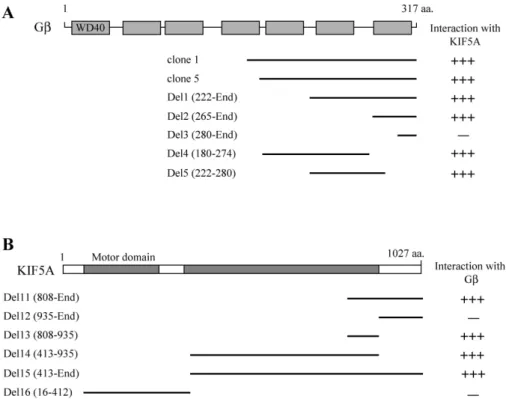
Two positive clones were turned out cDNA fragments con- taining Gβ (Fig. 1A). The two positive Gβ clones (clones 1, and 5) overlapped at the open reading frame (ORF) of Gβ (Fig. 1A). To identify the region of KIF5A required for the interaction with Gβ, a series of deletion mutants of KIF5A was constructed and analyzed their interactions with Gβ using the yeast two-hybrid assay (Fig. 1B). This experi- ment demonstrated that the minimal binding domain was located in a small region of KIF5A corresponding to amino
acids 808-935.
Gβ contains seven consensus WD40 repeat motifs, which seem to function as adaptors and enzyme regulators [19].
To identify the region of Gβ required for the interaction with KIF5A, a series of deletion mutants of Gβ was constructed and analyzed their interactions with KIF5A using the yeast two-hybrid assay. The only one WD40 motif of Gβ inter- acted with KIF5A in the yeast two-hybrid assay (Fig. 1A).
These results indicate that the binding domain was located in WD40 repeat motifs of Gβ.
To clarify whether Gβ interacts specifically with KIF5A or with other KIFs, the tails of KIF1A, KIF5A, KIF5B, KIF5C, and KIF17 were constructed and the interaction of KIFs were tested for binding with Gβ. There was no detectable binding between Gβ and the tail domains of other neuronal KIFs (KIF1A, and KIF17). Gβ interacted with the tail domains of the KIF5A, KIF5B, and KIF5C in the yeast two-hybrid system (Fig. 2A). This result was not surprising in view of the fact that the KIF5A, KIF5B, and KIF5C share extensive similarity in their primary structure (81%-83% identity in the minimal binding domain). These data indicate that Gβ binds specifi- cally to the C-terminal domain of KIF5s. To clarify whether KIF5A interacts specifically with Gβ or with other subunits of heterotrimeric G proteins, Gα, Gβ, and Gγ were tested for binding with KIF5A (Fig. 2B). There was no detectable binding between KIF5A and the other subunits of hetero- trimeric G proteins, such as Gα, and Gγ. These data indicate that KIF5A binds specifically to Gβ. To quantify the binding affinity of KIF5A to Gβ, the bait plasmid of KIF5A and Gβ was transformed to yeast and was measured using β -galactosidase activity in liquid cultures. The interaction of KIF5A with Gβ yielded approximately 412 units of β -galactosidase activity (Fig. 2C), reflecting a binding strength that is sufficient to mediate molecular sorting in vivo [36].
Gβ is associated with KIF5A at the protein level
As an additional demonstration for direct interaction be- tween KIF5s and Gβ, direct interaction between KIF5s and Gβ was assayed suing a GST pull-down experiments.
Recombinant GST-Gα, GST-Gβ or GST-Gγ fusion proteins were expressed in E. coli. The purified GST fusion proteins are allowed to interact with mouse brain extracts. Western blot analyses revealed that KIF5s interacted with GST- Gβ, but not with GST-Gα and GST-Gγ, consistent with the yeast two-hybrid assay results (Fig. 3A).
In order to determine whether the interaction between
Fig. 1. Identification of the proteins interacting with KIF5A by yeast two-hybrid screening. (A) Schematic diagram of Gβ.
The gray box corresponds to WD40 domain of Gβ. Clone 1 and 5 were isolated from the yeast two-hybrid screen and were overlapped at the C-terminal region of Gβ. Different truncations of Gβ were constructed by PCR. Several truncated forms of Gβ were tested in the yeast two-hybrid assay for interaction with KIF5A. aa, the amino acid residue number. +++, interaction with KIF5A; -, no interaction with KIF5A. (B) Minimal Gβ binding region in KIF5A.
KIF5A has motor domain and coiled-coil domain. Motor domain and coiled-coil domains are indicated in gray. Several truncated KIF5As were constructed by PCR and were tested in the yeast two-hybrid assay for interaction with Gβ.
aa, the amino acid residue number. +++, interaction with Gβ; -, no interaction with Gβ.
Fig. 2. Interaction between KIFs and Gβ. (A) The C-terminal regions of each KIF protein were fused to the pLexA DNA binding domain. Gβ specifically interacted with KIF5s but not with KIF1A and KIF17. +++, interaction with Gβ;
-, no interaction with Gβ. (B) Gα, Gβ, and Gγ were fused to the pLexA DNA binding domain. KIF5A specifically
interacted with Gβ but not with Gα or Gγ. +++, interaction with KIF5A; -, no interaction with KIF5A. (C) The strength
of interaction of Gα, Gβ, or Gγ and KIF5A was examined quantitatively using β–galactosidase activity in yeast
two-hybrid reporter assay.
Fig. 3. Association of KIF5s with Gβ in the GST pull-down assay and co-immunoprecipitation. (A) Proteins in the mouse brain lysate were allowed to bind to GST alone, GST- Gα, GST-Gβ and GST-Gγ fusion proteins. The elution fractions were resolved by SDS-PAGE, and Western blotting was performed us- ing an antibody to KIF5A or KIF5B. (B) Mouse brain lysates were immunoprecipitated with an anti-KIF5s antibody or preimmune serum, and then the precip- itates were immunoblotted with anti-Gα, Gβ or Gγ antibodies. Input: 10% of the mouse brain lysates used for each co-immunoprecipitation assay.
KIF5s and Gβ also takes place in vivo, immunoprecipitation analyses were performed. Lysates from mouse brain were incubated with an anti-KIF5s antibody. Protein G-agarose beads selectively precipitated the immuno-complexes, which were then subsequently separated by SDS-PAGE and im- munoblotted with anti-Gα, Gβ, and Gγ antibodies (Fig. 3B).
As shown in Fig. 3B, KIF5 was co-immunoprecipitated with Gβ. These results indicate that Gβ is a specific binding part- ner of KIF5s in vivo.
Discussion
The identification of kinesin-I binding proteins has greatly improved models of kinesin-dependent transport pathways.
Transmembrane and pheripheral membrane proteins serve
as cargo receptors [11,29]. In this study, yeast two-hybrid assays using the C-terminal domain of KIF5A isolated Gβ.
Furthermore, using a combination of yeast two-hybrid anal- ysis, in vitro and in vivo association assays, KIF5s was inter- acted directly with Gβ. The findings of this study provide evidence that Gβ is a candidate the cargo receptor of Kinesin-I. Therefore, it is tempting to speculate that kinesin-I transports the heterotrimeric G proteins attachment vesicle along microtubules to the plasma membrane through its in- teraction with Gβ.
Heterotrimeric G proteins, consisting of Gα, β, and γ subunits, function as signal transducers for the seven trans- membrane helix G protein-coupled receptors (GPCRs) [12,33,42]. In the classical model for G protein signaling, binding of GTP results in activation of the G protein and dissociation of the Gα subunit from the Gβγ subunits [35].
Gβ and Gγ subunits have no transmembrane hydrophobic domain and are synthesized in the cytoplasm. Gβ and Gγ subunits, not modified by lipid, are membrane bound by virtue of its interaction with Gγ subunit [34]. Gγ subunit is processed at the C-terminus to attach an isoprenoid moiety. Subsequently, the C-terminal three amino acids are proteolytically cleaved, and the C-terminus is carboxymethylated. This carboxymethylation is believed to be a membrane-associated event, and the insertion of the attached lipid into the cellular membrane completes the an- choring of Gβγ complex on the inner side of the cellular membrane [46]. Once modified with lipid Gβγ complex is targeted to the ER. Assembly with Gα may also occur on the cytoplasmic surface of the ER. The co-expression of Gβ and Gγ leads to localization to intracellular membranous structures, primarily ER. However, when co-expressed with Gα, Gβ and Gγ localizes primarily to the plasma membrane through an unknown mechanism [22,37,43].
The identification of proteins that bind to the C-terminal
domain of KHC (KIF5s) and KLC has suggested that there
are two mechanisms of linkage. KIF5s and KLC may bind
indirectly, through adaptor proteins linked in their turn to
transmembrane proteins such as GRIP1, Milton and β
-Dystrobrevin or they may bind directly to vesicles through
transmembrane proteins such as Sunday driver (SYD) and
amyloid precursor protein (APP) [1,3,14,20,24,31]. Although
this study did not show the interaction of kinesin-I with oth-
er heterotrimeric G protein isoforms, these observations sug-
gest a mechanism that KIF5s are linked directly to hetero-
trimeric G protein attachment vesicle and could transport
to the plasma membrane similar to SYD and APP in axonal transport. To address this issue, it would be worth to identi- fy the heterotrimeric G proteins containing. In this study, it is proposed that heterotimeric G proteins are a new kine- sin-I receptor protein. The direct interaction between Gβ and KIF5s sheds new light on the mechanisms of vesicle trans- port, giving at the same time one more example of kinesin-I binding to a vesicle receptor.
References
1. Bowman, A. B., A. Kamal, B. W. Ritchings, A. V. Philp, M.
McGrail, J. G. Gindhart, and L. S. Goldstein. 2000.
Kinesin-dependent axonal transport is mediated by the sun- day driver (SYD) protein.
Cell
103, 583-594.2. Brendza, R. P., L. R. Serbus, J. B. Duffy, and W. M. Saxton.
2000. A function for kinesin I in the posterior transport of oskar mRNA and Staufen protein.
Science
289, 2120-2122.3. Brickley, K., M. J. Smith, M. Beck, and F. A. Stephenson.
2005. GRIF-1 and OIP106, members of a novel gene family of coiled-coil domain proteins: association
in vivo
andin vi- tro
with kinesin.J. Biol. Chem.
280, 14723-14732.4. Carson, J. H. and E. Barbarese. 2005. Systems analysis of RNA trafficking in neural cells.
Biol. Cell
97, 51-62.5. Diefenbach, R. J., J. P. Mackay, P. J. Armati, and A. L.
Cunningham. 1998. The C-terminal region of the stalk do- main of ubiquitous human kinesin heavy chain contains the binding site for kinesin light chain.
Biochemistry
37, 16663- 16670.6. Diefenbach, R. J., M. Miranda-Saksena, E. Diefenbach, D.
J. Holland, R. A. Boadle, P. J. Armati, and A. L.
Cunningham. 2002. Herpes simplex virus tegument protein US11 interacts with conventional kinesin heavy chain.
J.
Virol
. 76, 3282-3291.7. Fichera, M., M. Lo Giudice, M. Falco, M. Sturnio, S. Amata, O. Calabrese, S. Bigoni, E. Calzolari, and M. Neri. 2004.
Evidence of kinesin heavy chain (KIF5A) involvement in pure hereditary spastic paraplegia.
Neurology
63, 1108-1110.8. Gindhart, J. G., C. J. Desai, S. Beushausen, K. Zinn, and L.
S. Goldstein. 1998. Kinesin light chains are essential for axo- nal transport in Drosophila.
J. Cell Biol.
141, 443-454.9. Gindhart, J. G. and L. S. Goldstein. 1996. Tetratrico peptide repeats are present in the kinesin light chain.
Trends Biochem.
Sci.
21, 52-63.10. Gorska-Andrzejak, J., R. S. Stowers, J. Borycz, R. Kostyleva, T. L. Schwarz, and I. A. Meinertzhagen. 2003. Mitochondria are redistributed in Drosophila photoreceptors lacking mil- ton, a kinesin-associated protein.
J. Comp. Neurol.
463, 372-388.11. Hirokawa, N. and R. Takemura. 2005. Molecular motors and mechanisms of directional transport in neurons.
Nat. Rev.
Neurosci.
6, 201-214.12. Huber, A. 2001. Scaffolding proteins organize multi- molecular protein complexes for sensory signal transduction.
Eur. J. Neurosci.
14, 769-776.13. Kamal, A. and L. S. Goldstein. 2000. Connecting vesicle transport to the cytoskeleton.
Curr. Opin. Cell Biol
. 12, 503-508.14. Kamal, A., G. B. Stokin, Z. Yang, C. H. Xia, and L. S.
Goldstein. 2000. Axonal transport of amyloid precursor pro- tein is mediated by direct binding to the kinesin light chain subunit of kinesin-I.
Neuron
28, 449-459.15. Kanai, Y., N. Dohmae, and N. Hirokawa. 2004. Kinesin trans- ports RNA: isolation and characterization of an RNA-trans- porting granule.
Neuron
43, 513-525.16. Kanai, Y., Y. Okada, Y. Tanaka, A. Harada, S. Terada, and N. Hirokawa. 2000. KIF5C, a novel neuronal kinesin en- riched in motor neurons.
J. Neurosci.
20, 6374-6384.17. Karcher, R. L., S. W. Deacon, and V. I. Gelfand. 2002.
Motor-cargo interactions: the key to transport specificity.
Trends Cell Biol
. 12, 21-27.18. Kelkar, N., S. Gupta, M. R. Dickens, and J. Divice. 2000.
Interaction of a mitogen-activated protein kinase signaling module with the neuronal protein JIP3.
Mol. Cell Biol.
20, 1030-1043.19. Lambright, D. G., J. Sondek, A. Bohm, N. P. Skiba, H. E.
Hamm, and P. B. Sigler. 1996. The 2.0 A crystal structure of a heterotrimeric G protein.
Nature
379, 311-319.20. Lazarov, O., G. A. Morfini, E. B. Lee, M. H. Farah, A.
Szodorai, S. R. DeBoer, V. E. Koliatsos, S. Kins, V. M. Lee, P. C. Wong, D. L. Price, S. T. Brady, and S. S. Sisodia. 2005.
Axonal transport, amyloid precursor protein, kinesin-1, and the processing apparatus: revisited.
J. Neurosci.
25, 2386- 2395.21. Li, X. J., S. H. Li, A. H. Sharp, F. C. Nucifora Jr, G. Schilling, A. Lanahan, P. Worley, S. H. Snyder, and C. A. Ross. 1995.
A huntingtin-associated protein enriched in brain with im- plications for pathology.
Nature
378, 398-402.22. Marraru, Y., M. Crouthamel, R. Irannejad, and P. B.
Wedegaertner. 2007. Assembly and trafficking of hetero- trimeric G proteins.
Biochemistry
46, 7665-7677.23. Patel, N., D. Thierry-Mieg, and J. R. Mancillas. 1993. Cloning by insertional mutagenesis of a cDNA encoding Caenorhabditis elegans kinesin heavy chain.
Proc. Natl.
Acad. Sci. USA
90, 9181-9185.24. Peters, M. F., K. F. OBrien, H. M. Sadoulet-Puccio, L. M.
Kunkel, M. E. Adams, and S. C. Froehner. 1997. Beta-dystro- brevin, a new member of the dystrophin family.
Identification, cloning, and protein associations.
J. Biol.
Chem.
272, 31561-31569.25. Reid, E., M. Kloos, A. Ashley-Koch, L. Hughes, S. Bevan, I. K. Svenson, F. L. Graham, P. C. Gaskell, A. Dearlove, M.
A. Pericak-Vance, D. C. Rubinsztein, and D. A. Marchuk.
2002. A kinesin heavy chain (KIF5A) mutation in hereditary spastic paraplegia (SPG10).
Am. J. Hum. Genet.
71, 1189-1194.26. Rietdorf, J., A. Ploubidou, I. Reckmann, A. Holmström, F.
Frischknecht, M. Zettl, T. Zimmermann, and M. Way. 2001.
Kinesin-dependent movement on microtubules precedes ac- tin-based motility of vaccinia virus.
Nat. Cell Biol.
3, 992-1000.27. Scholey, J. M., J. Heuser, J. T. Yang, and L. S. Goldstein.
1989. Identification of globular mechanochemical heads of kinesin.
Nature
338, 355-357.28. Seiler, S., J. Kirchner, C. Horn, A. Kallipolitou, G. Woehlke, and M. Schliwa. 2000. Cargo binding and regulatory sites in the tail of fungal conventional kinesin.
Nat. Cell Biol.
2, 333-338.29. Seog, D. H., D. H. Lee, and S. K. Lee. 2004. Molecular Motor Proteins of the Kinesin superfamily proteins (KIFs):
Structure, Cargo and Disease.
J. Korean Medical Science
19, 30. Setou, M., T. Nakagawa, D. H. Seog, and N. Hirokawa. 2000.1-7.Kinesin superfamily motor protein KIF17 and mLin-10 in NMDA receptor-containing vesicle transport.
Science
288, 1796-1802.31. Setou, M., D. H. Seog, Y. Tanaka, Y. Kanai, Y. Takei, M.
Kawagishi, and N. Hirokawa. Glutamate-receptor-interact- ing protein GRIP1 directly steers kinesin to dendrites.
Nature
417, 83-87.32. Skoufias, D. A., D. G. Cole, K P. Wedaman, and J. M.
Scholey. 1994. The carboxyl-terminal domain of kinesin heavy chain is important for membrane binding.
J. Biol.
Chem.
269, 1477-1485.33. Smrcka, A. V. 2008. G protein betagamma subunits: central mediators of G protein-coupled receptor signaling.
Cell Mol.
Life Sci.
65, 2191-2214.34. Sondek, J. and D. P. Siderovski. 2001. Ggamma-like (GGL) domains: new frontiers in G-protein signaling and beta-pro- peller scaffolding.
Biochem. Pharmacol
. 61, 1329-1337.35. Sprang, S. R. 1997. G protein mechanisms: insights from structural analysis.
Annu. Rev. Biochem.
66, 639-678.36. Takeda, S., H. Yamazaki, D. H. Seog, Y. Kanai, S. Terada, and N. Hirokawa. 2000. Kinesin superfamily protein 3 (KIF3) motor transports fodrin-associating vesicles im-
초록:Kinesin-I의 kinesin heavy chains과 직접 결합하는 heterotrimeric G protein의 β subunit의 규명 석 대 현*
(인제대학교 의과대학 생화학교실)
Kinesin-I은 4분자의 단백질로 구성되어 있으며, N-말단의 motor 영역과 C-말단영역을 가지는 장쇄(KHC, 또 한 KIF5s로도 통용) 2분자와 KIF5s (KIF5A, KIF5B와 KIF5C)의 줄기영역과 결합하는 단쇄(KLC) 2분자로 구성되 어 있다. KIF5A의 결합 단백질을 동정하기 위하여 효모 two-hybrid system을 사용하여 특이적으로 결합하는 heterotrimeric G 단백질의 β 단위체 단백질(Gβ)을 분리하였다. Gβ은 KIF5A의 808에서 935아미노산 부위와 결 합하며, 다른 KIF5들과도 결합함을 효모 two-hybrid assay로 확인하였다. 또한 Gβ의 WD40 반복 서열은 KIF5A 와의 결합에 필수영역임을 확인하였으며, 이러한 단백질간의 결합은 Glutathione S-transferase (GST) pull-down assay를 통하여 확인하였다. 생쥐의 뇌 파쇄액에 KIF5들의 항체로 면역침강을 행하여 heterotrimeric G 단백질을 확인한 결과, KIF5들은 heterotrimeric G 단백질과 특이적으로 같이 침강하였다. 이러한 결과들은 kinesin-I는 het- erotrimeric G 단백질이 포함된 소포를 미세소관을 따라 이동시킴을 시사한다.
portant for neurite building.
J. Cell Biol.
148, 1255-1265.37. Takida, S. and P. B. Wedegaertner. 2003. Heterotrimer for- mation, together with isoprenylation, is required for plasma membrane targeting of Gbetagamma.
J. Biol. Chem.
278, 17284-17290.38. Tanaka, Y., Y. Kanai, Y. Okada, S. Nonaka, S. Takeda, A.
Harada, and N. Hirokawa. 1998. Targeted disruption of mouse conventional kinesin heavy chain, kif5B, results in abnormal perinuclear clustering of mitochondria.
Cell
93, 1147-1158.39. Tekotte, H. and I. Davis. 2002. Intracellular mRNA local- ization: motors move messages.
Trends Genet
18, 636-642.40. Vale, R. D. 2003. The molecular motor toolbox for intra- cellular transport.
Cell
112, 467-480.41. Verhey, K. J., D. Meyer, R. Deehan, J. Blenis, B. J. Schnapp, T. A. Rapoport, and B. Margolis. 2001. Cargo of kinesin identified as JIP scaffolding proteins and associated signal- ing molecules.
J. Cell Biol.
152, 959-970.42. Wall, M. A., D. E. Coleman, E. Lee, J. A. Iñiguez-Lluhi, B.
A. Posner, A. G. Gilman, and S. R. Sprang. 1995. The struc- ture of the G protein heterotrimer Gi alpha 1 beta 1 gamma 2.
Cell
83, 1047-1058.43. Wedegaertner, P. B., P. T. Wilson, and H. R. Bourne. 1995.
Lipid modifications of trimeric G proteins.
J. Biol. Chem.
270, 503-506.44. Welte, M. A. 2004. Bidirectional transport along microtubules.
Curr. Biol.
14, 525-537.45. Yu, H., I. Toyoshima, E. R. Steuer, and M. P. Sheetz. 1992.
Kinesin and cytoplasmic dynein binding to brain microsomes.
J. Biol. Chem.
267, 20457-20464.46. Zhang, F. L. and P. J. Casey. 1996. Protein prenylation: mo- lecular mechanisms and functional consequences.