APP Tail 1 (PAT1) Interacts with Kinesin Light Chains (KLCs) through the Tetratricopeptide Repeat (TPR) Domain
Won Hee Jang
1, Sang-Jin Kim
2, Young Joo Jeong
1, Hee Jae Jun
3, Il Soo Moon
4and Dae-Hyun Seog
1*
1
Departments of Biochemistry,
2Departments of Neurology,
3Departments of Thoracic & Cardiovascular Surgery, College of Medicine, Inje University, Busan 614-735, Korea
4
Departments of Anatomy & Dongguk Medical Institute, College of Medicine, Dongguk University, Gyeongju 780-714, Korea
Received November 6, 2012 /Revised November 20, 2012 /Accepted November 21, 2012A conventional kinesin, KIF5/Kinesin-I, transports various cargoes along the microtubule through in- teraction between its light chain subunit and the cargoes. Kinesin light chains (KLCs) interact with many different cargoes using their tetratricopeptide repeat (TPR) domain, but the mechanism under- lying recognition and binding of a specific cargo has not yet been completely elucidated. We used the yeast two-hybrid assay to identify proteins that interact with the TPR domain of KLC1. We found an interaction between the TPR domain of KLC1 and an amyloid precursor protein (APP)-binding protein PAT1 (protein interacting with APP tail 1). The yeast two-hybrid assay demonstrated that the TPR domain-containing region of KLC1 mediated binding to the C-terminal tail region of PAT1. PAT1 also bound to KLC2 but not to kinesin heavy chains (KIF5A, KIF5B, and KIF5C) in the yeast two-hy- brid assay. These protein-protein interactions were also observed in the glutathione S-transferase (GST) pull-down assay and by co-immunoprecipitation. Anti-PAT1 antibody as well as anti-APP anti- body co-immunoprecipitated KLC and KHCs associated with PAT1 from mouse brain extracts. These results suggest that PAT1 could mediate interactions between Kinesin-I and APP containing vesicles.
Key words : Microtubule motors, Kinesin-I, PAT1, APP, adaptor proteins, protein-protein interaction
*Corresponding author
*Tel:+82-51-890-6974, Fax:+82-51-894-5801
*E-mail : [email protected]
This is an Open-Access article distributed under the terms of the Creative Commons Attribution Non-Commercial License (http://creativecommons.org/licenses/by-nc/3.0) which permits unrestricted non-commercial use, distribution, and reproduction in any medium, provided the original work is properly cited.
Journal of Life Science 2012 Vol. 22. No. 12. 1608~1613 DOI : http://dx.doi.org/10.5352/JLS.2012.22.12.1608
Introduction
Molecular motors (kinesin, myosin, and dynein) drive ac- tive transport processes within neuron [5,7]. Kinesins are a family of molecular motor proteins that move along micro- tubules to transport membrane vesicles, organelles and pro- tein complexes. Kinesin-I is the well-studied member of ki- nesin superfamily (KIF) [6]. Kinesin-I performs multiple dis- tinct transport functions within the same cell. Kinesin-I is a tetrameric protein composed of two heavy chains (KHCs) and two light chains (KLCs) [6,7]. The amino (N)-terminal heptad repeat regions of KHCs bind KLCs. In mice, both the KHCs (KIF5A, KIF5B, and KIF5C) and the KLCs (KLC1, KLC2) are encoded by different genes and distinct ex- pression patterns [12,17]. KIF5A, KIF5C, and KLC1 are ex- pressed in neuron, whereas KIF5B and KLC2 are ubiq- uitously expressed [6,12]. The KHCs consist of three do-
mains: the N-terminal motor domain that contains the mi- crotubule binding domain, the central coiled-coil domain re- sponsible for dimerization of KHC and KLC, and the carbox- yl (C)-terminal tail domain [6,7]. Also, the KLC consists of three domains: the N-terminal coiled-coil domain that binds to KHC, the central tetratripeptide repeat (TPR) domain, and the C-terminal domain. The TPR domain of KLC is known to be involved in the binding of cargoes [2,17]. Two isoforms of KLC exist in mice: KLC1, and KLC2. The KLC1 isoform is highly expressed in neuron and binds to several proteins [17]. The TPR domains shows very high sequence similarity between the two isoforms [17] and understandably, the KLC2 isoform shares several, but not all, cargo proteins with KLC1 [6,10,21].
The TPR domain is known as a protein-protein interaction
motif, which consists of multiple tandem repeats of 34 amino
acids [21]. The structure of TPR domain reveals a he-
lix-turn-helix arrangement for each TPR repeat and a super-
helical conformation of multiple TPR repeats [2]. The first
protein identified to bind to Kinesin-I through the TPR do-
main was the c-Jun NH
2-terminal kinase (JNK)-interacting
protein (JIP) group of scaffold proteins [16,19]. Many differ-
ent types of cargoes moved by Kinesin-I have been identi-
fied, including apolipoprotein E receptor 2 (ApoER2), amy-
loid precursor protein (APP), mitochondria, and mRNA granules [3,6,11]. In some cases, these cargoes bind to soluble adaptor proteins/scaffolding proteins that mediate the at- tachment of kinesins to cargoes [6,18]. Understanding how Kinesin-I becomes linked to particular cargoes and decipher- ing the regulatory mechanism for vesicle transport remain to be unsolved question. In this study, we screened for pro- teins that interact specifically with KLC1, and found protein interacting with APP tail 1 (PAT1), which interacts with APP intracellular domain [20]. The Kinesin-I and PAT1 inter- action suggests that PAT1 contributes as an adaptor pro- tein/scaffolding protein between Kinesin-I and APP contain- ing vesicles.
Materials and Methods Plasmid constructs
Full-length mouse KLC1 (accession NM_001081959), KLC2 (accession NM_008451), and PAT1 (accession NM_025825) were amplified by polymerase chain reaction (PCR) from Marathon-Ready
TMcDNA library (Clontech, Palo Alto, CA, USA) and cloned into pGEM T-easy vector (Promega Corp, Madison, WI, USA). The TPR domain of KLC1 was utilized as a template to amplify the region cod- ing for amino acids 80-541 using the appropriate primers [17]. The amplified fragment was cloned into pGEM T-easy vector (Promega). The resulting recombinant plasmid was then cut with EcoRI and XhoI and the insert was subcloned into pLexA (Clontech) and pJG4-5 (Clontech).
Screening of KLC-binding proteins by yeast two-hybrid assay
The Matchmaker LexA two-hybrid system was used for screening according to the manufacturer’s manual (Clontech). In brief, pLexA-KLC1-TPR was transformed into yeast strain EGY48 carrying the p8op-lacZ gene.
Transformed cells were transformed with the mouse brain cDNA library [13] and grown on synthetic dextrose (SD) plates supplemented with glucose but with no histidine, tryptophan, or uracil (SD/-His/-Trp/-Ura). The selection of positive clones was performed on an SD/-His/-Trp/-Ura/
-Leu plate containing galactose, raffinose, X-gal, and BU salts. Plasmids from positive clones were analyzed by re- striction digestion. Unique inserts were sequenced and pro- tein sequence analysis was performed with the BLAST algo- rithm at the National Center for Biotechnology Information
(NCBI). Sequence-verified clones were tested again for inter- actions of with the bait in yeast by the retransformation.
β -Galactosidase activity in liquid cultures of yeast
The β-galactosidase activity of yeast was assayed as de- scribed previously [13]. Mid-log phase yeast cells were col- lected and permeabilized with 0.1% sodium dodecyl sul- phate (SDS) and chloroform. An excess amount of o-nitro- phenyl-β-D-galactoside (ONPG) was added to yeast lysate, and the mixture was incubated at 30℃, and then the reaction was stopped by increasing pH to 11 by the addition of 1 M Na
2CO
3. The formation of the reaction product, o-nitro- phenol, was determined by measuring absorbance at 420 nm on a spectrophotometer and normalizing for the reaction time. The units of enzyme activity were calculated by the following equation: units=1000×[(OD
420-1.75×OD
550)]/
(reaction time x culture volume x OD
600). All experiments were independently performed at least three times [1].
Co-immunoprecipitation and Immunoblot analysis
Mouse brains were homogenized in ice-cold homoge- nization buffer (0.32 M sucrose, 4 mM HEPES, pH 7.3) sup- plemented with protease inhibitor cocktail (Sigma-Aldrich, St. Louis, MO, USA). For immunoprecipitation, mouse brain lysate was diluted in the same volume of 2X binding buffer (50 mM HEPES, 200 mM KCl, 0.2% Triton X-100, pH 7.0) and incubated with anti-APP antibody (Santacruz Biotechnology, Santa Cruz, CA, USA), monoclonal an- ti-PAT1 antibody (BD Transduction Laboratories, San Jose, CA, USA), or control IgG overnight at 4
oC, followed by pre- cipitation with protein-A Sepharose (Amersham Pharmacia, Piscataway, NJ, USA). The beads were collected by brief cen- trifugation and washed three times with TBS-T (20 mM Tris-HCl, pH 7.5, 0.2 M NaCl, 0.1% Tween 20). The washed beads were resuspended with Laemmli
’s loading buffer and the proteins were eluted and denatured by boiling for 2 mi- nutes and then separated by SDS-PAGE. The proteins were transferred from the gel to a nitrocellulose membrane and incubated with anti-KLC (Millipore, Billerica, MA, USA), an- ti-KIF5B, and anti-KIF5C antibodies [12].
Glutathione S-transferase (GST) pull-down assays
cDNA encoding the full length PAT1 [20] was cloned in
pET41a. The recombinant GST-PAT1 fusion protein was ex-
pressed in bacterial strain BL21 GOLD (Stratagene, La Jolla
CA, USA) after induction with 0.5 mM isopropyl thio-β
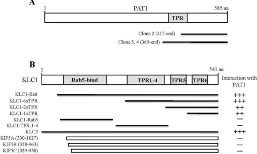
Fig. 1. Identification of the proteins interacting with KLC1 by yeast two-hybrid assay. (A) Schematic diagram of PAT1. The gray box corresponds to TPR domain of PAT1. Clones 2, 3, and 4 were isolated from the yeast two-hybrid screen and were overlappedat the C-terminal region of PAT1. aa, the amino acid residue number. (B) Minimal PAT1 binding region in KLC1.
KLC1 has Rab5-bind domain and the TPR domains. Rab5-bind domain and the TPR domains are indicated in gray. Several truncated forms of KLC1, KLC2, and KHCs were constructed by PCR and were tested in the yeast two-hybrid assay for interaction with PAT1. aa, the amino acid residue number.++ or +++, interaction with PAT1; -, no interaction with PAT1.
-D-galactopyranoside (IPTG) for 3 hr. The fusion proteins were purified using glutathione-agarose beads (Sigma- Aldrich) according to the manufacturer’s protocol. The mouse brain S2 fraction was incubated overnight at 4
oC with the GST fusion protein-coupled glutathione beads. The beads were pelleted by centrifugation, washed three times with the extraction buffer (1% Triton X-100 in PBS containing 10 μg/ml each aprotinin, leupeptin, and pepstatin and 1 μM phenylmethanesulfonyl fluoride), and once with PBS. The bound proteins were eluted from the glutathione beads with 100 μl of Laemmli
’s loading buffer. The pulled-down pro- teins were analyzed by Western blotting with anti-KIF5B, anti-APP, and anti-KLC antibodies [18].
Results
Identification of KLC1 interacting proteins by yeast two-hybrid assay
To identify KLC1-interacting proteins, we used the TPR domain containing region (aa 80-541) of KLC1 fused to the DNA-binding domain of pLexA as a bait (Fig. 1B) and iso- lated positive clones from a mouse brain cDNA library.
From 5×10
6colonies screened, we obtained 4 positive clones. Three positive clones (clone 2, 3, and 4) turned out
to possess PAT1 cDNA fragments (Fig. 1A). The clones overlapped at the open reading frame (ORF) of PAT1 and possessed cDNA fragments corresponding to the C-termi- nal region of PAT1 (Fig. 1A). PAT1 has been reported to interact with the APP, the precursor protein of amyloid be- ta peptide (Aβ), which is the component of plaques in Alzheimer’s disease [20]. The C-terminal region of PAT1 is contained the TPR domain that is also found in KLCs from all the species studied to date. From the isolated positive clones, KLC1 was found not to interact with the TPR do- main of PAT1 (Fig. 1A). KLC1 is composed of several pro- tein-protein interaction domains, six TPR domains and one Rab5-binding domain (Fig. 1B) [17]. To determine the mini- mal binding domain of KLC1 that is required for the inter- action with PAT1, we constructed several deletion mutants of KLC1. Yeast two-hybrid assays showed that the minimal domain required for binding was dependent on the sixth TPR domain of KLC1 (Fig. 1B).
Next we investigated whether KHCs (KIF5A, KIF5B, and
KIF5C) and KLC2 interact with PAT1. As shown in Fig. 1B,
the C-terminal fragments of KHCs did not interact with
PAT1 but KLC2 bound to PAT1. Together, these results
show that the interaction between KLCs and PAT1 is medi-
ated through the TPR domain containing region of KLC1.
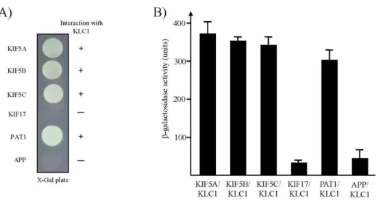
Fig. 2. Interaction between KHCs or APP and KLC1. (A) The C-terminal regions of each KHC protein and APP were fused to the pLexA DNA binding domain. KLC1 specifically interacted with PAT1 and KHCs but not with APP and KIF17. +, inter- action; -, no interaction. (B) The strength of interactions between KHCs, KIF17, PAT1, or APP and KLC1 were examined quantitatively using β-galactosidase activity in yeast two-hybrid reporter assay.
When the cytoplasmic region of APP was tested for KLC1-binding by the color assay, there was no detectable binding between KLC1 and APP (Fig. 2A). A quantitative β -galactosidase assay showed that KLC1 directly bound to PAT1 and KHCs (Fig. 2B). These data indicate that the inter- action of KLC1 with PAT1 is specific.
PAT1 is associated with Kinesin-I
PAT1 has been known to interact with APP [20].
Therefore, we next determined whether PAT1 interact spe- cifically with KLC and whether the interaction includes APP at the protein level using GST pull-down experiments.
Recombinant GST-PAT1 fusion protein was expressed in E.
coli. The purified GST fusion protein was allowed to interact with mouse brain lysates. Immunoblotting analyses revealed that KLC and APP interacted with GST-PAT1, but not with GST (Fig. 3A).
To address the question whether the direct binding of KLC to PAT1 mediates the interaction between Kinesin-I and APP, we performed co-immunoprecipitation analyses.
Lysates from mouse brain were incubated with anti-PAT1 antibody or anti-APP antibody. Protein G-agarose beads selectively precipitated the immuno-complexes, which were then subsequently separated by SDS-PAGE and immunoblotted with anti-KLC, anti-KIF5B, and anti-KIF5C antibodies (Fig. 3B). As shown in Fig. 3B, both anti-PAT1 and anti-APP antibodies efficiently precipitated the
Fig. 3. Association of KLCs with PAT1 in the GST pull-down assay and co-immunoprecipitation. (A) Proteins in the mouse brain lysate were allowed to bind to GST alone and GST-PAT1 fusion proteins. The elution fractions were resolved by SDS-PAGE, and Western blotting was performed using anti-KLC, KIF5B, or APP antibodies. (B) Mouse brain lysates were immunoprecipitated with an- ti-PAT1, APP antibodies, or preimmune serum, and then the precipitates were immunoblotted with anti-KLC, KIF5B, or KIF5C antibodies.
Kinesin-I complex. This result suggests that the attach- ment of APP to Kinesin-I is mediated by KLC-PAT1 interaction.
Discussion
In this study, we have shown that KLC1 can associate
with PAT1, a protein that specifically binds to APP. Using
the TPR domain containing region of KLC1 as bait, we
identified PAT1 in a yeast two-hybrid assay of a mouse
brain cDNA library. Only sixth TPR domain of KLC1 in- teracts with PAT1. Furthermore, using a combination of GST pull-down assay and co-immunoprecipitation, we confirmed that PAT1 interacted with KLC at the protein level. Moreover, we showed that Kinesin-I complex can be co-precipitated with APP. Although we did not de- termine the specific character of APP containing vesicles, these results suggest that Kinesin-I transports APP con- taining cargoes through the interaction between KLCs and PAT1.
PAT1 was first identified in human cells. It has been re- ported that PAT1 was identified to be a cytoplasmic protein, associated with membranes, cofractionated with APP-con- taining vesicles, and interacted with microtubule in an ATP-sensitive manner. PAT1 was shown to enhance endocy- tosis of APP in neurons [5,20]. In light of the homology be- tween the TPR domains of PAT1 and KLCs, KLCs-like func- tion of PAT1 has been proposed [20]. However, the sequence and structure similarities between the N-terminal regions of PAT1 and KLCs are low [20]. As described in this work, PAT1 did not directly interact with KHCs (KIF5A, KIF5B, and KIF5C) by yeast two-hybrid assay. These data indicated that PAT1 has no KLC-like function.
The TPR domains of KLCs are highly conserved across species, and the TPR domains are known to be involved in protein-protein interactions [2]. Several Kinesin-I cargo mol- ecules have been shown to interact with the TPR domain of KLCs [3,18]. For example, the c-Jun N-terminal kinase-in- teracting protein 1 (JIP1), a Kinesin-I cargo-adaptor mole- cule, interacts with the TPR domain of KLC [16,19]. In this study, it was shown that PAT1 interacted with the TPR do- main of KLC1.
In recent reports, many cargoes that bind to Kinesin-I through the TPR bundle have been identified, including Huntington-associated protein-1, Calsyntenin, collapsing re- sponse mediator protein-2, APP, torsinA, 14-3-3 and Kidins220 [8,9,10,14,15]. Many intracellular cargoes are transported by Kinesin-I. How can one motor mediate the transport of many cargoes? One possibility is that these dif- ferent cargoes may use different soluble adaptor proteins that mediate the attachment of Kinesin-I to mem- brane-bound cargoes. Based on the ability of PAT1 to bind both APP and KLCs, we favor model that PAT1 functions as an adaptor that mediates the attachment of APP contain- ing vesicles to KLCs for transport by Kinesin-I. Although we did not determine the characteristics of KLCs-PAT1 asso-
ciated vesicles, the available data strongly suggest that Kinesin-I mediate the transport of Golgi-derived APP-con- taining vesicles to the cell surface.
Acknowledgment
This research was supported by Basic Science Research Program though the National Research Foundation of Korea (NRF) funded by the Ministry of Education, Science and Technology (2011-0026579).
References
1. Ausubel, F. M., Brent, R., Kingston, R. E., Moore, D. D., Seidman, J. G., Smith, J. A. and Struhl, K. 1998.
Current Protocols in Molecular Biology
. John Wiley & Sons.2. Blatch, G. L. and Lässle, M. 1999. The tetratricopeptide re- peat: a structural motif mediating protein-protein interactions.
Bioessays
21, 932-939.3. Bowman, A. B., Kamal, A., Ritchings, B. W., Philp, A. V., McGrail, M., Gindhart, J. G. and Goldstein, L. S. 2000.
Kinesin-dependent axonal transport is mediated by the sun- day driver (SYD) protein.
Cell
103, 583-594.4. Bracale, A., Cesca, F., Neubrand, V. E., Newsome, T. P., Way, M. and Schiavo, G. 2007. Kidins220/ARMS is trans- ported by a kinesin-1-based mechanism likely to be in- volved in neuronal differentiation.
Mol. Biol. Cell
18, 142-152.5. Brady, S. T. 1985. A novel brain ATPase with properties expected for the fast axonal transport motor.
Nature
317, 73-75.6. Hirokawa, N. and Takemura, R. 2005. Molecular motors and mechanisms of directional transport in neurons.
Nat. Rev.
Neurosci
. 6, 201-214.7. Hirokawa, N. 1998. Kinesin and dynein superfamily pro- teins and the mechanism of organelle transport.
Science
279, 519-526.8. Ichimura. T., Wakamiya-Tsuruta, A., Itagaki, C., Taoka, M., Hayano, T., Natsume, T. and Isobe, T. 2002. Phosphorylation- dependent interaction of kinesin light chain 2 and the 14-3-3 protein.
Biochemistry
41, 5566-5572.9. Kamal, A., Stokin, G. B., Yang, Z., Xia, C. H. and Goldstein, L. S. 2000. Axonal transport of amyloid precursor protein is mediated by direct binding to the kinesin light chain sub- unit of kinesin-I.
Neuron
28, 449-459.10. Kamm, C., Boston, H., Hewett, J., Wilbur, J., Corey, D. P., Hanson, P. I., Ramesh, V. and Breakefield, X. O. 2004. The early onset dystonia protein torsinA interacts with kinesin light chain 1.
J. Biol. Chem
. 279, 19882-19892.11. Kanai, Y., Dohmae, N. and Hirokawa, N. 2004. Kinesin transports RNA: isolation and characterization of an RNA-transporting granule.
Neuron
43, 513-525.12. Kanai, Y., Okada, Y., Tanaka, Y., Harada, A., Terada, S. and Hirokawa, N. 2000. KIF5C, a novel neuronal kinesin en-
초록:APP tail 1 (PAT1)과 kinesin light chains (KLCs)의 tetratricopeptide repeat (TPR) domain을 통한 결합
장원희
1․김상진
2․정영주
1․전희재
3․문일수
4․석대현
1*
(인제대학교 의과대학
1생화학교실,
2신경과학교실,
3흉부외과학교실,
4동국대학교 의과대학 해부학교실)
KIF5/Kinesin-I는 경쇄(light chain)를 통하여 결합함으로써 다양한 운반체들을 미세소관을 따라 운반한다. Kinesin light chains (KLCs)은 tetratricopeptide repeat (TPR) 영역을 매개로 운반체와 결합한다. 현재까지 KLCs와 결합하는 많 은 운반체들이 확인되었으나 KLCs가 어떻게 특정운반체를 인식하여 결합하는지는 아직 확실히 밝혀지지 않았다. 본 연구에서 KLC1의 TPR 영역과 결합하는 단백질을 분리하기 위하여 효모 two-hybrid system을 이용하여 탐색한 결과 amyloid precursor protein (APP)과 결합하는 것으로 보고된 protein interacting with APP tail 1 (PAT1)을 분리하였다.
KLC1은 PAT1의 C-말단 부위와 결합하며, PAT1은 KLC1의 TPR 영역을 포함한 부위와 결합함을 효모 two-hybrid as- say로 확인하였다. 또한 PAT1는 KLC2와도 결합하였지만 kinesin heavy chains (KHCs)인 KIF5A, KIF5B, KIF5C와는 결합하지 않았다. 단백질간 결합은 glutathione S-transferase (GST) pull-down assay와 공동면역침강으로도 확인하였 다. 생쥐의 뇌 파쇄액을 PAT1 항체와 APP 항체로 면역침강을 행한 결과 KLC와 KHCs가 같이 침강하였다. 이러한 결과 들은 PAT1이 Kinesin-I와 APP 포함 소포간의 상호작용을 매개한다는 것을 시사한다.
riched in motor neurons.
J. Neurosci
. 20, 6374-6384.13. Kim, S. J., Lee, C. H., Park, H. Y., Yea, S. S., Jang, W. H., Lee, S. K., Park, Y. H., Cha, O. S., Moon, I. S. and Seog, D. H. 2007. JSAP1 interacts with kinesin light chain 1 through conserved binding segments.
J. Life Sci.
17, 889-895.14. Konecna, A., Frischknecht, R., Kinter, J., Ludwig, A., Steuble, M., Meskenaite, V., Indermühle, M., Engel, M., Cen, C., Mateos, J. M., Streit, P. and Sonderegger, P. 2006.
Calsyntenin-1 docks vesicular cargo to kinesin-1.
Mol. Biol.
Cell
17, 3651-3663.15. McGuire, J. R., Rong, J., Li, S. H. and Li, X. J. 2006.
Interaction of Huntingtin-associated protein-1 with kinesin light chain: implications in intracellular trafficking in neurons.
J. Biol. Chem.
281, 3552-3559.16. Muresan, Z. and Muresan, V. 2005. Coordinated transport of phosphorylated amyloid-beta precursor protein and c-Jun NH2-terminal kinase-interacting protein-1.
J. Cell Biol
. 171, 615-625.17. Rahman, A., Friedman, D. S. and Goldstein, L. S. 1998. Two kinesin light chain genes in mice. Identification and
characterization of the encoded proteins.
J. Biol. Chem
. 273, 15395-15403.18. Setou, M., Seog, D. H., Tanaka, Y., Kanai, Y., Takei, Y., Kawagishi, M. and Hirokawa, N. 2002. Glutamate-re- ceptor-interacting protein GRIP1 directly steers kinesin to dendrites.
Nature
417, 83-87.19. Verhey, K. J., Meyer, D., Deehan, R., Blenis, J., Schnapp, B. J., Rapoport, T. A. and Margolis, B. 2001. Cargo of kinesin identified as JIP scaffolding proteins and associated signal- ing molecules.
J. Cell Biol.
152, 959-970.20. Zheng, P., Eastman, J., VandePol, S. and Pimplikar, S. W.
1998. PAT1, a microtubule-interacting protein, recognizes the basolateral sorting signal of amyloid precursor protein.
Proc. Natl. Acad. Sci. USA
95, 14745-14750.21. Zhu, H., Lee, H. Y., Tong, Y., Hong, B. S., Kim, K. P., Shen, Y., Lim, K. J., Mackenzie, F., Tempel, W. and Park, H. W.
2012. Crystal structures of the tetratricopeptide repeat do- mains of kinesin light chains: insight into cargo recognition mechanisms.