pISSN 1229-3008 eISSN 2287-6251
Progress in Superconductivity and Cryogenics
Vol.19, No.1, (2017), pp.9~12 https://doi.org/10.9714/psac.2017.19.1.009
```
1. INTRODUCTION
In this study, a new gene transfer method in combination of jet injection and magnetic transfection that can be used for DNA vaccine was examined.
Vaccine is a biological preparation to improve immunity toward particular pathogens. Recent research on vaccination has expanded from infections to chronic diseases [1]. Originally antigens derived from viral and bacterial pathogens were used in vaccination for infections.
In treatment for diseases such as cancer, hypertension and Alzheimer's disease, some important mediators that aggravates the diseases are targeted as antigens for example angiotensin II to hypertension [2].
Recently, researches and developments of DNA vaccines have been extensively conducted. It is a new approach to immunotherapy in some clinical trials. In DNA vaccination, genes encoding antigens are injected into the tissues, and then the antigens expressed in patients’ bodies induces immunity. DNA vaccines have unique advantages toward peptide vaccines. DNA vaccines using plasmid DNA (pDNA) is free from infection risk, and also easy to develop, manufacture and transport. In this study, we aim to apply DNA vaccination into treatment for rheumatoid arthritis. Rheumatoid arthritis is one of the autoimmune diseases. Interleukin 17A (IL-17A) is supposed to be one of the important mediators that aggravate rheumatoid arthritis [3]. We examined DNA vaccination to improve
IL-17A-specific immune response.
The effectiveness of DNA vaccines depends on gene transfer efficiency. In order to establish safe, simple and effective gene transfer method, we proposed a hybrid method of jet injection and magnetic transfection. The jet injection is a method to inject medical liquid by momentary high pressure without needle. The injected liquid diffuses in the bio tissue and the endocytosis is considered to be improved by the diffusion. In Jet injection, slight and recoverable cell membrane injuries are given to the cells by injection power, and thus genes can permeate into cells through the injuries. More injuries were given on cell membranes after jet injection. The magnetic transfection, on the other hand, is a method to deliver the conjugates of plasmid DNA and magnetic particles to the desired site by external magnetic field. Genes combined with magnetic particles are pulled by the magnetic field, which can increase permeation of the genes into the cells. In practical use, the superconducting bulk magnet will be useful, because DNA vaccines are required to diffuse in the horizontal direction in the skin and the superconducting magnet can generate strong magnetic field in wide area.
From above features of two methods, it is expected that jet injection of the conjugates causes slight membrane disruptions and the traction of the conjugates by magnetic field induces the efficient gene transfer. In this study, we examined gene transfer efficiency and immune inductivity using the hybrid method of jet injection and magnetic transfection.
Fundamental study on gene transfer utilizing magnetic force and jet injector
T. Hasegawaa, H. Nakagamib, Y. Akiyama*, a, and S. Nishijimaa
a Graduate School of Engineering, Osaka University, Yamadaoka 2-1, Suita, Osaka 565-0871, Japan
b Graduate School of Medicine, Osaka University, Yamadaoka 2-2, Suita, Osaka 565-0871, Japan
(Received 22 March 2017; revised or reviewed 27 March 2017; accepted 28 March 2017)
Abstract
Recently, DNA vaccination is attracting attentions as a new therapeutic method for lifestyle diseases and autoimmune diseases.
However, its clinical applications are limited because a safe and efficient gene transfer method has not been established yet. In this study, a new method of gene transfer was proposed which utilizes the jet injection and the magnetic transfection. The jet injection is a method to inject medical liquid by momentary high pressure without needle. The injected liquid diffuses in the bio tissue and the endocytosis is considered to be improved by the diffusion. The magnetic transfection is a method to deliver the conjugates of plasmid DNA and magnetic particles to the desired site by external magnetic field. It is expected that jet injection of the conjugates causes slight membrane disruptions and the traction of the conjugates by magnetic field induces the efficient gene transfer. In conclusion, the possibility of improvement of the gene expression by the combination of jet injection and magnetic transfection was confirmed.
Keywords: DNA vaccination, jet injection, magnetic force control, gene transfer
* Corresponding author: [email protected]
Fundamental study on gene transfer utilizing magnetic force and jet injector
2. METHODS
2.1. Magnetic field sources and magnetic particles
We used three kinds of magnets; ferrite magnet, neodymium magnet and Halbach array magnet as magnetic field sources for magnetic transfection. TABLE I shows surface magnetic flux density and geometry of the magnets.
In use of ferrite magnet, 3 magnets of φ8 × 5mm were stacked so as to be the same geometry of the neodymium magnet. Surface magnetic flux density was measured by a handy tesla meter (TM-701, KANETEC CO., LTD., Japan).
Fig. 1 shows the arrangements of the magnets in the magnetic transfection experiment. The magnet was placed on the injection site immediately after injection of the conjugates of plasmid DNA and magnetic particles by jet injection as the procedure shown in section 2.3 and 2.4.
As the magnetic particles, magnetite (Fe3O4) nanoparticles were used in this study. The magnetite nanoparticles are suitable for clinical application because it is used as the main component of contrast agent for MRI (Magnetic Resonance Imaging). Here, positively-charged
TABLE I
MAGNETIC PROPERTYES AND GEOMETRIES OF MAGNETS. Magnetic field sourse Surface magnetic
flux density [T] Size [mm]
Ferrite magnet 0.15 φ8 × 15
Neodymium magnet 0.5 φ8 × 15
Halbach array magnet 0.9 230 × 128 × 48
Fig. 1. The arrangement of the magnets in the magnetic transfection experiments; (a)Ferrite or Neodymium magnet was placed in a hole in a wooden board and rat was laid on it. (b)Rat was laid on a particular position of Halbach array magnet.
magnetite particles by coating with amino groups that were commercially produced (SPM-NH2, Opto Science. Inc) were used in order to form conjugates with negatively-charged plasmid DNA. The primary particle diameter of magnetite was 170 nm. The particle size distribution was measured by dynamic light scattering method (FOQELS, Nihon Rufuto Co., Ltd.)
2.2. Treatment of animals in experiment
Female SD (Sprague Dawley® ) rats aged 7 weeks were purchased from Charles River Laboratories Japan, Inc.
(Yokohama, Japan) and were housed in a temperature- and light cycle-controlled environment. Animals had food and water ad libitum in facility. All experiments were conducted according to the protocol approved by the Ethical Committee for Animal Experiments of the Osaka University Graduate School of Medicine.
In order to minimize suffering, experimental treatments including hair removal, injection of DNA and sacrifice were conducted humanely under anesthesia. Animals were given inhalation anesthesia and injected an anesthetic.
Isoflurane (Pfizer Inc., USA) was used as inhalation anesthesia. The anesthetic was prepared by mixing 2 ml of Dormicam (Astellas Pharma Inc., Japan), 5 ml of Vetorphale (Meiji Seika Pharma Co., Ltd., Japan), 2 ml of Domitor (Nippon Zenyaku Kogyo Co., Ltd., Japan) and 9 ml of physiological saline (Otsuka Pharmaceutical Factory, Inc., Japan). The anesthetic was injected into rats’
abdominal cavities in the amount of 5 μl per 1 g of body weight.
2.3. Evaluation of the gene expression
Firstly, the gene expression was evaluated by the activity of luciferase, a luminous enzyme. Prior to the experiment, the body hair of rats on their backs were removed by the depilatory cream and razor. Luciferase-coding pDNA (pGL3) solutions including magnetite particles (SPM-NH2, NANOBRICK Co., Ltd, Korea) were injected into rats.
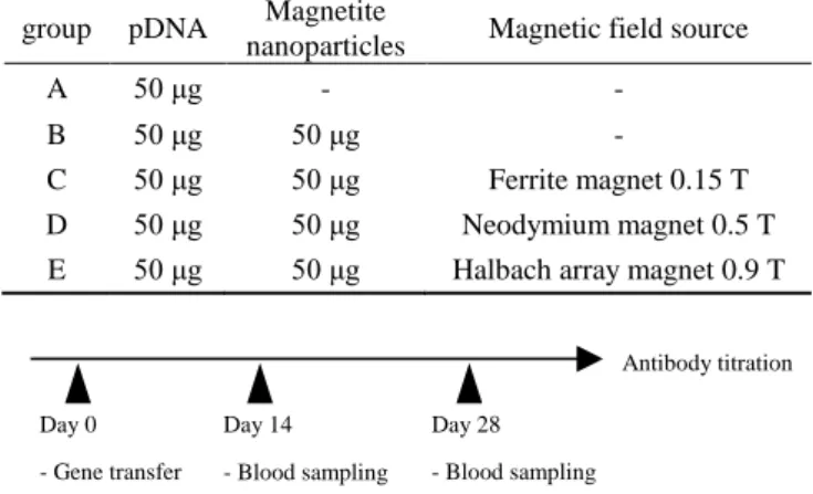
Immediately after injection, magnet was placed on the injection site for 30 minutes to apply magnetic field, so as to the magnetic field applying point is just under the administration point. The experiments were conducted for 5 conditions shown in TABLE II (N=5 for each condition;
A to E). After 1 day of culture, rats were sacrificed under anesthesia and skins of the injection point were harvested into 1 cm square. Harvested skin was cut into small pieces and dissolved into 1.5 ml of lysis buffer, repeated freezing-thawing at 3 times, and then centrifuged (4°C, 12000 rpm, 5 min). The supernatants were assayed with a commercial luciferase assay kit (luciferase assay system, Promega, USA). Luminescence was measured by a luminometer (Lumat LB9507, Berthold Japan K.K., Japan).
Protein in supernatant was determined with DC protein assay kit (Bio-Rad, USA). Luciferase activity was normalized to each protein mass in supernatant and evaluated as relative light unit (RLU), i.e. luminescent strength per mg protein.
(a)
(b) 10
T. Hasegawa, H. Nakagami, Y. Akiyama, and S. Nishijima
TABLE II
CONDITIONS OF REAGENT COMPOSITION AND MAGNETIC FIELD. group pDNA Magnetite
nanoparticles Magnetic field source
A 50 μg - -
B 50 μg 50 μg -
C 50 μg 50 μg Ferrite magnet 0.15 T
D 50 μg 50 μg Neodymium magnet 0.5 T
E 50 μg 50 μg Halbach array magnet 0.9 T
Day 0 - Gene transfer
Day 14 - Blood sampling - Gene transfer
Day 28 - Blood sampling
Antibody titration
Fig. 2. Immunization schedule.
TABLE III
CONDITIONS ABOUT REAGENT COMPOSITION AND MAGNETIC FIELD. pDNA Magnetite
nanoparticles Magnetic field Number of samples
50 μg - - 3
50 μg 50 μg - 4
50 μg 50 μg 0.15 T 4
50 μg 50 μg 0.5 T 4
2.4. Evaluation of immune response
Next, induction of immune response by the hybrid method of jet injection and magnetic transfection was examined. The immunization schedule is shown in Fig. 2.
Female SD rats aged 8 weeks were immunized with 50 μl of reagents containing pDNA encoding HBc-IL-17A and magnetite nanoparticles through the skin utilizing jet injector (ShimaJET® ) at two sites per one time, two times each two weeks. Immediately after injection, magnet was placed on the injection site for 30 minutes to apply magnetic field. Experimental conditions of reagent composition per one injection and magnetic field are shown in TABLE III. A 400 μl blood sample per a rat was collected in a capillary blood collection tube (CAPIJECT® , TERUMO CORPORATION). Blood sampling was conducted three times for each two weeks from the first vaccination to day 28. Serums were separated by centrifugation (25°C, 10000 rpm, 5 min) and stored at -80°C until assay.
The serum samples were assayed for anti-IL-17A antibodies titer by ELISA. BSA-IL-17A conjugate peptide (10 μg/ml) in carbonate buffer was dispensed in each well of 96-well flat-bottom polystyrene plates (50 μl/well). After overnight incubation at 4°C, the solution was removed and all wells were blocked with 150 μl of 5% skim milk in PBS for 2 hours at 25°C followed by removal of the liquid. After removal of the liquid, 50 μl of the serum samples gradually diluted from 10 to 31250 times with 5% skim milk in PBS were dispensed. Following overnight incubation at 4°C, liquid in wells were removed and wells were washed 7 times with 0.05% Tween-20 PBS (0.05% PBS-T). Liquid was removed and 50 μl of anti-IgG-horseradish peroxidase
conjugate diluted 1000 times with 5% skim milk in PBS was added in each well. After 3 hours incubation at 25°C, wells were washed 4 times with 0.05% PBS-T. Liquid was removed and 50 μl of TMB solution was dispensed in each well. Following 30 minutes incubation in dark place, 50 μl of 0.5 N H2SO4 was added. After 15 minutes incubation, absorbance was measured at 450 nm by iMark Microplate Absorbance Reader (Bio-Rad, USA).
3. RESULTS AND DISCUSSIONS
3.1 Particle size distribution of conjugate of magnetite nanoparticle and plasmid DNA
Fig.3 shows the particle size distribution of magnetite nanoparticle before and after mixing with plasmid DNA. To prepare the conjugate of magnetite nanoparticle and plasmid DNA, pGL3 solution and magnetite suspension were mixed so that the weight fraction between pGL3 and magnetite is 1:1. pGL3-magnetite mixture showed the median size of 400 nm in addition to small peak of 60 nm, whereas simple magnetite suspension showed a single peak whose median diameter was 240 nm. It indicates that pGL3 and magnetite particle is successfully combined with electrostatic interaction, while small amount of pGL3 is still remaining in the solution. The conjugate was used for the following gene expression and immune response experiments.
3.2. Gene expression administered by jet injection and magnetic transfection
Luciferase activity in rat skin transfected by jet injection and magnetic transfection is shown in Fig. 4. All values represent the trimmed means ± S.E., i.e. the maximum and minimum values in each group were excluded and the average and S.E. of 3 values were calculated.
Luciferase activity after jet injection of pDNA and magnetite particles (group B) without magnetic field was about 1.6-fold greater than that by injection of pDNA. It is supposed that cell injuries were promoted by injection of magnetic particles and cell permeation of genes. Luciferase activity after jet injection and magnetic transfection with 0.15 T of magnetic field (group C) was about 3.8-fold
0 0.05 0.1 0.15 0.2 0.25 0.3 0.35
0 200 400 600 800
Number rate[-]
particle diameter [nm]
Magnetite Magnetite + DNA
Fig. 3. The particle size distributions of magnetite nanoparticles before and after mixing with pDNA.
11
Fundamental study on gene transfer utilizing magnetic force and jet injector
Fig. 4. Luciferase activity in rat skin after jet injection and magnetic transfection. Luciferase activity was determined after intradermal administration with jet injection and applying magnetic field. Each bar represents the trimmed mean ± S.E.
Fig. 5. Anti-IL-17 antibody titer in rat serum after jet injection and magnetic transfection. Anti-IL-17A antibodies induced by DNA vaccination were evaluated with ELISA. Each bar represents the mean ± S.E.
greater than that by simple jet injection (group A). The result show that permeation of the gene was improved by magnetic field. It is confirmed that the hybrid method of jet injection and magnetic transfection is a promising approach on gene transfer. However, luciferase activity decreased as magnetic field increased from 0.15 T to 0.9 T.
Increase in particle size due to magnetic aggregation in stronger magnetic field might inhibit gene transfer through endocytosis. In order to realize more efficient gene transfer, optimization of magnetic field applying conditions and control of size of magnetite nanoparticles would be needed.
3.3. Immune response in rat
Anti-IL-17 antibody titer in rat serum after jet injection and magnetic transfection was evaluated by ELISA.
Optical density (OD) at 450 nm (Day 28, 10-fold diluted) is
TABLE IV
THE NUMBER OF IL-17A-SPECIFIC IMMUNE RESPONDER IN RATS.
Groups Number of rats,
responder / total magnetite magnetic field
A - - 1 / 3
B 50 μg - 0 / 4
C 50 μg 0.15 T 2 / 4
D 50 μg 0.5 T 0 / 4
shown in Fig. 5. Each bar represents the mean ± S.E. The number of IL-17A-specific immune responders in rats is shown in TABLE IV.
Anti-IL-17 antibody titer in rat serum after jet injection and magnetic transfection with 0.15 T of magnetic field (group C) was the highest. In this condition, two of four rats had certain immune responses. We confirmed that the hybrid gene transfer method would be applicable into DNA vaccination. In order to improve immune inductivity, gene transfer has to be more efficient by efforts to prevent magnetic aggregation such as optimization of magnetic field applying conditions and control of size of magnetite nanoparticles, as mentioned in the section 3.1.
4. CONCLUSION
In this study, we examined gene transfer efficiency and immune inductivity of hybrid method of jet injection and magnetic transfection in order to establish safe, easy-to-use and effective gene transfer method. Gene transfer efficiency and immune inductivity were evaluated respectively by luciferase assay and antibody titration.
Luciferase assay revealed feasibility of efficient gene transfer by jet injection and magnetic transfection, and possibility of decrease of the efficiency due to magnetic aggregation of magnetite particles. Antibody titration also revealed applicability of the hybrid gene transfer method into DNA vaccination. We confirmed that the hybrid gene transfer method combining needle-free jet injection and magnetic transfection is a promising approach for DNA vaccination. To apply this method to DNA vaccination, detailed control of magnetic field such as intensity and distribution, and of magnetic particles such as surface modification and particle size.
REFERENCES
[1] T. Kanazawa, Y. Takashima, T. Tamura, M. Tsuchiya, H. Udagawa and H. Okada, “Local gene expression and immune responses of vaginal DNA vaccination using a needle-free injector,”
International Journal of Pharmaceutics, vol. 396, pp. 11-16, 2010.
[2] H. Koriyama, H. Nakagami, F. Nakagami, M. K. Osako, M.
Kyutoku, M. Shimamura, el al., “Long-Term Reduction of High Blood Pressure by Angiotensin II DNA Vaccine in Spontaneously Hypertensive Rats,” Hypertension, vol. 66, pp. 167-174, 2015.
[3] Y. Iwakura, H. Ishigame, S. Sajio and S. Nakase, “Functional specialization of interleukin-17 family members,” Immunity, vol.
34, pp. 149-162, 2011.
×106
12