Biodegradation pathway of dibenzofuran (DBF) of Novosphingobium pentaromativorans US6-1, a high-molecular-weight polycyclic aromatic hydrocarbons degrading strain, was investigated via analysis of metabolic intermediates and transcriptome. As a result, 3(2H)-benzofuranone, a basic skeleton of the metabolic intermediates produced by lateral dioxygenation process, was detected as an intermediate. RNA-Seq analysis confirmed that most of the expressed genes upon exposure to DBF were related to the lateral degradation pathway. Based on these results, the biodegradation pathway of DBF by N. pentaromativorans US6-1 was proposed.
Keywords: Novosphingobium pentaromativorans US6-1, degra- dation pathway, dibenzofuran (DBF), metabolite, transcriptome
다이옥신류란 다이벤조-파라-다이옥신(dibenzo-p-dioxin, DD), 다이벤조퓨란(dibenzofuran, DBF)와 바이페닐(biphenyl) 을 기본 구조로 하여 1개에서 8개의 염소 원자를 함유할 수 있
는 2 또는 3고리 방향족 화합물이다(Hong et al., 2004; WHO, 2010). 두 개 이상의 염소가 결합된 형태를 다중염화 다이벤조 -파라-다이옥신(polychlorinated dibenzo-p-dioxin, PCDD), 다 중염화 다이벤조퓨란(polychlorinated dibenzofuran, PCDF) 또 는 다중염화 바이페닐(polychlorinated biphenyl, PCB)이라고 하며 자연에 유출되면 잘 분해되지 않고 오랜 기간 동안 존재하 고(Pollitt, 1999) 지방조직에 축적되어 먹이사슬을 통하여 농 축되기 때문에 잔류성 유기오염물(persistent organic pollutants, POPs) 중 하나에 속한다(Bordajandi et al., 2004; Fiedler et al., 2013). 다이옥신류 중 가장 독성이 강한 물질은 2,3,7,8 위치에 염소가 붙은 2,3,7,8-TCDD이며(Van den Berg et al., 1994) UN 산하 국제암연구소(IARC)와 한국 노동환경건강연구소는 다 이옥신을 1급 발암물질로 지정하였다(Jensen et al., 2003; Gai et al., 2007). 또한 호르몬 조절기능에 영향을 일으켜 돌연변이 유발 및 면역독성을 일으키는 물질이다(Mandal, 2005; Schecter et al., 2006).
다이옥신 오염의 대표적인 사건으로 베트남 전쟁(1962년~
1971 년)이 있는데 당시 미군은 적군의 근거지를 노출시키기 위해서 다이옥신이 포함되어 있는 고엽제(대표제품 암호명 Agent Orange)를 살포하였다(Hay, 1979). 고엽제 후유증은 당
전사체와 대사물질 구조분석을 통한 Novosphingobium pentaromativorans US6-1의 dibenzofuran 분해 경로 해석
나혜윤1,2,3 ・ 권개경1,2*
1해양생명공학연구센터 한국해양과학기술원(KIOST), 2해양생명공학전공 과학기술연합대학원대학교(UST), 3(주)비제이씨
Investigation of biodegradation pathway of dibenzofuran by
Novosphingobium pentaromativorans US6-1 via transcriptomic and mass-spectrometric analysis
Hyeyun Na1,2,3 and KaeKyoung Kwon1,2*
1Marine Biotechnology Center, KIOST, Busan 49111, Republic of Korea
2Major of Marine Biotechnology, UST, Daejeon 34113, Republic of Korea
3BJC Inc. Incheon 21990, Republic of Korea
(Received November 9, 2017; Revised January 9, 2018; Accepted January 23, 2018)
*For correspondence. E-mail: [email protected];
Tel.: +82-51-664-3240; Fax: +82-51-955-3981
시 참전 용사들뿐만 아니라 그 후세에게도 유전되어 수많은 피해자들이 현재까지 고통 받고 있다.
다이옥신의 생물학적 정화처리는 비교적 저렴한 비용으로 현장에 적용할 수 있고 미생물의 생명력과 환경 적응력 및 환경 에 미치는 영향이 적다는 특징으로 인해 물리・화학적 처리의 희망적 대안으로 주목 받고 있다(Fukuda et al., 2001; Chang, 2008). 따라서 다이옥신을 분해하는 미생물의 분해활동과 경 로에 대한 연구가 미생물 분해를 이해하고 분해효율을 증가시 키는데 필요하다.
DBF 는 PCDFs의 기본 모듈이며 다이옥신의 생분해 연구 를 위한 모델 화합물 중 하나로 연구되어왔다(Jin et al., 2006;
Li et al., 2009). DBF를 분해할 수 있는 다수의 미생물들이 분 리되었고(Fortnagel et al., 1990; Monna et al., 1993; Wilkes et al., 1996; Schmid et al., 1997; Le et al., 2013) 이들의 연구를 통해 DBF는 분해 첫 단계에서 방향족 고리의 어느 탄소 원자 에서 반응이 시작 되는지에 따라 꼭짓점 분해 경로(angular pathway)와 측면 분해 경로(lateral pathway)로 구분된다는 사 실이 밝혀졌다. 꼭짓점 분해 경로에서는 방향족 고리의 4,4a 위치에서 공격이 일어나서 불안정한 시스-다이하이드로다이 올(cis-dihydrodiols)이 생성되고 이 헤미아세탈(hemiacetal) 은 자발적으로 분리되어 2,2',3-trihydroxybiphenyl을 생성한 다(Xu et al., 2006). Sphingomonas wittichii RW1이 가장 대표 적인 균주로써 현재까지 많은 연구가 진행되었다(Wittich et al., 1992; Chai et al., 2016). 반면에, 측면 분해 경로는 방향족 고리의 1-4 위치의 탄소원자에 공격이 일어난다. 주로 나프탈 렌(naphthalene)이나 바이페닐(biphenyl)을 탄소원으로 사용 하는 미생물의 대사과정과 비슷하며 Pseudomonas putida와 Sphingomonas yanoikuyae가 대표적 균주이다(Hiraishi, 2003; Li et al., 2009). 측면 분해과정에서는 2-oxo-4-(3'-hydroxybenzofuran- 2'-yl)-but-3-enoic acid나 2-hydroxy-4-(3'-oxo-3'H-benzofuran- 2'-yliden)-but-2-enoic acid 와 같은 오렌지색 중간대사물질이 생성되는 특징이 있다(Hiraishi, 2003).
고분자 방향족 화합물(polycyclic aromatic hydrocarbon, PAH) 분해 미생물인 Nonosphingobium pentaromativorans US6-1 (Sohn et al., 2004) 의 PAH 분해효소들의 유전자는 주로 plasmid 에 존재한다는 사실이 보고되었으나(Choi et al., 2015) 다이옥신류의 분해에 관해 연구된 바가 없었다. 본 연구에서 는 단일 탄소원으로 DBF를 제공해 준 다음 배양시간에 따른 전사체(Transcriptome) 분석과 더불어 가스 크로마토그래피/
질량분석기(gas chromatography/mass spectrometry ,GC/MS) 를 이용하여 중간대사산물을 확인함으로써 균주 US6-1의 DBF 분해경로와 분해 관련 주요 유전자를 규명하고자 하였다.
재료 및 방법
배지 및 균주
실험에는 N. pentaromativorans US6-1과 비교균주로 Sphingo- monas wittichii RW1 (Wilkes et al., 1996) 이 사용되었는데, 균 주 RW1은 포항공대에서 제공 받아 사용되었다. 실험에 사용 된 모든 균주들은 Marine Broth 2216 (MB; BD Difco™) 배지에 서 130 rpm, 30°C로 36시간 전배양 한 후 1% 배양액을 새 MB 배지에 재접종하여 같은 조건으로 18시간 동안 배양하여 준비 하였다. DBF 분해여부를 확인하기 위한 모든 실험의 기본 배지 로는 DBF의 수용성을 높여주기 위해 β-HPCD (2-hydroxypropyl- β-cyclodextrin)를 10% 농도로 첨가해준 MM2 무기영양배지 를 사용하였다(Lyu et al., 2014). 20 ml MB 배지에서 키운 균 주 US6-1과 RW1의 배양액 각각 1 ml을 1분 동안 17,000 × g으 로 원심분리하여 균주를 모은 뒤 1 ml의 여과 해수로 세척하는 과정을 2회 반복하였다. 세척이 끝난 뒤 1 ml MM2 배지에 현 탁하여 균주를 준비하였다.
형광광도계를 이용한 DBF 분해능 측정
DBF의 분해도는 형광측정 방법을 이용하여 분석하였다 (Zhang et al., 2004). 형광광도계 (Model F-20000, Hitachi)는 전압 400 V, EX:280 nm와 EM:630 nm로 설정하였다. 실험조 건은 1) 5 ppm 농도의 DBF가 포함된 대조구, 2) 대조구에 1%
농도로 균주 배양액이 접종된 실험구, 3) 균주에 의한 형광간 섭을 배제하기 위해 DBF 없이 균주만 접종된 균주 대조구로 구성되었다.
중간대사산물 확인
DBF를 840 ppm의 농도로 첨가한 MM2 배지에 600 nm에 서 흡광도 5의 농도가 되도록 균주를 접종하여 배양하면서 시 간에 따라 형광값을 측정하였다. DBF 분해 과정 중 생성되는 중간대사물질을 확인하기 위해서 분해 초기, 중간 그리고 완전 분해가 일어난 시간대의 배양액 1 ml를 채취하였다. 시료는 25 분 동안 2,539 × g (GYROZEN 1236MG)으로 원심분리하여 상 층액만 얻은 후 동량의 에틸 아세트산(ethyl acetate)으로 추출 한 다음 Jin 등(2006)이 보고한 바와 같이 GC-MS 분석을 진행 하였다.
DBF 분해 관련 유전자의 발현 분석
DBF 최종 농도는 10 ppm이 되도록 하였고 균주 농도 A
660=3
으로 접종한 것 외에는 중간대사산물 확인 과정과 동일하게
진행하였다. 균주 대조구와 실험구에서 각각 0분, 20분, 60분 과 120분째에 DBF의 분해도를 측정하는 한편, RNA 추출용 으로 배양액 10 ml씩을 채취하였다. 채취된 배양액은 20분간 2,539 × g (GYROZEN 1236MG)에서 원심분리 시킨 다음 1 ml RNA 고정액(RNA stabilization reagent) (Qiagen, cat no. 76104) 을 넣고 다시 한번 같은 조건으로 원심분리 시켰다. 상층액을 깨끗하게 제거해준 뒤 Applied Biosystems에서 제공하는 TRI Reagent® Solution Protocol에 따라 1 ml Trizol reagent (Ambion, cat no. 15596026) 을 넣어 상온에서 5분간 반응 시킨 후 4°C, 17,000 × g 에서 15분간 원심분리 시켰다. 상층액을 취하여 100 µl BCP 용액을 넣고 섞어준 후 상온에서 3분간 반응시킨 후 4°C, 17,000 × g에서 15분간 원심분리하고 투명층을 취하여 동량의 클로로포름 아이소아밀 알코올(chloroform-isoamyl alcohol) 을 넣어준 후 4°C, 17,000 × g에서 15분간 원심분리 시 켰다. RNA pellet을 건드리지 않게 주의하여 상층액을 제거하 고 1 ml 70% 에탄올(ethanol)을 넣고 17,000 × g에서 7분간 원 심분리한 후 에탄올을 제거하고 10~15분 건조시켰다. 에탄올 이 제거되면 30 µl RNase-free water (Qiagen, cat no. 129112) 를 넣어 RNA pellet을 녹이고 RNase-Free DNase Set (Qiagen, cat no. 79254)를 이용하여 DNA를 제거하였다.
준비된 샘플은 ㈜천랩에 RNA 염기서열분석(RNA sequencing, RNA-seq) 서비스를 의뢰하였으며 분석 결과는 CLT 파일로 받 아서 CLRNAseq™ (ChunLab) 소프트웨어 프로그램을 이용하 여 각 유전자에 대한 발현량을 확인하고 발현 양상 및 다양한 통 계적 수치를 이용하여 결과를 분석하였다. 또한, CLRNAseq™
프로그램에서 제공되는 분석 browser 중 main browser 기능을 사 용하여 DBF 분해와 관련 높은 유전자를 선별하였으며 추가적인 고급분석 기능인 Kyoto encyclopedia of genes and genomes (KEGG Browser)를 통하여 DBF 분해경로를 분석하였다.
결과 및 고찰
균주 US6-1의 DBF 분해능 평가
단일 탄소원으로 5 ppm의 DBF를 넣어준 배지에 우수 PAHs 분해균주인 US6-1과 DD, DF의 우수분해 균주로 알려진 RW1 를 접종하고 형광광도계를 이용하여 DBF 분해능을 비교하였 다(Fig. 1). 균주 RW1은 약 10시간 이후 DBF를 분해하기 시작 하는 반면에 균주 US6-1은 접종 2시간 이내부터 분해를 시작 하여 24시간째에 완전분해 하였다. 이러한 결과는 균주 RW1 은 DBF 분해 관련 유전자 발현이 기질에 의해 유도(induction) 되지만 균주 US6-1은 항상 DBF 분해에 관련하는 유전자가 발 현되고 있음을 의미한다.
GC/MS 분석을 통한 DBF 분해 중간대사산물 확인
단일 탄소원으로 840 ppm의 DBF를 넣어준 배지에 균주 US6-1 (A
660= 5)을 접종하고 일정 시간 마다 형광광도계로 DBF 분해패턴을 확인하였을 때, 19시간 이내에 대부분의 DBF가 분해되는 것을 확인할 수 있었다. 실험 중 배양액 색깔이 초기 옅은 노란색에서 시간이 지날수록 붉은색으로 변하는 것을 관 찰하였다(Fig. 2). 이와 같은 결과는 측면 이산소화 경로(lateral
Fig. 1. Comparison of dibenzofuran degradation (DBF) by Sphingomonas wittichii RW1 and Novosphingobium pentaromativorans US6-1 measured by fluorescence spectrophotometer at 30°C for 24h. (○, control [MM2 + 5 ppm DBF]; □, MM2 + 1% strain RW1; ■, MM2 + 5 ppm DBF + 1%
strain RW1; ◇, MM2 + 1% strain US6-1; ◆, MM2 + 5 ppm DBF + 1%
strain US6-1).
Fig. 2. Degradation pattern of DBF (A) and color of the culture broths (B) during incubation of N. pentaromativorans US6-1 supplemented with DBF. Broths in (B) were designated as follows; (a), MM2 + DBF; (b), MM2 + strain US6-1; (c), MM2 + DBF + strain US6-1.
dioxygenation pathway) 에서 중간대사물질로 오렌지색을 띠는 화합물들이 생성된다는 연구결과와 유사하다(Hiraishi, 2003).
DBF의 분해패턴과 시간에 따른 배양액의 색깔 변화(Fig. 2) 를 고려하여 10분, 5시간, 19시간째의 시료를 선정하여 중간 대사산물을 분석하기 위한 GC/MS 분석을 진행하였다. 그 결 과, 19시간 후 DBF의 약 98%가 감소되었으며 19시간 배양액 에서만 3(2H)-벤조퓨라논 (3(2H)-benzofuranon)이 검출되었 다(Fig. 3). 이와 같은 결과는 측면 이산소화 반응을 통해 DBF 를 분해하는 Pseudomonas putide strain B6-2에 의한 DBF 분 해 중간대사물질들의 기본 골격이 3(2H)-벤조퓨라논으로 이 루어져있고 이 물질이 중간대사산물로 검출 될 수 있다는 연 구 결과(Li et al., 2009)와 일치된다.
RNA-Seq 분석을 통한 균주 US6-1의 DBF 분해 경로와 관 련 유전자 확인
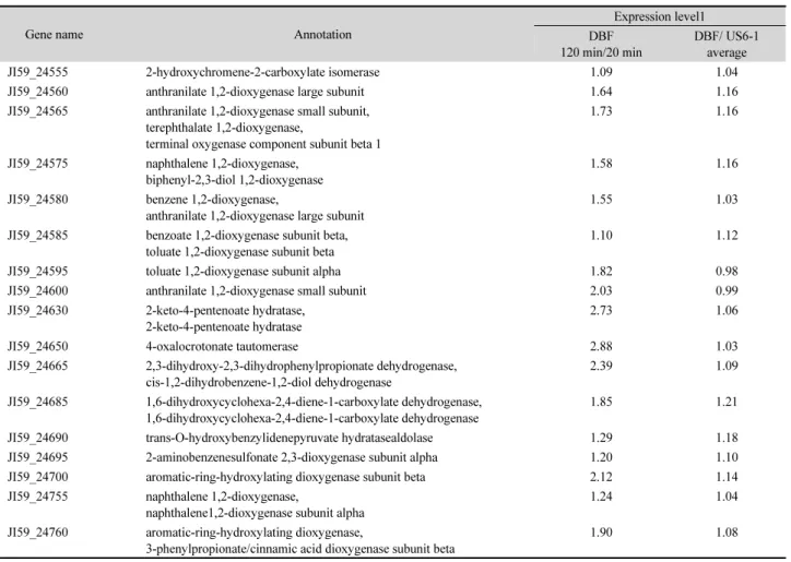
CLRNSeq™ (ChunLab) 프로그램을 이용하여 RNA 염기서 열분석(RNA-Sequencing) 결과를 분석하였다. DBF 분해과정 의 하부경로(lower pathway)는 페난트렌(phenanthrene) 분해 과정(Yun et al., 2014)과 동일하기 때문에 상부 경로(upper pathway)에 초점을 두고 분석하였다. KEGG browser에 올라있 는 다이옥신 분해 경로(dioxin degradation pathway)는 꼭짓점 분 해 경로에 해당되는데 RNA-Seq으로 확인한 균주 US6-1의 발 현 유전자들과는 일치하지 않았다. 이에 따라 상부 경로 관련 유전자를 찾기 위해서 플라스미드 1 (plasmid 1)에 위치하면 서 방향족 고리 분해에 관련되는 유전자들의 시간과 DBF 첨 가 여부에 따른 발현변화를 중점적으로 분석하였다(Table 1, Fig. 4). 그 결과, 다이벤조퓨란이 1,2-다이하이드록시-1,2-다이하
이드로벤조퓨란(1,2-dihydroxy-1,2-dihydrobenzofuran)으로 전 환되는 과정에는 나프탈렌 이산소화효소(naphthalene dioxy- genase) 로 추정된 JI59_24575와 JI59_24755가 작용하며 1,2- 다이하이드록시-1,2-다이하이드로벤조퓨란(1,2-dihydroxy- 1,2-dihydrobenzofuran)이 2-옥소-4-(3'-하이드록시벤조퓨란
Fig. 3. GC-MS analysis of the DBF metabolites by strain US6-1. 3(2H)-benzofuranone was detected only in the 19 h culture broth.
Fig. 4. The expression of genes encoded in plasmid 1 during DBF degradation by strain US6-1 (DBF, cultivation with DBF; US6-1, cultivation without DBF, front number [20, 60, and 120] means cultivation time [min]).
-2'-일)-뷰트-3-엔산(2-oxo-4-(3'-hydroxybenzofuran-2'-yl)- but-3-enoic acid)과 2-하이드록시-4-(3'-옥소-3'H-벤조퓨란-2'- 일리덴)-뷰트-2-엔산(2-hydroxy-4-(3'-oxo-3'H-benzofuran- 2'-yliden)-but-2-enoic acid) 으로 전환될 때는 바이페닐-2,3-다 이올 1,2-이산소화 효소(biphenyl-2,3-diol 1,2-dioxygenase) 로 추정된 JI59_24575와 2-하이드록시크로멘-2-카복실산 이성화 효소(2-hydroxychromene-2-carboxylate isomerase) 로 추정된 JI59_24555 유전자가 관여한 것으로 판단하였다 (Mohammadi and Sylvestre, 2005). 다음 분해 단계에서는 살리 실알데하이드 탈수소화 효소(salicylaldehyde dehydrogenase) (JI59_05885)에 의해 살리실산염(salicylate)으로 전환되는데 이 유전자는 플라스미드 1에 존재하지 않고 염색체에 존재하 였다. 전환된 살리실산염(salicylate)은 다시 두 개의 소단위체 (subunit)로 이루어진 살리실산염 1-수산화 효소(salicylate 1- hydroxylase)에 의해 카테콜(catechol)로 전환되는데 유전자 JI59_24700 은 20분 대비 120분 배양 후에 약 2배 정도 발현량 이 증가하였으며 정렬검색(basic local alignment search tool,
BLAST) 결과, 방향족-고리-수산화 이산소화 효소(aromatic- ring-hydroxylating dioxygenase)의 베타 소단위체(beta subunit) 로 추정되었다. 이에 따라 2-아미노벤젠설폰산 2,3-이산소화 효 소 알파 소단위체(2-aminobenzenesulfonate 2,3-dioxygenase alpha subunit)로 추정된 바로 앞의 유전자(JI59_24695)가 실 제로는 살리실산염 1-수산화 효소 알파 소단위체(salicylate 1-hydroxylase alpha subunit) 일 것으로 판단하였다(Fig. 4).
결 론
PAHs의 분해균주로 알려진 Novosphingobium pentaroma- tivorans US6-1 은 다이벤조-파라-다이옥신(dibenzo-p-dioxin, DD) 과 다이벤조퓨란(dibenzofuran, DBF)의 우수분해균주인 Sphingomonas wittichii RW1와는 달리 초기부터 DBF를 분해 하였다(Fig. 1). 또한 시간이 흐름에 따라 배양액의 색깔이 옅 은 노란색에서 붉은색으로 변하였는데 이는 DBF가 측면 이산
Table 1. Genes in plasmid 1 involved in aromatic ring degradation which is identified by RNA-Seq analysis and its relative expression level
Gene name Annotation
Expression level1 DBF
120 min/20 min
DBF/ US6-1 average
JI59_24555 2-hydroxychromene-2-carboxylate isomerase 1.09 1.04
JI59_24560 anthranilate 1,2-dioxygenase large subunit 1.64 1.16
JI59_24565 anthranilate 1,2-dioxygenase small subunit, terephthalate 1,2-dioxygenase,
terminal oxygenase component subunit beta 1
1.73 1.16
JI59_24575 naphthalene 1,2-dioxygenase, biphenyl-2,3-diol 1,2-dioxygenase
1.58 1.16
JI59_24580 benzene 1,2-dioxygenase,
anthranilate 1,2-dioxygenase large subunit
1.55 1.03
JI59_24585 benzoate 1,2-dioxygenase subunit beta, toluate 1,2-dioxygenase subunit beta
1.10 1.12
JI59_24595 toluate 1,2-dioxygenase subunit alpha 1.82 0.98
JI59_24600 anthranilate 1,2-dioxygenase small subunit 2.03 0.99
JI59_24630 2-keto-4-pentenoate hydratase, 2-keto-4-pentenoate hydratase
2.73 1.06
JI59_24650 4-oxalocrotonate tautomerase 2.88 1.03
JI59_24665 2,3-dihydroxy-2,3-dihydrophenylpropionate dehydrogenase, cis-1,2-dihydrobenzene-1,2-diol dehydrogenase
2.39 1.09
JI59_24685 1,6-dihydroxycyclohexa-2,4-diene-1-carboxylate dehydrogenase, 1,6-dihydroxycyclohexa-2,4-diene-1-carboxylate dehydrogenase
1.85 1.21
JI59_24690 trans-O-hydroxybenzylidenepyruvate hydratasealdolase 1.29 1.18
JI59_24695 2-aminobenzenesulfonate 2,3-dioxygenase subunit alpha 1.20 1.10
JI59_24700 aromatic-ring-hydroxylating dioxygenase subunit beta 2.12 1.14
JI59_24755 naphthalene 1,2-dioxygenase,
naphthalene1,2-dioxygenase subunit alpha
1.24 1.04
JI59_24760 aromatic-ring-hydroxylating dioxygenase,
3-phenylpropionate/cinnamic acid dioxygenase subunit beta
1.90 1.08
소화에 의해 분해되는 과정에서 오렌지색 중간대사산물이 생 산되는 결과로 보인다(Fig. 2)(Hiraishi, 2003). 실제로 측면 분 해(lateral degradation) 반응이 진행될 때 생성될 가능성이 큰 3(2H)- 벤조퓨라논(3(2H)-benzofuranone)이 중간대사산물로 검출됨으로써 균주 US6-1의 DBF 분해가 측면 이산소화에 의해 진행될 가능성을 뒷받침해준다(Fig. 3)(Li et al., 2009). 이때 관여하는 유전자들은 전사체(Transcriptome) 분석 결과로 볼 때 다이벤조퓨란(dibenzofuran)이 JI59_24575, JI59_24755와 JI59_24555에 의해 2-옥소-4-(3'-하이드록시벤조퓨란-2'-일)- 뷰트-3-엔산(2-oxo-4-(3'-hydroxybenzofuran-2'-yl)-but-3- enoic acid)을 거쳐 2-하이드록시-4-(3'-옥소-3'H-벤조퓨란-2'- 일리덴)-뷰트-2-엔산(2-hydroxy-4-(3'-oxo-3'H-benzofuran- 2'-yliden)-but-2-enoic acid)으로 전환되며 JI59_05885에 의해 살리실알데하이드(salicylaldehyde)가 살리실산염(salicylate)이 되고 JI59_24700와 JI59_24695이 작용하여 카테콜(catecho)l로 전환되며 최종적으로 아세트알데하이드(acetaldehyde)와 피 루브산염(pyruvate)로 분해될 것으로 추측된다(Fig. 5).
적 요
다환 방향족 탄화수소(polycyclic aromatic hydrocarbon, PAH) 우수 분해균주인 Novosphingobium pentaromativorans US6-1의 dibenzofuran (DBF) 분해경로를 밝히기 위하여 중간
대사물질 분석과 전사체 분석을 진행하였다. GC/MS로 중간 대사물질을 분석한 결과, 3(2H)-벤조퓨라논이 검출되었는데 이 화합물은 측면 이산소화에 의해 생성된 중간대사산물들의 기본 골격이 되는 물질로써 균주 US6-1에 의한 DBF의 분해 가 측면 이산소화로 진행될 가능성을 시사한다. RNA-Seq 분 석 결과, 균주 US6-1이 DBF에 노출되었을 때 발현되는 유전 자들의 대부분이 lateral dioxygenation과 관련이 있다는 것을 확인하였다. 이상의 결과로부터 N. pentaromativorans US6-1 에 의해 일어나는 측면 이산소화를 통한 DBF 분해경로와 관련 유전자들을 제시하였다.
감사의 말
이 논문은 KIOST (PE99622)와 MOF (PJT200620)의 연구 비 지원을 받았습니다. 초기 연구 진행에 조언을 해주신 김상 진 박사님과 RNA 추출 실험에 큰 도움을 주신 이경원 박사님 께 감사드립니다.
References
Bordajandi, L.R., Gomez, G., Abad, E., Rivera, J., Del Mar Fernandez- Baston, M., Blasco, J., Gonzalez, M.J. 2004. Survey of persistent organochlorine contaminants (PCBs, PCDD/Fs, and PAHs), Fig. 5. Suggested degradation pathway of dibenzofuran (DBF) and related genes in strain US6-1.
heavy metals (Cu, Cd, Zn, Pb, and Hg), and arsenic in food samples from Huelva (Spain): levels and health implications. J.
Agric. Food Chem. 52, 992–1001.
Chai, B., Tsoi, T.V., Iwai, S., Liu, C., Fish, J.A., Gu, C., Johnson, T.A., Zylstra, G., Teppen, B.J., Li, H., et al. 2016. Sphingomonas wittichii strain RW1 genome-wide gene expression shifts in response to dioxins and clay. PLoS One 11, e0157008.
Chang, Y.S. 2008. Recent developments in microbial biotransformation and biodegradation of dioxins. J. Mol. Microbiol. Biotechnol.
15, 152–171.
Choi, D.H., Kwon, Y.M., Kwon, K.K., and Kim, S.J. 2015. Complete genome sequence of Novosphingobium pentaromativorans US6-1T. Stand. Genomic Sci. 10, 107.
Fiedler, H., Abad, E., van Bavel, B., de Boer, J., Bogdal, C., and Malisch, R. 2013. The need for capacity building and first results for the Stockholm Convention Global Monitoring Plan. Trends Anal. Chem. 46, 72–84.
Fortnagel, P., Harms, H., Wittich, R.M., Krohn, S., Meyer, H., Sinnwell, V., Wilkes, H., and Francke, W. 1990. Metabolism of dibenzofuran by Pseudomonas sp. strain HH69 and the mixed culture HH27.
Appl. Environ. Microbiol. 56, 1148–1156.
Fukuda, K., Nagata, S., and Taniguchi, H. 2001. Isolation and charac- terization of dibenzofuran-degrading bacteria. FEMS Microbiol.
Lett. 208, 179–185.
Gai, Z., Yu, B., Li, L., Wang, Y., Ma, C., Feng, J., Deng, Z., and Xu, P. 2007. Cometabolic degradation of dibenzofuran and dibenzo- thiophene by a newly isolated carbazole degrading Sphingomonas sp. strain. Appl. Environ. Microbiol. 73, 2832–2838.
Hay, A. 1979. Dioxin: the 10-year battle that began with agent orange.
Nature 278, 108–109.
Hiraishi, A. 2003. Biodiversity of dioxin-degrading microorganisms and potential utilization in bioremediation. Microbes Environ.
18, 105–125.
Hong, H.B., Nam, I.H., Murugesan, K., Kim, Y.M., and Chang, Y.S.
2004. Biodegradation of dibenzo-p-dioxin, dibenzofuran, and chlorodibenzo-p-dioxins by Pseudomonas veronii PH-03.
Biodegradation 15, 303–313.
Jensen, A.M., Finster, K.W., and Karlson, U. 2003. Degradation of carbazole, dibenzothiophene, and dibenzofuran at low temperature by Pseudomonas sp. strain C3211. Environ. Toxicol. Chem. 22, 730–735.
Jin, S., Zhu, T., Xu, X., and Xu, Y. 2006. Biodegradation of dibenzofuran by Janibacter terrae strain XJ-1. Curr. Microbiol. 53, 30–36.
Le, T.T., Murugesan, K., Nam, I.H, Jeon, J.R., and Chang, Y.S. 2013.
Degradation of dibenzofuran via multiple dioxygenation by a newly isolated Agrobacterium sp. PH-08. J. Appl. Microbiol.
116, 542–553.
Li, Q., Wang, X., Yin, G., Gai, Z., Tang, H., Ma, C., Deng, Z., and Xu, P. 2009. New metabolites in dibenzofuran cometabolic degradation by a biphenyl-cultivated Pseudomonas putida strain B6-2. Environ.
Sci. Technol. 43, 8635–8642.
Lyu, Y., Zheng, W., Zheng, T., and Tian, Y. 2014. Biodegradation
of polycyclic aromatic hydrocarbons by Novosphingobium pentaromativorans US6-1. PLoS One 9, e101438.
Mandal, P. 2005. Dioxin: a review of its environmental effects and its aryl hydrocarbon receptor biology. J. Comp. Physiol. B. 175, 221–230.
Mohammadi, M. and Sylvestre, M. 2005. Resolving the profile of metabolites generated during oxidation of dibenzofuran and chlorodibenzofurans by the biphenyl catabolic pathway enzymes.
Chem. Biol. 7, 835–846.
Monna, L., Omori, T., and Kodama, T. 1993. Microbial degradation of dibenzofuran, fluorene, and dibenzo-p-dioxin by Staphylococcus auriculans DBF63. Appl. Environ. Microbiol. 59, 285–289.
Pollitt, F. 1999. Polychlorinated dibenzo-p-dioxins and polychlorinated dibenzofurans. Regul. Toxicol. Pharm. 30, S63–S68.
Schecter, A., Birnbaum, L., Ryan, J., and Constable, J. 2006. Dioxins:
An overview. Environ. Res. 101, 419–428.
Schmid, A., Rothe, B., Altenbuchner, J., Ludwig, W., and Engesser, K.
1997. Characterization of three distinct extradiol dioxygenases involved in mineralization of dibenzofuran by Terrabacter sp.
strain DPO360. J. Bacteriol. 179, 53–62.
Sohn, J.H., Kwon, K.K., Kang, J.H., Jung, H.B., and Kim, S.J. 2004.
Novosphingobium pentaromativorans sp. nov., a high-molecular- mass polycyclic aromatic hydrocarbon-degrading bacterium isolated from estuarine sediment. Int. J. Syst. Evol. Microbiol.
54, 1483–1487.
Van den Berg, M., De Jongh, J., Poiger, H., and Olson, J.R. 1994. The toxicokinetics and metabolism of polychlorinated dibenzo-p- dioxins (PCDDs) and dibenzofurans (PCDFs) and their relevance for toxicity. Crit. Rev. Toxicol. 24, 1–74.
WHO. 2010. Preventing disease through healthy environments. exposure to dioxins and dioxin-like substances: a major public health concern. World Health Organization. http://www.who.int/ipcs/
features/dioxins.pdf..
Wilkes, H., Wittich, R.M., Timmis, K.N., Fortnagel, P., and Francke, W. 1996. Degradation of chlor-inated dibenzofurans and dibenzo- p-dioxins by Sphingomonas sp. strain RW1. Appl. Environ.
Microbiol. 62, 367–371.
Wittich, R.M., Wilkes, H., Sinnwell, V., Francke, W., and Fortnagel, P. 1992. Metabolism of dibenzo-p-dioxin by Sphingomonas sp.
strain RW1. Appl. Environ. Microbiol. 58, 1005–1010.
Xu, P., Yu, B., Li, F.L., Cai, X.F., and Ma, C.Q. 2006. Microbial degradation of sulfur, mitrogen and oxygen heterocycles. Trends Microbiol. 14, 398–405.
Yun, S.H., Choi, C.W., Lee, Y.G., Kwon, J., Leem, S.H., Chung, Y.H., Kahng, H.Y., Kim, S.J., Kwon, K.K., and Kim, S.I. 2014.
Proteomic characterization of plasmid pLA1 for biodegradation of polycyclic aromatic hydrocarbons in the marine bacterium, Novosphingobium pentaromativorans US6-1. PLoS One 9, e90812.
Zhang, Y., Zhu, Y.X., Kwon, K.K., Park, J.H., and Kim, S.J. 2004.
Novel method for determining pyrene biodegradation using synchronous fluorimetry. Chemosphere 55, 389–394.