마황이 LPS투여 흰쥐의 면역조절능에 미치는 영향
이 은*
상지대학교 보건과학대학 제약공학과
Effect of Ephedrae Herba on Immunomodulatory Activity in Lipopolysaccharide-Exposed Rats and Raw 264.7 Cells
Eun Lee*
Department of pharmaceutical Engineering, College of Health Sciences, Sangji University, Wonju 220-702, Korea
Abstract - To investigate the anti-inflammatory effect of Ephedrae Herba in vivo and in vitro acute inflammation was induced by lipopolysaccharide (LPS) shock in rats fed Ephedrae Herba extracts and inflammatory cytokine concentrations were examined. In addition, the effect of Ephedrae Herba extracts on the production of inflammatory cytokines was examined in LPS-stimulated Raw 264.7 cells. In an in vivo experiment, plasma interleukin-1β (IL-1β), interleukin-6 (IL-6), interleukin-10 (IL-10) and tumor necrosis factor-α (TNF-α) concentrations were increased at 2 h and reached to maximal levels at 5 h after LPS treatment in all groups. Compared with control group, plasma IL-1β, IL-6, and TNF-α levels were lowered at 5 h after LPS treatment, but plasma IL-10 level was higher in at 2 and 5 h after LPS treatment in Ephedrae Herba extract group. In an in vitro experiment using Raw 264.7 macrophages, IL-1β, IL-6 and TNF-α concentrations in the Ephedrae Herba extract group were lower than those in control group. Compared with control group, IL-10 concentration appeared to be higher in the Ephedrae Herba extract group, but this trend was not significant. In conclusion, these results suggested that functional compound (s) in Ephedrae Herba extract may play a role in alleviating inflammatory response.
Key words -Ephedrae Herba extract, Lipopolysaccharide, Anti-inflammatory, Cytokine
*
교신저자(E-mail) : [email protected]서 언
최근 들어 치료가 잘 되지 않거나, 장기간의 치료를 요 하는 염증성 질환들이 많아, 현재 개발된 염증치료제들로 서는 이러한 염증성 질환들을 치료하는데 한계가 있다. 또 한 현재 임상에서 응용되고 있는 염증 치료제들은 부작용 으로 인해 투약기간과 환자의 상태를 고려하여 임상적 대 응을 해야 하는 어려움이 있다. 따라서 전 세계적으로 염증 치료효과를 보다 더 높이고, 부작용이 없는 새로운 염증치 료제의 개발을 위한 많은 연구가 진행되고 있다(Eduard et al., 2004). 그러나 그간의 많은 연구에도 불구하고 만족할 만한 연구결과는 아직 없으며, 보다 더 다양한 분야에서 체 계적인 연구가 필요함을 인식시켜 주었다.
마황은 한방에서 널리 사용하는 약재로 중풍, 상한, 두
통, 발한, 해열, 진해 및 항염증 등에 응용되고 있다(생약 연구회, 1985). 약리적인 면에서는 adrenaline과 유사한 작용을 하는 ephedrine에 의하여 중추흥분작용, 진해작 용, 발한작용, 항염증작용, 이담, 항알르레기 등에 대한 효 과를 나타낸다고 보고되었다(이 등, 1982; 채, 1987; 왕, 1988; 용, 1989; 조, 1989; 여, 1993; 문, 1994). 따라서 본 연구는 새로운 염증치료제를 개발하기 위한 기초연구의 일환으로 마황의 면역조절능을 보다 더 구체적으로 알아보 기 위하여 LPS 처리를 한 흰쥐 및 Raw 264.7 macrophage 에서 마황이 전염증성 cytokines들의 생산에 미치는 영향 을 검토했다.
재료 및 방법
실험동물 및 시험군
평균체중이 218.17±5.94 g의 Sprague-Dawley계 흰
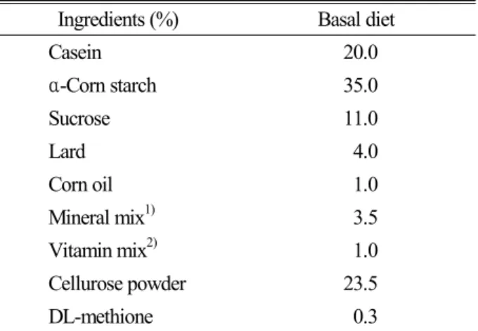
Table 1. Composition of experimental diet Ingredients (%) Basal diet
Casein 20.0
α-Corn starch 35.0
Sucrose 11.0
Lard 4.0
Corn oil 1.0
Mineral mix1) 3.5 Vitamin mix2) 1.0 Cellurose powder 23.5
DL-methione 0.3
1) Mineral mix. (g/kg diet) : CaCO3, 29.29; CaHPO4・2H2O, 0.43;
KH2PO4, 34.30; NaCl, 25.06; MgSO4・7H2O, 9.98; Feric citrate hexahydrate, 0.623; CuSO4・5H2O, 0.516; MnSO4・H2O, 0.121;
ZnCl2, 0.02; KI, 0.005; (NH4)6 MO7O24・4H2O, 0.0025.
2) Vitamin mix. (mg/kg diet) : Thiamine-HCl, 12; Riboflavin, 40;
Pyrodoxin-HCl, 8; Vitamin-B12, 0.005; Ascorbic acid, 300; D- biotin, 0.2; Menadione, 52; Folic acid, 2; D-calcium pantothenate, 50; P-aminobenzoic acid, 50; Nicotinic acid, 60; Cholin choloride, 2000 (IU/kg diet); Rethinyl acetae, 5000 (IU/kg diet); Cholecal- ciferol, 250 (IU/kg diet).
쥐(수컷) 32두를 1주일간 시험식이에 적응시킨 후, 평균체 중이 유사하게 대조군(생리식염수 100 mg/kg), 처리1군(마 황추출액 100 mg/kg), 처리2군(마황추출액 200 mg/kg) 및 처리 3군(마황추출액 300 mg/kg)으로 나누어, 각 처리군 당 8두 씩 임의 배치했다.
식이 및 물
식이(Table 1) 및 물은 시험기간 6주 동안 자유 급여하 였다.
마황추출물 및 급여
시중에서 구입한 양질의 마황 500 g(건조중량)을 적량 으로 나누어 수조상에서 냉각수 환류하에 5시간씩 3회 추출하고, 여과, 감압농축하여 EtOH 추출물 127 g을 만들 었다. 급여는 매일 오후 5시경에 죤대를 이용하여 경구 투 여했다. 대조군은 동일한 방법으로 생리식염수를 급여했다.
Lipopolysaccharide 처리
LPS 처리는 6주간의 사양기간이 종료된 후, 5 mg/kg 의 수준으로 각 처리군 모두 동일하게 복강 주사하였다.
혈액 및 간장 채취
혈액채취는 시험 최종일에 LPS 처리 직전(0h), LPS 처 리 후, 2시간(2h) 및 5시간(5h)째에 각 처리군 별로 심장천 자법에 의해 채혈했다. 간장채취는 LPS 처리 후, 5h에 혈 액채취가 끝난 후 적출했다.
Raw264.7 cells 배양, LPS 및 마황추출물 처리 Raw 264.7 cells은 한국세포주은행(서울)에서 구입하였 으며, Dulbecco’s modified Eagle’s medium(DMEM)에 10% fetal bovine serum(FBS), penicillin(100U/ml) 및 streptomycin(100 μg/ml)이 첨가된 배지를 사용하여 37℃, 5% CO2 incubator에서 배양하였다. 시험과정의 모든 cells 는 80~90%의 confluency에서 실험하였고, 20 passages 를 넘기지 않은 cell만 사용하였다.
LPS shock시 마황 추출물이 Raw 264.7 cells의 전염증 성 cytokines 생산에 미치는 영향을 검토하기 위하여 4 well dish 에 cells을 분주하고(106/ml), 마황 에탄올추출 물을 농도별(0 μg, 10 μg, 30 μg, 100 μg/ml)로 처리한 다음, 1시간 후에 각각 LPS 1 μg/ml를 첨가, LPS shock 후, 6시간째에 시료를 채취하였다.
Cytokines 정량
혈장 cytokine정량용 시료는 채혈 직후, 혈장을 분리하 여 -80℃에 냉동 보관하였다. 간장 cytokine정량용 시료는 1 g의 간장을 채취하여 5 ml의 cold phosphate buffered saline(PBS, pH7.4, containing a protease inhibitors cocktail)과 함께 혼합하여 얼음위에서 분쇄(homogenized) 하였다. 분쇄혼합물을 4℃, 15,000 rpm, 15분간 원심분리 한 후, 상층부를 0.45 ㎛ 필터로 여과하고, 다시 원심분리 해서 상층부를 -80℃에 냉동 보관했다.
Raw 264.7 cells들이 생산한 cytokines 농도측정을 위 해 배양액을 -80℃에 냉동 보관했다. 각종 cytokines(IL -1β, TNF-α, IL-6 및 IL-10)정량은 Kit(Biosource Inter- national, USA)를 이용했다. TNF-α의 최저 측정농도는 0.7 pg/ml이며, 다른 cytokines들은 3-8 pg/ml이다. 간 장 cytokines 정량은 5 ml의 PBS에 생 간장 1g을 혼합한 조정액으로 측정하였으며, pg/mg 단위로 나타내었다.
통계처리
실험결과는 SPSS package를 이용하여 one-way ANOVA 검정을 수행하였으며, 각 처리군 간의 유의성 검정은 Duncan’s
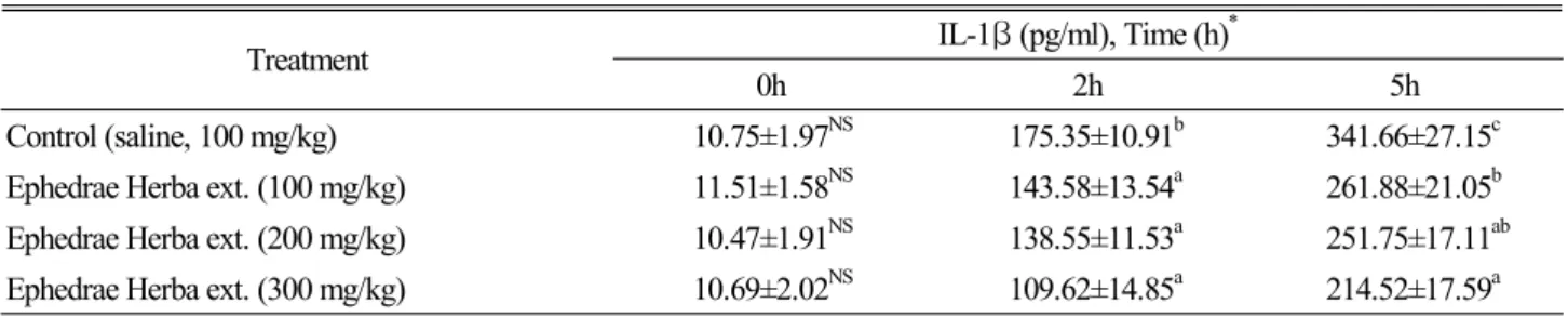
Table 2. Effect of Ephedrae Herba extract on plasma IL-1β concentration in lipopolysaccharide-exposed rats
Treatment IL-1β (pg/ml), Time (h)*
0h 2h 5h
Control (saline, 100 mg/kg) 10.75±1.97NS 175.35±10.91b 341.66±27.15c Ephedrae Herba ext. (100 mg/kg) 11.51±1.58NS 143.58±13.54a 261.88±21.05b Ephedrae Herba ext. (200 mg/kg) 10.47±1.91NS 138.55±11.53a 251.75±17.11ab Ephedrae Herba ext. (300 mg/kg) 10.69±2.02NS 109.62±14.85a 214.52±17.59a
*: 0h, 2h and 5h after LPS injection.
a,b: Means in the same column with different superscripts are significantly different (p<0.05). NS: Not significantly different (P>0.05).
Table 3. Effect of Ephedrae Herba extract on plasma IL-6 concentration in lipopolysaccharide-exposed rats
Treatment IL-6 (pg/ml), Time (h)*
0h 2h 5h
Control (saline, 100 mg/kg) 18.51±4.19NS 271.41±22.69NS 581.17±38.54c Ephedrae Herba ext. (100 mg/kg) 21.79±3.85NS 302.59±27.48NS 511.27±29.58b Ephedrae Herba ext. (200 mg/kg) 19.05±3.55NS 259.83±31.52NS 397.44±30.27a Ephedrae Herba ext. (300 mg/kg) 20.76±3.42NS 278.41±23.85NS 405.49±26.28a
*: 0h, 2h and 5h after LPS injection.
a,b,c: Means in the same column with different superscripts are significantly different (p<0.05). NS: Not significantly different (P>0.05).
multiple range test 에 의해 P<0.05 수준에서 실시했다.
결과 및 고찰
염증반응의 적절한 제어는 질환치료의 기본이며, 수술 환자의 회복속도 및 생존율과 불가분의 관계를 가지고 있 다. 그러나 기존의 항염증제들은 여러 부작용을 나타내어 임상현장에서 제한적으로 사용되고 있다(Eduard et al., 2004). 따라서 항염증효과가 우수하고 부작용이 없는 새로 운 항염증제의 개발이 필요하다. 본 연구는 항염증제의 개 발을 위한 기초연구로, 생약재인 마황의 항염증 효과를 in vivo 및 in vitro에서 검토해 보기위하여 마황 추출물을 급 여한 흰쥐에게 LPS로 급성기 염증반응을 유발시킨 후, 혈 액 및 간장의 전염증성 cytokines들의 농도를 경시적으로 조사하였으며, 한편으로는 Raw 264.7 cell에 LPS shock 를 가한 후, 마황 추출물이 각종 전염증성 cytokines들의 생산량에 미치는 영향을 조사했다.
LPS처리 후, 혈장 및 간장의 cytokine 측정시간은 염증 반응으로 나타나는 각종 cytokines의 농도에 영향을 미친 다. 본 연구에서는 여러 연구자들의 실험결과(Mathiak et al., 2000; Eduard et al., 2004)를 참고하여 흰쥐의 내독
소 쇼크를 검토하기위한 시료채취시간으로 적당하다고 생 각되어, LPS 처리 전(0h), 처리 후 2시간째(2h) 및 5시간 째(5h)에 채혈을 실시하여, 혈장 cytokine을 측정하였으 며, 간장 cytokine측정은 LPS 처리 후 5시간째(5h)에 실 시하였다. 또한 LPS 처리농도도 단시간에 rat와 마우스에 내독소의 쇼크를 주어 간장과 혈액내의 cytokine농도를 높 였다는 다른 연구자의 실험결과를 참고하여, 5 mg/kg 수 준에서 실시했다(Aono et al., 1997; Barton et al., 2001;
Corral et al., 1996; Sang et al., 1999; Harry et al., 1999). 각 처리군 별 plasma IL-1β, IL-6, TNF-α 및 IL-10 농도의 경시적 변동(Table 2,3,4,5)은 전 처리군 모 두가 LPS 처리 후, 2h째 급격하게 증가하여, 5h째에 최고 치를 나타내었다. 이러한 결과는 LPS 처리후 4-6h째에 혈 장 전염증성 cytokines 농도가 최고치였다는 다른 연구결 과(Mathiak et al., 2000)와 유사했다. IL-1β는 생물학 적 기능이 TNF-α와 유사하며, 이들 두 싸이토카인들은 여 러 형태의 실험에서 상호 상가효과를 나타내며, 급성기 염 증반응에서 급격히 증가한다(Mathiak et al., 2000)고 보 고되었다. 본 연구에서 IL-1β의 농도(Table 2)는 LPS 처 리 후, 2h 및 5h에서 마황 처리군 들이 대조군보다 낮은 수 치를 보여, 염증반응이 대조군과 비교하여 완화되었음을
Table 4. Effect of Ephedrae Herba extract on plasma TNF-α concentration in lipopolysaccharide-exposed rats
Treatment TNF-α (pg/ml), Time (h)*
0h 2h 5h
Control (saline, 100 mg/kg) 15.47±3.25NS 595.37±32.98c 647.27±39.16b Ephedrae Herba ext. (100 mg/kg) 13.63±3.44NS 451.22±30.11b 508.32±34.57a Ephedrae Herba ext. (200 mg/kg) 12.71±3.51NS 372.39±31.47a 512.71±28.45a Ephedrae Herba ext. (300 mg/kg) 13.68±3.79NS 411.85±34.17ab 484.75±25.38a
*: 0h, 2h and 5h after LPS injection.
a,b,c: Means in the same column with different superscripts are significantly different (p<0.05). NS: Not significantly different (P>0.05).
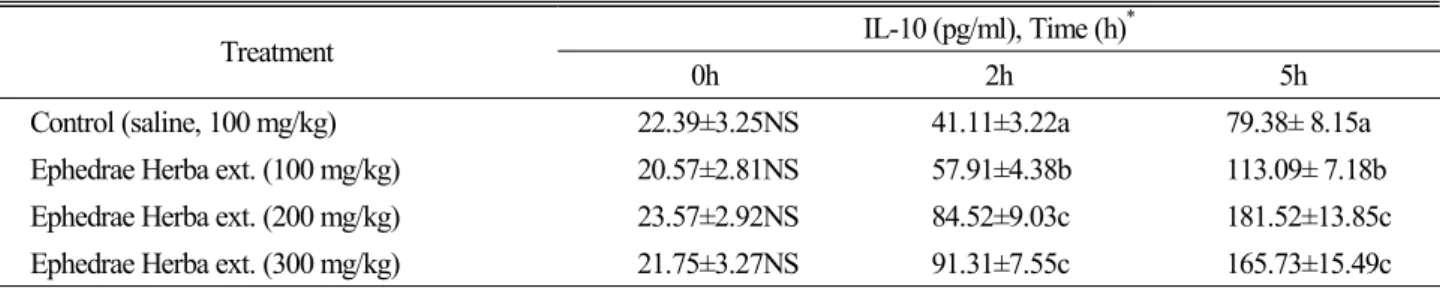
Table 5. Effect of Ephedrae Herba extract on plasma IL-10 concentration in lipopolysaccharide-exposed rats
Treatment IL-10 (pg/ml), Time (h)*
0h 2h 5h
Control (saline, 100 mg/kg) 22.39±3.25NS 41.11±3.22a 79.38± 8.15a Ephedrae Herba ext. (100 mg/kg) 20.57±2.81NS 57.91±4.38b 113.09± 7.18b Ephedrae Herba ext. (200 mg/kg) 23.57±2.92NS 84.52±9.03c 181.52±13.85c Ephedrae Herba ext. (300 mg/kg) 21.75±3.27NS 91.31±7.55c 165.73±15.49c
*: 0h, 2h and 5h after LPS injection.
a,b,c: Means in the same column with different superscripts are significantly different (p<0.05). NS: Not significantly different (P>0.05).
시사해 주었다. Plasma IL-6(Table 3)의 농도는 LPS 처 리 후 2h째에는 전 처리군 간에 유의한 차이를 나타내지 않 았으나, 5h째에는 마황처리군 들이 대조군보다 낮은 값을 나타내었다. 또한 마황 처리군들 간에서는 100 mg/kg 처 리군보다 200 및 300 mg/kg 처리군들이 낮은 값을 나타내 었다. IL-6는 monocytes/macrophages 및 간장의 Kupper cell에서 생산되는 중요한 전염증성 cytokine으로, 염증반 응의 경우 급격하게 증가한다(Eduard et al., 2004). 따라 서 LPS 처리 후 5h째에서 마황처리군들에서의 IL-6의 농 도하락은 마황추출물에 염증반응을 완화시키는 기능성 물 질이 내재하고 있음을 시사해 준다. TNF-α는 LPS를 비롯 한 여러 가지의 자극에 반응하여 monocytes와 macro- phage에 의해 방출되는 peptide mediator이며(Chamuli- trat et al., 1995), endotoxin의 제거효과를 가지는 가장 중요한 mediator로 가정되었다(Harbrecht et al., 1994).
그러나 TNF-α는 LPS의 shock에 의해 Kuffer cell로부터 방출되며, 간장에 상처를 주고 간세포의 사멸을 일으키며 (Hamada et al., 1999), TNF-α의 과잉 생산은 광범위의 병 적상태를 유발한다. 따라서 생체 내에서 효율적으로 TNF-α 의 생산을 억제하는 방법에 대해 연구가 진행되고 있다
(Marriot et al., 1998). 본 연구에서는 plasma TNF-α농 도(Table 4)는 LPS처리 후, 2h째 및 5h째 모두에서 마황추 출물 처리군들이 대조군보다 낮은 값을 나타내었다. 이러한 결과는 IL-1β 및 IL-6 농도의 변동결과와 잘 부합되며, 마 황추출물이 면역조절기능에 관여하고 있음을 시사했다.
Plasma IL-10(Table 5)의 농도는 LPS처리 후, 2h째에 증 가하여 5h째에 최고치를 나타내었다. 각 처리군 별 변동경향 은 LPS처리 후, 2h째 및 5h째 모두에서 마황추출물 첨가군들 이 대조군보다 높은 값을 나타내었다. IL-10은 lymphocytes 와 macrophages에 의해 생산되는 강력한 항염증 cytokine 이다(Thompson et al., 1998). 또한 T helper type 1 cells, mono/macrophages 및 polymorphonuclear cells에 의해서 IL-6 및 TNF-α 등의 전 염증성 싸이토카인들의 합성을 억 제하며, in vitro와 in vivo에서 T-cell활성화를 감소시켰다 (Sang et al., 1999; Moreira., 1993). 본 실험의 결과에서 는 LPS처리 후 2h 및 5h째, 모두에서 마황추출물 처리군 들의 IL-10농도는 대조군보다 높은 값을 나타내었는데, 이 러한 결과가 IL-6 및 TNF-α 농도의 변동경향에 영향을 주 었을 것으로 생각된다.
LPS shock시에 간장의 활성화된 Kuffer cell들은 IL-1
Table 6. Effects of Ephedrae Herba extract on the liver concentration of inflammatory cytokines in lipopolysaccharide-exposed rats Treatment IL-1β (pg/mg) IL-6 (pg/mg) TNF-α (pg/mg) IL-10 (pg/mg) Control (saline, 100 mg/kg) 25.39±3.15b 13.75±1.88c 1.51±0.77NS 1.53±0.77NS Ephedrae Herba ext. (100 mg/kg) 18.75±3.08ab 11.42±1.55bc 1.45±0.83NS 1.91±0.71NS Ephedrae Herba ext. (200 mg/kg) 16.33±3.16a 8.91±1.37ab 1.51±0.94NS 2.13±0.84NS Ephedrae Herba ext. (300 mg/kg) 17.25±3.91a 7.55±1.25a 1.38±0.71NS 2.89±0.92NS
a,b,c: Means in the same column with different superscripts are significantly different (p<0.05). NS: Not significantly different (P>0.05).
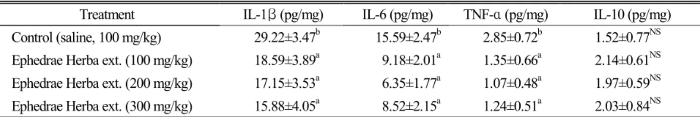
Table 7. Effect of Ephedrae Herba extract on the concentration of inflammatory cytokines in lipopolysaccharide induced Raw 264.7 macrophages
Treatment IL-1β (pg/mg) IL-6 (pg/mg) TNF-α (pg/mg) IL-10 (pg/mg) Control (saline, 100 mg/kg) 29.22±3.47b 15.59±2.47b 2.85±0.72b 1.52±0.77NS Ephedrae Herba ext. (100 mg/kg) 18.59±3.89a 9.18±2.01a 1.35±0.66a 2.14±0.61NS Ephedrae Herba ext. (200 mg/kg) 17.15±3.53a 6.35±1.77a 1.07±0.48a 1.97±0.59NS Ephedrae Herba ext. (300 mg/kg) 15.88±4.05a 8.52±2.15a 1.24±0.51a 2.03±0.84NS
a,b: Means in the same column with different superscripts are significantly different (p<0.05). NS: Not significantly different (P>0.05).
β를 지속적으로 방출하며(Simpson et al., 1997), LPS 처 리 후 30분 이내에 간장 IL-1β 농도는 급격하게 증가한다 고 보고되었다(Sang et al., 1999; Eduard et al., 2004).
또한 Sang et al(1999)은 LPS 처리 후 30분 이내에 IL-6 mRNA가 급격하게 증가하였으며, 3시간 후에도 이러한 현 상은 지속되었다고 보고하였다. 이와 같은 여러 연구 결과 들을 참고해 보면 본 연구에서의 간장 cytokines 측정 시 간은 다소 차이가 있으나, LPS shock가 완전히 회복된 상 태는 아니었음을 짐작하게 한다. 따라서 이와 같은 상태에 서 마황추출물 처리군들의 간장 IL-1β 및 IL-6 농도 (Table 6) 모두가 대조군보다 낮은 값을 보여, 마황추출물 이 간장 IL-1β 및 IL-6 생산을 저해하였음을 시사해 주었다.
TNF-α는 LPS shock에 의해 간장 Kuffer cell로부터 방출되며, 간장 세포의 사멸을 일으키고(Hamada et al., 1999), LPS 처리 후 2시간째에 급격하게 증가하는 것으로 보고되었다(Beutler et al., 1985; Klapproth et al., 1980).
본 연구의 결과에서는 간장 TNF-α농도(Table 6)가 대조 군과 마황추출물 처리군들 간에 유의한 차이를 나타내지 않았다. 이와 같은 결과는 측정시간이 다소 지연되어 간장 에서 생산된 TNF-α가 혈액으로 유입되어 소량으로 잔류 하였는데 기인한 것으로 생각된다.
IL-10은 간장에서 주로 생산되며, 간장에서 IL-10을 생 산하는 주요 세포들은 macrophages, Kupffer cells, T와
B lymphocytes 및 hepatocytes이다(Louis et al., 1997).
특히 설치류에서 IL-10은 LPS처리 시에 Kufer cell에서 TNF-α 및 IL-6의 생산을 억제한다(Simpson et al., 1997;
Thompson et al., 1998). 본 연구의 결과에서는 IL-10의 농도는 대조군과 마황처리군 간에 유의한 차이를 나타내지 않았다(Table 6). 이와 같은 결과는 TNF-α와 마찬가지로 간장에서 생산된 IL-10이 혈류로 유출되고 난 후의 휴지기 상태로, 소량의 cytokines들 만이 잔류하였기 때문인 것으 로 생각된다.
마황추출물이 LPS shock를 준 Raw 264.7 macrophages 의 전염증성 cytokines 생산에 미치는 영향을 Table 7에 나타내었다. IL-1β, IL-6 및 TNF-α의 농도들은 대조군 보다 마황처리군들 모두가 낮은 값을 나타내었다. IL-10 의 농도는 대조군보다 마황처리군 들이 다소 높은 경향을 보였으나 유의한 차이는 아니었다. 이와 같은 결과들은 in vivo 실험에서 나타난 결과들과 잘 부합되었으며, 마황의 염증반응 완화효과를 시사해 주었다.
적 요
in vivo 및 in vitro에서 마황의 항염증효과를 검토하기 위하여 마황추출물을 급여한 흰쥐에게 LPS shock로 급성 기 염증반응을 유발시킨 후, 혈액 및 간장의 전염증성
cytokines들의 농도를 경시적으로 조사하였으며, 한편으 로는 Raw 264.7 cell에 LPS shock를 가한 후, 마황추출 물이 각종 전염증성 cytokines들의 생산량에 미치는 영향 을 조사했다.
in vivo 실험에서, 각 처리군 별 plasma IL-1β, IL-6, TNF-α 및 IL-10 농도의 경시적 변동은 전 처리군 모두가 LPS 처리 후, 2h째 급격하게 증가하여, 5h째에 최고치를 나타내었다. Plasma IL-1β, IL-6 및 TNF-α의 농도는 LPS처리 후 5h째에서 마황 첨가군 모두가 대조군보다 낮 은 값을 나타내었다. Plasma IL-10의 농도는 LPS처리 후, 2h째 및 5h째 모두 에서 마황추출물 첨가군 들이 대조 군보다 높은 값을 나타내었다.
Raw 264.7 macrophages를 이용한 in vitro 실험에서, IL-1β, IL-6 및 TNF-α의 농도들은 대조군보다 마황처리 군들 모두가 낮은 값을 나타내었다. IL-10의 농도는 대조 군보다 마황처리군들이 다소 높은 경향을 보였으나, 유의 한 차이는 아니었다.
이상의 결과들을 종합해보면 마황에 내재하는 기능성 물 질들이 염증반응을 완화하는 효과를 가지고 있음을 시사해 준다.
사 사
본 연구는 2008년도 상지대학교 학술연구비 지원에 의 해 수행되었으며 이에 감사드립니다.
인용문헌
Aono, K., K. Isobe, K. Kuichi, Z. Fan, M. Ito and A. Takeuchi.
1997. In vitro and in vivo expression of inducible nitric oxide synthase during experimental endotoxemia: involvement of other cytokines. J. Cell Biochem. 65:349-358.
Barton, C.C., E.X. Barton, P.E. Ganey, S.L. Kunkel and R.A.
Roth. 2001. Bacterial lipopolysaccharide enhances aflatoxin B1 hepatotoxicity in rats by a mechanism that depends on tumor necrosis factor-α. Hepatology 33:66-73.
Beutler B., B. Greenwald, J.D. Hulmes, M. Chang, Y.C. Pan and J. Mathison. 1985. Identity of tumor necrosis factor and the macrophage secreted factor cachectin. Nature. 316:
552-556.
Chamulitrat, W., M.E. Blazka, S.J. Jordan, M.I. Luster and R.
P. Mason. 1995. Tumor necrosis factor-α and nitric oxide
production in endotoxin-primed rats administered carbon tetrachloride. Life Sci. 24:2273-80.
Corral, L. G., G. W. Muller, A. L. Moreira, X. Chen, M. Wu and D. Stirling. 1996. Selection of novel analogs of thalidomide with enhanced tumor necrosis factor-α inhibitory activity.
Mol med. 25:964-969.
Eduard, F.M., S.M.R. Martha, P.A. Victor and M. Pablo. 2004.
Immunomodulatory effects of thalidomide analogs on LPS- induced plasma and hepatic cytokines in the rat. Biochemical pharmacology 68:1321-1329.
Harbrecht, B.G., M. DiSilvio, A.J. Demetris, R.L. Simmons and T.R. Billiar. 1994. Tumor necrosis factor-α regulates in vivo nitric oxide synthesis and induces liver injury during endotoxemia. Hepatology 20:1055-1060.
Hamada, E., T. Nishida, Y. Uchiyama, J. Nakamura, K . Isihara and H. Kazuo. 1999. Activation of Kupffer cells and caspases-3 involved in rat hepatocyte apoptosis induced by endotoxin. J. Hepatol. 30:807-818.
Harry, D., R. Anand, S. Holt, S. Davies, R. Marley and B.
Fernando. 1999. Increased sensitivity to endotoxemia in the bile duct-ligated cirrhotic rat. Hepatology 30:1198-205.
Klapproth J., T. Geiger, T. Andus and P.C. Heirich. 1980. Fate and biological action of human recombinant interleukin 1β in the rat in vivo. Eur. J. Immunol. 19:1485-1490.
Louis H., O. LeMoine, M.O. Peny, E. Quertinmont, D. Fokan and M. Goldman. 1997. Production and role of interleukin- 10 in concanavalin A- induced hepatitis in mice. Hepatology 25:1382-1389.
Mathiak, G., G. Grass, T. Herzmann, T. Luebke, C. Cu-Zetina and S.A. Boehm. 2000. Capase-1-inhibitor ac-YVAD-cmk reduces LPS-lethality in rats without affecting haematology or cytokine responses. Br. J. Pharmacol. 131:383-386.
Marriot, J.B., M. Westby, S. Cookson, M. Guckian, S. Good- bourn and G. Muller. 1998. CC-3052: a water-soluble analog of thalidomide and potent inhibitor of activation- induced TNF-α production. J. Immunol. 161:4236-4243.
Moreira, A. L., E.P. Sampaio, A. Zmuidzinas, P. Frindt, K. A.
Smith and G. Kaplan. 1993. Thalidomide exerts its inhibitory action on tumor necrosis factor alpha by enhancing mRNA degradation. J. Exp. Med. 177:1675-1680.
Sang, H., G. L. Wallis, C.A. Stewart and K. Yashige. 1999.
Expression of cytokines and activation of transcription factors in lipopolysaccharide-administered rats and their inhibition by phenyl N-tert-butylnitrone (PBN). Arch. Biochem.
Biophys. 363:341-348.
Simpson, K.J., N. W. Lukacs, L. Colletti, R.M. Strieter and S.L.
Kunkel. 1997. Cytokines and the liver. J. Hepatol. 27:
1120-1232.
Thompson, K.C., A. Trowern, A. Fowell, M. Marathe, C.
Haycock and M.J.P. Arthur. 1998. Primary rat and mouse hepatic stellate cells express the macrophage inhibitor cytokine interleukin-10 during the course of activation in vitro.
Hepatology 28:1518-1524.
문관심. 1994. 약초의 성분과 이용. 서울, 일월서각. 124-125.
생약연구회. 1985. 개정판 현대생약학. 한국학습교재사. 292-294.
여명천, 유문발. 1993. 중약적중독여방치. 중광대학출판사. 27-29.
용준충. 1989. 임상중약학. 북경. 인민위생출판사. 109-110.
이상인, 안덕균, 신민교. 1982. 한약임상응용. 서울, 성보사. 44-52.
왕병묵, 왕령. 신농본초경경고증. 길림. 길림과학기술출판사. 316-317.
조강준. 1989. 중초약중독급구, 사천성. 성도전파공정학원출판 사. 274-275.
채인식. 1987. 상한록역전. 서울. 고문사. 298-299.
(접수일 2009.*.*; 수락일 2009.*.*)